LASERS in OMFS
•Als PPTX, PDF herunterladen•
11 gefällt mir•1,399 views

LASER PRINCIPLES, CHARACTERISTICS, CLASSIFICATION AND APPLICATION OF LASERS IN OMFS

Empfohlen
Weitere ähnliche Inhalte
Was ist angesagt?
Was ist angesagt? (20)
Ähnlich wie LASERS in OMFS
Ähnlich wie LASERS in OMFS (20)
Kürzlich hochgeladen
Kürzlich hochgeladen (20)
LASERS in OMFS
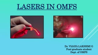
- 1. Dr. VIJAYA LAKSHMI G Post-graduate student Dept. of OMFS LASERS IN OMFS
- 2. • Introduction • Historical background • Components of Lasers • Generation of Laser energy • Mechanism of action of dental Lasers • Classification of Lasers • Laser safety • Clinical Applications • Recent advances • Conclusion • References CONTENTS-
- 3. INTRODUCTION
- 4. “Laser” -“Light Amplification by Stimulated Emission of Radiation” Lasers are heat producing devices converting electromagnetic energy into thermal energy.
- 6. HISTORICAL BACKGROUND Albert Einstein (1917)– Theory of stimulated emission Javan (1961) – HeNe Continuous mode of laser Patel (1964) – CO2 Laser Shfir (1977) – First documented use in OMFS Anderson RR & Parrish JA (1983) – Selective Photothermolysis
- 7. COMPONENTS OF LASER The inner part of laser, or the components of laser, are as follows: (A)Optical cavity B) Two mirrors- one at each end of optical cavity (C) Excitation sources (D) Cooling system (E) Focusing lenses (F) Other controls
- 9. LASER CAVITY/RESONATOR CAVITY Actual optical cavity can be of different shapes and design Cylindrical or rectangle (simplest design) Elliptical– rod and lamp at the ellipse foci Circular– rod and lamp parallel to one other Optical cavity is constructed of a reflecting or scattering material for e.g. ceramic or polished metal. Core of the cavity comprised of chemical elements, molecules or compounds is called active medium.
- 10. ACTIVE MEDIUM - Most lasers are named after the substance that is used to create the actual laser light – LASANT Lasant can be a gas, a crystal or a solid-state semiconductor; Neon gas in He:Ne laser Argon ions in argon laser Gas molecules in CO2 laser Although these characteristic elements or molecules are the main source of laser there may be other ingredients with in the resonator cavity that assist in the alteration of the quantum state of the species. E.g in CO2 laser helium and nitrogen assist in the process of activation and deactivation
- 12. TWO MIRRORS- • One at each end of optical cavity, placed parallel to each other. • One mirror is reflective, which allows photons to be reflected back and forth to allow further stimulated emission. • The other mirror is partially transmissive thus allowing light of sufficient energy to exit the optical cavity.
- 13. The mirrors are separated by a fixed distance (d) forming a Fabry-Perot interferometer. Principle of interference- Two or more waves simultaneously penetrate some material, forms a combined wave. Resulting a larger wave. Constructive interference – when the waves are in phase Destructive interference – when waves not in phase
- 14. Dielectric mirrors: alternative layers of high refractive index materials, such as titanium oxide, and low refractive index material, such as silicon oxide. These materials are deposited on a glass substrate and each layer is designed to be one quarter of the wavelength thick.
- 15. Alternating pattern of high and low RI materials– light waves are reflected from successive surfaces and undergoes phase change equivalent to one half wavelength on reflection from high RI materials, But no phase change occurs on reflection from low RI materials; thus all the reflected rays are interfered constructively, and light in this particular wave length is kept inside the laser chamber and rest leak out of the system. Depending on the number of layers in the mirror – 20 or more layers – the reflectance is 100% for atleast one particular wave length
- 16. EXCITATION SOURCES This applies to the energy or excitation level of a laser medium. The energy possessed by the lasant at a given time is related directly to the application of external or internal energy from a source. E.g. Electrical discharge – CO2 laser, He:Ne, Krypton Chemical reaction – External high powered radiant source as xenon or krypton flash lamp – Nd:YAG, Ruby laser. Alternating magnetic fields – X-ray lasers Excitation sources - either a flash lamp strobe device or an electrical coil, which provides energy into active medium;
- 17. This causes a process called OPTICAL PUMPING, in which energy is driven into the resonant chamber. This energy is used to change the energy level or quantum state.
- 18. GENERATION OF LASER ENERGY- Certain laser medium or LASANT with in the resonator space is energized by internal or external energy to produce an excited population of atoms, molecules and rare gases (SPECIES). The energy with in the resonator reaches a population inversion in which the greatest cohort of species is in an excited state and in which photos are emitted and amplified within a laser cavity. The radiant energy is released as a laser beam.
- 19. MECHANISM OF DENTAL LASER ACTION • Laser light • Amplification • Stimulated Emission • Radiation • Tissue interaction
- 20. LASER LIGHT Distinctive features laser light are- 1. Monochromatic- one specific colour, thus of a single wavelength 2. Coherent- each wavelength is identical in physical size and shapes 3. Collimate- photons can be collimated into an intensely focused energy beam that interacts with the target tissues
- 21. AMPLIFICATION As the photons are produced, they continue to travel within the laser chamber, exciting more atoms, which then produce additional photons. The intensity of energy increases within the laser chamber as the chain reaction continues. This is called amplification of the laser beam. When the laser cavity is opened, a beam of light of a singular wavelength is released from the chamber and delivered as laser beam.
- 22. The smallest unit of energy is absorbed by the electrons of an atom or molecule (of the active medium), creating a short excitation; then a quantum is released, a process called spontaneous emission. The mirrors at each end of the active medium return the photons back and forth to permit the emission of the laser beam. STIMULATED EMISSION
- 23. Depending on how the laser active medium is energized, the laser photonic emission can occur inherently in a continuous wave (CW) or free-running pulsed (FRP) emission mode. CW means that energy is emitted constantly for as long as the laser is activated. A “gated” or “super-pulsed” laser is a variation of CW.
- 24. The length of each pulse is called “pulse width” or “pulse duration”. On the other hand, FRP is a characteristic seen in lasers whose pulses have peak powers in the 1000w range.
- 25. It refers to the light waves produced by laser as a specific form of electromagnetic energy. The very short wavelength below approximately 300nm is called ionizing. Non ionizing radiations are those with wavelengths larger than 300nm and they have lesser frequency and less photon energy. They cause excitation and heating of tissues with which they interact. All dental lasers are non-ionizing. RADIATION
- 26. NON - IONISINGIONISING The wavelength used in medicine and dentistry generally ranges from 193 to 10.600nm, representing a broad spectrum from ultraviolet to the far infra-red range.
- 27. 1. Fiber optic delivery system: use optic strands, by and large made of quartz, 2. Hollow Fiber: Er: YAG and CO 2 lasers utilize a hollow tube with reflective internal walls which transmit laser energy along its internal axis. 3. Articulated arm delivery system: This delivery system utilizes a progression of verbalized mirrors (generally 7) associated one to each other, prompting transmission of vitality. Disadvantage: requires a precise system for alignment of mirrors. 4. Handpieces-close contact and non-contact handpiece TYPES OF LASER LIGHT DELIVERY
- 30. Energy density (Fluency)- Energy density is defined as energy (Joules) per square centimetre of spot size (J/cm2).
- 31. The laser beam spot size can be focused or defocused. Depending on the degree of beam focus, the laser beam spot size can be altered and fluency will accordingly change. Effect of distance of laser beam on the spot-size at target tissue surface Effect of focused and defocused laser beam on target tissue
- 32. The exposure time is the amount of time the operator keeps the laser light directed at the tissue. The time allowed for the energy to be absorbed by the target tissue and dissipate is the thermal relaxation time.
- 33. When the fluence, or the energy given to a particular area , is greater than the tissue vaporisation threshold, and the pulse width is less than the thermal relaxation time of tissue, the tissue is vaporised with each pulse and no significant and no thermal damage occurs beyond the site of laser impact. Selective Photothermolysis This was first theorised by Anderson and Parrisch in 1983.
- 34. The action of lasers on dental tissues and bacteria depends on the absorption of laser by tissue chromophores (water, apatite minerals and various pigmented substances) within the target tissue. The principle action of laser energy on tissue is photo-thermal, and any mechanisms may be secondary to this process or may be totally independent. In general, lasers have four different interactions with the tissues. 1. Photo-thermal interaction 2. Photo-chemical interaction 3. Photo-mechanical interaction 4. Photo-electrical interaction TISSUE INTERACTION
- 37. 1. Photo-thermal interaction- This occurs with high powered lasers. The radiant energy absorbed by tissue substances are transformed into heat energy, which produce the tissue effect.
- 40. 2. Photo-chemical interaction- The basic principle of photochemical process is that specific wavelengths of laser light are absorbed by naturally occurring chromophores, which are able to induce certain biochemical reactions.
- 41. 3. Photo-mechanical interaction- This includes photo-disruption or photo-dissociation and photo-acoustic interactions. In photo-acoustic effects, the pulse of laser energy on the dental tissues can produce a shock wave. When this shock wave explodes the tissue, it creates an abraded crater.
- 42. 4. Photo-electrical interaction– This includes photo-plasmolysis, which describes how the tissue is removed through formation of electrically charged ion.
- 43. Lasers used in dental practice can be classified into several categories according to: (1)the range of wavelength, (2) the lasing medium, such as gas laser and solid laser (3) tissue penetration - soft tissue and hard tissue lasers (4) The risk related to laser application, and (5) potential hazards. CLASSIFICATION OF LASERS
- 44. The most commonly used nowadays are the following.
- 46. Developed by Patel in 1964 Emits light in the invisible mid infrared portion of the spectrum at a wavelength of 10600 nm. Uses a mixture of carbon dioxide, nitrogen, and helium as its medium. Excited by a high-voltage electrical current. Invisible, a red helium-neon laser is often used in parallel, as an aiming beam. Chromophore that absorbs the carbon dioxide wavelength is water The depth of penetration can be as shallow as 0.2 mm, with very little scatter, reflection, or transmission. Used ideally for soft tissue incision and ablation, sub-gingival curettage, superficial lesions and removal of sialoliths. CARBON DIOXIDE LASERS
- 48. ADVANTAGES OF CO2 LASER 1. Sterile surgical field, bactericidal, viricidal 2. Minimal cicatrix formation 3. Access to difficult areas by reflection 4. Ability to coagulate, vaporize and incise 5. Good haemostasis 6. Reduced local tissue trauma and edema 7. Precise delivery of energy to diseased tissue vis microscopes 8. Reduced pain – neuron sealing, decreased pain mediator release. 9. Minimized tumor cell dispersion by lymphatic sealing.
- 49. Erbium Laser - The erbium “family” laser has two wavelengths: • Er: YAG (yttrium aluminium garnet) laser - (2.940nm) • (Er,Cr):YSGG (yttrium scandium gallium garnet) laser - (2.780nm) ERBIUM LASERS
- 50. Erbium: YAG (Er: YAG) laser An active medium of a solid crystal of yttrium aluminium garnet that is doped with erbium. For facial resurfacing Incision and ablation of soft tissue. The presumed advantage of the Er: YAG laser system is its ability to remove superficial skin layers ER: YAG LASERS
- 51. ER, Cr: YSGG LASERS • (Er,Cr):YSGG (yttrium scandium gallium garnet) laser - (2.780nm). • Active medium of a solid crystal of yttrium scandium gallium garnet that is doped with erbium and chromium. • There is absence of melting, charring and carbonization. • Absorption in water is two to three times lower than Er:YAG laser • Thermal effects on the tissue are much higher if not administered correctly. • The erbium wavelengths have a high empathy for hydroxyapatite and the highest absorption of water compared to other dental laser wavelengths. • This is the preferable laser for treatment of dental hard tissue, but also, in contact mode with special surgical tips, it can be used to cut soft tissues.
- 52. Benefits- • Bactericidal effects, which can sterilize the area, • Analgesic effect on the target tissues, similar to the Nd:YAG devices. • Erbium laser energy applied to bone releases growth factors that enhance regeneration of bone. The difference between CO2 and Er:YAG laser lies in their differing absorption coefficients: Er:YAG lasers are much more strongly absorbed in the water. On the other hand, CO2 lasers show very high absorption on the tissue surface.
- 53. Developed by Bridges in 1964. Delivers a green-blue light beam in the 488- or 514-nm range, placed in the visible spectrum. Active medium -argon gas that is energized by a high-current electrical discharge. It is fibre optically delivered with fibre diameter 300μm in continuous wave and gated pulsed modes. Because the argon beam is highly absorbed by hemoglobin and melanin, it has excellent hemostatic capabilities. Neither wavelength is well absorbed in dental hard tissues or in water. ARGON LASERS
- 54. Argon LASER These lasers are useful in the treatment of- • pigmented lesions, • vascular anomalies and • soft tissue incisions and ablations.
- 55. Neodymium: Yttrium -Aluminium-Garnet (Nd: YAG) Geusic and coworkers in 1964, with wavelength of 1064 nm It belongs to invisible near- infrared portion of the electromagnetic spectrum. A flashlamp is used as the energy source to activate Nd: YAG crystal Nd: YAG with a very long pulse duration (90-150µs) penetrates water upto 6mm depth before it is attenuated to 10% of its original strength. Energy is scattered rather than absorbed. Used as a contact laser scalpel or ablation tool, with excellent hemostasis and cutting abilities ND: YAG LASERS
- 56. Excellent for the treatment of- • vascular lesions • intraoral and extraoral pigmented lesions • achieving hemostasis. • open TMJ arthroplasty, • malignant lesion excision, • black and blue tattoo pigment removal, With ND: YAG laser procedures anesthesia is required in less than 50% of cases.
- 57. The wide-spread belief that Nd:YAG lasers have the highest penetration depths in the soft tissue is only partly correct. A study conducted at the RWTH Aachen proved that a free-running pulse Nd:YAG laser has a penetration depth of approximately 0.1mm to 0.3mm, whereas a continuous wave mode Nd:YAG laser has a penetration depth of up to 6mm. State of the art in lasers for dentistry. Gutknecht N. Journal of the laser and health academy Vol.2008;No3/1.
- 58. This laser is a modified version of the Nd: YAG laser. With the addition of a frequency- doubling crystal, this laser emits laser light at the 532-nm wavelength Used in the treatment of vascular and pigmented lesions, tattoo removal, blepharoplasty, and some endoscopic procedures KTP LASER
- 59. • Wavelength range from about 800nm to 980nm, 1-10W power • Solid active medium laser that includes semi-conductor crystals using some combination of aluminium or indium, arsenic and gallium. • The light energy is placed at the starting of the near-infrared portion of the invisible non- ionizing spectrum. • Employs a flexible optic fibre (300μm diameter) to deliver the treatment beam to the desired area. • They run in either CW or pulsed mode. • Diode laser is one of the most versatile with regard to the number of possible treatments options and can be effectively used in the field of soft tissue surgery. DIODE LASER
- 60. In oral surgery, these machines can be used in numerous clinical procedures, • soft tissue surgery, • second stage implant recovery, in peri-implantitis, • sub-gingival curettage etc. ADVANTAGES- • Disinfects the treated area. • Ease of operation, the sub-millimetre dimension and their extreme compactness.
- 61. The holmium: yttrium-aluminum-garnet (Hol:YAG) laser emits laser light at 2140 nm Extensively used in endoscopic orthopedic surgery. It is also extensively used in the TMJ for lysis of adhesions and sculpting of fibrocartilaginous disk tissue. HOL: YAG LASER
- 62. The Q-switched ruby laser produces visible, pulsed red light at the 694-nm wavelength To treat some pigmented lesions and tattoos effectively. The slightly longer wavelength allows for greater depth of penetration and is more effective in the removal of deeper lesions Q-SWITCHED RUBY LASER
- 63. This laser produces yellow visible light in the 400- to 1000-nm range pigmented and hemopigmented lesions, tattoo removal. scar revision, achieving hemostasis, photodynamic cancer therapy, ablation of salivary gland and kidney stones, FLASHLAMP-PUMPED PULSED DYE (FLPPD) LASER
- 64. At wavelengths of 511 and 578 nm, Effective in treating hemangiomas such as port-wine stains or large superficial telangiectasias,. used to ablate some pigmented lesions such as lentigines, ephelides, lentiginous nevi, and tattoos COPPER VAPOR LASER
- 65. These lasers emit ultraviolet light at 193 to 351 nm, Active medium- Halide gas It is currently used for keratotomy to reshape corneal tissues and to correct poor vision. EXCIMER LASER
- 66. Personnel safety Drapes: Not recommended Paper Plastic Recommended Cloth saturated with water around the field Laser resistant drapes for personnel, anesthetic circuit. LASER SAFETY IN SURGERY AND ANESTHESIA
- 67. Field preparation Alcohol as a part field is to be avoided If not the alcohol should vaporize completely before draping. Protection of patient’s throat and delicate oral tissues from accidental beam impact Use of wet gauze packs or towels to avoid reflection from shiny metal surfaces Adequate high speed evacuation should be used to capture laser plume, which is biohazard Specular reflection The surgical beam should be tested for alignment prior to each use of the machine. No instruments are passed across the intended path of laser.
- 68. Speed of movement of the laser beam over the target tissue in order not to occur thermal damage – • Exposure of bone to heating at levels equal to or more than 47°C is reported to include cellular damage leading to osseous resorption.
- 69. Anesthetic agents • Inflammable agents like ether and cyclopropane is absolutely contraindicated in laser surgeries. • Instead halothane, enflurane, isoflurane and sevoflurane • If surgery along the airway – helium and oxygen can be used
- 70. Viral particles • When used to remove viral lesions (warts) the fumes can carry viral particles so proper face mask is a must. • Also the fumes can have carcinogens.
- 71. Eye Retinal damage Even if eyes closed it can penetrate the eye lids Only normal saline is used to lubricate the eye, petroleum based is avoided.
- 72. Skin Avoid alcohol preparation Hairs near the field can ignite. It can be kept moist. Teeth Etching and disfigurement of enamel Dental splints fabricated from laser resistant material.
- 73. Endotracheal tubes Nonmetallic Red rubber, PVC, silicon – red rubber is overall better. Tubes wrapped with metallic foil – mucosal injury Wrapped with metallic tape of copper or silver. Silver anode sheet that has spongy water-absorbant material outside and adhesive inside Ceramic coated endotracheal tube by Xomed (Florida) Metallic Norton and Devos endotracheal tubes , Porch tube are used through oropharynx or trachea. Cuffed metallic tubes are available. Water is injected in to the cuff to inflate
- 74. 1. Post signs that lasers are being used at all possible entry points 2. Eye shields must be worn by all personnel at all times 3. Safety shields must be used 4. A bucket of sterile water should be immediately available in the operating room 5. Credentialing of surgeons for the use of each type of laser and laser apparatus is needed. 6. Short bursts, intermittent lasing, and changing from area of the lesion to other sequentially 7. Cooled irrigation to keep the tissues from heating
- 75. Protocol for treatment of Airway Fire
- 76. The advantages of lasers in comparison to other conventional dental equipment are reported by various authors include: ● Increased coagulation ● Reduction in bacteraemia ● Tissue surface sterilization ● Faster healing response ● Decreased swelling, oedema and scarring ● Reduced pain and discomfort after ● Minimally invasive surgical procedures, compared to conventional techniques; ● Increased patient acceptance ● Reduced surgical time. ADVANTAGES OF LASERS-
- 77. COMPLICATIONS OF LASERS- 1. Herpes Simplex 2. Dyschromias 3. Scarring 4. Eye and Teeth Injuries
- 80. Soft Tissue Clinical Applications- The most popular and effective lasers nowadays for soft tissue procedures are CO2, Nd:YAG and Diode lasers. There are many categories of soft tissue procedures that can be treated by lasers, such as • gingivectomy and gingivoplasty, • frenectomy, • de-epithelialization of reflected flaps, • depigmentation, • second stage exposure of dental implants, • sub-gingival debridement curettage, • incisional and excisional biopsies of both benign and malignant lesions, • removal of granulation tissue, • coagulation of free gingival graft donor site, • irradiation of apthous ulcers, • removal of diseased tissue around the implants etc.
- 81. FACIAL SKIN RESURFACING Indications: 1. Photo damage: Dyschromias & Rhytides 2. Atrophic (depressed) scars : Post acne Chromophore: water Mechanism: Thermal ablation of Epidermis & papillary dermis Lasers a) Single pass CO2 b) Modulated Er : YAG
- 83. PHOTO DAMAGE
- 84. DEPRESSED SCARS
- 85. VASCULAR LESIONS Chromophore– Oxyhaemoglobin Absorption wavelengths – 418, 542, 577 nm Laser of Choice: FPPDL – wavelength – 585, 590, 595, 680 nm Fluence –5-14 J/cm2 Spot Size – 2-10 mm Density – Less than 10% Pulse Duration: 1.5-40 milliseconds Delay between pulses – 10-500 milliseconds
- 88. HYPERTROPHIC SCARS, KELOIDS & STRIAE DISTANSAE FPPDL (585nm) – Laser of Choice Fluence – 3 J/cm2 Spot Size – 10 mm Sessions – 4-6 weekly intervals Atrophic scars : Non-ablative lasers
- 91. NASOLABIAL SCAR
- 92. PIGMENTED LESIONS QS Nd: YAG QS ALEXANDRITE
- 95. TATTOOS 1. Black pigment QS Nd:YAG (1046NM) QS ALEXANDRITE (755 NM Versa pulse coherent) 2. Blue & green pigments QS ALEXANDRITE (755 nm) 3. Red, orange & yellow QS Nd:YAG (532nm) FPPDL (510nm)
- 96. AMATEUR TATTOO
- 99. HAIR REMOVAL Hair follicle thermal relaxation time : 10-100 milli seconds Cooling system: Decreases epidermal injury Lasers & IPL (600-1200nm) QS & LP Nd:YAG (1064 nm) LP Alexandrite (775 nm) Pulsed Diode (800 nm)
- 100. HAIR REMOVAL
- 101. HAIR REMOVAL
- 102. Biopsy margins: An additional 0.5 mm should be added to a normal margin to allow for the lateral zone of necrosis associated with laser action.
- 103. Fibroma of the right cheek (left) and removal of the lesion using Er: YAG laser with X-Runner handpiece (right)
- 104. Fibroma of the hard palate (left) and removal of the lesion using Er: YAG laser with X-Runner handpiece (right) 3 weeks after surgery
- 105. Leukoplakia of the right cheek 3 weeks Post-op
- 106. Leukoplakia of the alveolar ridge (left) and removal using Er: YAG laser 6 weeks Post-op
- 107. The results study confirmed that treatment of leukoplakia by Er: YAG laser had less edema, post- operative bleeding and pain, in comparison to the conventional surgical methods of treatment such as scalpel. The procedure was easily tolerated and postoperative pain was low or absent. Evaluation of Er:YAG laser for surgical treatment of precancerous lesion (leukoplakia) Clinical cases of soft-tissue surgery with X-Runner in QSP mode. Gabrić Pandurić D, Katanec D, Filipović Zore I Journal of the laser and health academy
- 108. Gingival melanin pigmentation 6 weeks Post-op
- 109. Frenectomy
- 110. Exposure of an impacted tooth (soft tissue impaction) can be done using Laser HF, (Gingivectomy mode 975nm, 3W, CW).
- 111. Hard Tissue Clinical Applications • removal of impacted teeth under bone, • apicoectomies, • osseous re-contouring, • implant and bone osteotomies, • bone grafting, • jaw continuity defects, • removal of inflammatory tissues around implants, • crown lengthening, • uncovering of permanent teeth for orthodontic purposes etc. Erbium (Er) family of lasers can be the lasers of choice. Er lasers use extremely short pulse durations and can easily ablate layers of calcified tissue with minimal thermal effects.
- 112. Bone removal prior to apicectomy
- 113. Removal of the cortical plate of the maxilla using Er: YAG laser (X-Runner, QSP mode, 750 mJ, 10 Hz, 10 ml/ min) to show the implant within the sinus and to allow implant removal
- 114. Er:YAG laser usage for preparation of the dental implant site in the lateral part of the right mandible;
- 115. Comparison between Er: YAG laser (lateral implants) and scalpel for second stage surgery 3 days Post-op
- 116. Holmium laser fiber with aluminium case over quartz fiber proved suitable for removal of fragmented synovium within the TMJ. It also replaced cautery instruments used for hemostasis, synovectomy and anterior release procedures.
- 117. Low level laser therapy LLLT works through photo biomodulation, which is based on metabolic activation through through stimulation of the cellular respiratory chain in mitochondria that in turn increases vascularization and enhances the supply of oxygen in hypoxic cells.
- 118. 1. Improvements & Combinations 2. Laser Phototherapy : Vitiligo-eximer laser : 308 nm 3. Non – ablative lasers Nd :YAG (1320 nm) Diode (1450 nm) Er-glass laser (experimental) 4. Optical imaging a) Confocal Scanning Laser Microscope - Diagnosis & Marginal Clearance without biopsy b) Optical Coherence Tomography - Skin Tumors and Bullous disease RECENT ADVANCES
- 119. Photodynamic therapy (PTD) is currently being evaluated for the treatment of head and neck, skin, intra-abdominal, and other types of cancers. carbon dioxide laser and other lasers have also been used in the micro anastomosis of nerve and vascular tissue with some success.
- 121. • Reviewed the effectiveness of PDT in the treatment of early SCC of the head and neck, with similar response rate to surgery. • The treatment is relatively easy to perform, can be repeated and has minimal scarring.
- 122. 65 year old female with T2 SCC of lower lip and vestibule. PDT- 2 days prior to surgery 2mg/kg of porfimer sodium was injected iv and laser at 639 nm wavelength and light dose of 50 J/cm2 was used
- 123. As Dr Theodore Maiman, the inventor of the first laser stated: “The medical application of the laser is fascinating for two reasons. It is optimistic mission on the one hand while, on the other, it counteracts the original impression of the laser being a death ray”. CONCLUSION
- 124. REFERENCES 1. Fonseca, oral and maxillofacial surgery, vol. 1 2. Clinics of North America, LASERS in OMFS, vol.16, May2004 3. Fundamentals of LASER dentistry, Kripa Johar 4. Theodoros Tachmatzidis, Nikolaos Dabarakis ,Technology of Lasers and Their Applications in Oral Surgery: Literature Review, BALKAN JOURNAL OF DENTAL MEDICINE ISSN 2335-0245 5. Dragana Gabrić, Advanced Applications of the Er:YAG Laser in Oral and Maxillofacial Surgery, A Textbook of Advanced Oral and Maxillofacial Surgery Volume 2, chapter 34
- 125. Thank you…..