1. CHAPTER 1
ANALYTICAL CHEMISTRY OVERVIEW
Analytical Chemistry can be defined as the marriage of qualitative and quantitative
analyses, that is, the identification of an analyte (the substance sought for) and then the
determination of the concentration of that analyte. Simply put, qualitative analysis
answers the question “What is in the sample?” and quantitative analysis answers the
question “How much is in the sample?”. Usually, the emphasis in analytical chemistry is
the determination of the quantity of a pure substance in a sample.
Clinical Analytical Chemistry is concerned with the identification and quantitation of
substances related to living organisms, especially human beings. This sub-discipline is
concerned with the sampling and analysis of body fluids, such as urine, blood serum and
blood plasma. Most clinical chemistry laboratories are located in hospitals and medical
centers; although today some are located in stand-alone testing facilities.
Modern physicians are very dependent on the analytical results that are provided by the
clinical laboratory. Health care specialists order many lab tests in an effort to ascertain
what illnesses or conditions exist for any particular patient. The lab tests may also rule
out the presence of certain diseases, and are also used to track the levels of certain
medications in the body. Life-and-death decisions may be based on these clinical
laboratory results. For these reasons, great care must be exercised when taking samples,
preparing the samples, analyzing the samples, and then interpreting the results. This
course will provide a foundation for these very important steps in the clinical analytical
chemistry discipline.
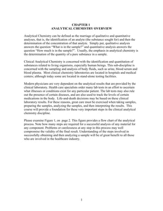
Please examine Figure 1, on page 2. This figure provides a flow chart of the analytical
process. Note how many steps are required for a successful analysis of any material for
any component. Problems or carelessness at any step in this process may well
compromise the validity of the final result. Understanding of the steps involved in
successfully obtaining and then analyzing a sample will be of great benefit to all those
who are involved in the healthcare industry.
1
2. Figure 1: Process Flow for Analytical Chemistry
Collect sample Preserve Deliver Sample to lab Log In
Sample custody
Sample
Sample Preparation Sample Analysis Record Data
Unacceptable? Re-analyze sample
Evaluate Sample Data
Acceptable? Write Lab Report
There are eight main steps in the analytical chemistry process:
1. The sampling step. Before a sample is analyzed, it must be obtained. Selection of an
analytical sample from the gross sample requires that the sample be representative of the
whole sample. Representative means that the portion of the gross sample that is collected
and analyzed should provide the same results as other portions of the gross sample
collected and analyzed. If one is sampling a patient’s urine for protein, for example, the
urine sample must be representative. The physician’s assistants give specific directions
for obtaining such a sample. Some samples are, by their nature, homogeneous, or well-
mixed. Obtaining blood from a living patient usually represents a homogeneous sample.
Some samples may not be homogeneous, and the sampling personnel must take
2
3. additional measures to attempt to achieve this condition. Taking several samples over a
period of time (then mixing) may provide an acceptable degree of homogeneity.
2. Sample Preservation. After a sample is successfully obtained from a source (usually a
living being), the sample must be preserved before delivery to the lab. Because sample
can undergo chemical and even physical changes soon after sampling, steps must be
taken to preserve the sample so that it reaches its destination essentially unchanged.
Preservation may be as simple as refrigeration of the sample, or some preservation agent
may have to be added to the samples just after it is collected (such as acid or base or a
chemical salt).
3. Sample Delivery. Samples must be delivered to the testing facility in an expedient
manner. Many samples, even when preserved, begin to degrade or decay within 24 hours.
Many samples are shipped by Federal Express or some other rapid service.
4. Sample Log-In. Once the sample is received at the testing facility, it must be registered
(logged) as having been received. Sloppy receiving procedures cause samples to be
mixed up or otherwise poorly identified. Incorrect tests may be performed, which may
result in costly re-sampling and re-analyses.
5. Sample Preparation. Once properly received, samples often must be sub-sampled to
obtain a quantity that can be tested in the lab. Most analyses today are accomplished
through the use of sophisticated instrumentation, and only a few milliliters or milligrams
of sample are required. The aliquot (sub-sample) must also be representative.
In the case of blood specimens obtained for clinical tests, a specimen of whole blood is
collected in a glass tube (a vial). When the specimen is allowed to stand for several
minutes, the soluble protein fibrinogen is converted by the coagulation mechanism to
fibrin, which forms a clot that entraps blood cells. After the clot forms, it shrinks and
squeezes out a straw-colored liquid which is known as serum. The serum contains all the
constituents of whole blood except the fibrinogen.
An aliquot of the unclotted whole blood (obtained by the addition of an anticoagulant
[anti-clotting agent] such as heparin) may be used as the sample, or the blood cells may
be separated by centrifugation. The supernatant fluid obtained from centrifugation is
known as the plasma. However, plasma may still contain a small quantity of fibrinogen,
(which could clot) so serum is often used instead.
The preparation step is not always simple. A sample is not usually introduced directly
into an instrument or analyzed as-is. It must often be prepared for the analysis steps. The
analyte of interest must be amenable to the analysis- that is, it must be in a form where it
can be measured. Preparation may be as simple as adjusting the pH of the sample or as
complicated as extracting the analytes of interest through a series of extraction and
separation steps. Today, many of these extractions are automated, so there is not a lot of
intensive hands-on work required. Once the analytes are separated / isolated, the analyte
may require purification so no interferences are present during the analysis. A common
3
4. interference is the presence of serum proteins, which can be removed by precipitation and
filtration. Most of the lab investigations we will perform require a minimum of sample
preparation.
6. The Sample Analysis / Measurement Step. Once the sample is properly prepared,
actual chemical analysis can be performed. This step involves the measurement of some
chemical or physical property of the analyte which can then be transformed into a
quantitative value (concentration). It is most common to measure a chemical property of
the analyte- such as absorbance of some form of energy (atomic absorbance, UV or IR
absorbance), reactivity with certain substances (as in a titration), retention on a separation
column (GC or HPLC), and similar techniques. Most of these techniques yield relative
responses that must be converted to absolute responses. This is done by comparison to
standards, or by calculation of results using stoichiometric factors.
6a. The Calculation Step. The results must be in the correct units, and must be interpreted
with the proper statistical considerations (error range, for example). The accuracy of a
given procedure is determined through the analysis of standards and reference check
samples.
The weakest link in performing calculations deals with the issue of significant figures or
digits. The precision of a result is directly related to the number of significant figures
contained in that result. There are two components to significant figures. The first
component deals with the precision of the instruments used in the analysis. The second
component deals with the actual values themselves. Let’s look at these in more detail.
Precision of instruments. Instruments not only include actual analytical instrumentation,
but also include measuring tools such as balances, glassware, and thermometers. The
following table provides some typical precision values for various instruments:
Instrument typical precision
Platform balance ± 0.5 g
Triple beam balance ± 0.01 g
Top-loading semimicro balance ± 0.001 g
Analytical Balance ± 0.0001 g
10-ML graduated cylinder ± 0.1 mL
100- ML graduated cylinder ± 0.2 mL
25-ML buret ± 0.01 mL
50-ML buret ± 0.02 mL
10-ML pipette ± 0.01 mL
110o C Thermometer ± 0.2 0C
2. Actual values involved in calculations. In general, the significant figures that are
associated with a result are based on the least amount of significant figures associated
with the calculations that lead to that result. These calculations can be based on the
masses of materials, volumes of materials, the instrument readings themselves, and other
factors.
4
5. Guidelines to determining significant figures:
1. Zeros between non-zero digits are always significant. For example, in the number
302045, there are 6 significant figures.
2. Leading zeros (in front of non-zero digits) are never significant. They may be used to
locate a decimal point, but are not significant. For example, in the value 0.000345, there
are only three sig. figs.
3. Trailing zeros may or may not be significant. The value 7000 mL may have four
sig.figs, or may have one sig. fig. if it can be written as 7 x 10³ mL. Some scientists place
a line over the significant zeros- 7000 means there are 4 sig. figs.
4. If a value is greater than 1, then all zeros to the right of the decimal point are
significant. For example, 5.000 contains 4 sig. figs. 4.0605 contains 5 sig. figs.
If the value is less than one, than zeros following nonzero digits are significant. For
example, 0.0034500 contains 5 sig. figs.
5. In the addition or subtraction of a set of values, the number of significant figures is
governed by the value containing the least amount of sig. figs. after the decimal point.
For example, given this series of operations: 101.27 + 2.336 – 10.5 + 0.2973 = 93.4
Three significant figures are determined as the correct precision because of the 10.5
value, which contains 1 sig. fig. after the decimal point.
6. In the multiplication and division of a series of values, the precision of the answer is
determined by the value with the lowest number of significant figures.
For example, in this series of operations: (2.8 * 108.7)/(0.0373 * 5298.3), the correct
result is 1.5 because 2.8 only contains 2 sig. figs.
7. Logarithmic terms: Quantities such as pH should be expressed with the same number
of sig. figs. to the right of the decimal point as the number of sig. figs. of the non-
exponential digits. For example, a solution with a [H+] of 6.6 x 10⁻¹¹ has a pH of 10.18
(2 sig. figs to the right of the decimal point) because 6.6 has 2 sig. figs. If the [H+] is
3.75 x 10⁻⁵ then the pH will be 4.426 (3 sig. figs to the right of the decimal point) because
3.75 has 3 sig. figs.
Conversely, for a solution of pH 10.7, the [H+] = 2 x 10-11 because 10.7 has only one sig.
fig after the decimal point.
7. Data Reporting
What happens after results are obtained and checked? That depends on who needs the
data and in what form they need it. A simple e-mail or fax or phone call may be all that
is required. However, for many institutions and programs, a written report may be
5
6. required, especially if the data were generated as a part of a research project. Whatever
the end use of the data is, it is imperative that everything is properly recorded, so that the
records can be evaluated and the values verified at some later time.
8. The Data Evaluation Step. The data and results should always be reviewed to ensure
that the correct analyses were done, that the calculations are correct, and that the units are
correct. In the event that the data are found to be incorrect, this situation must be quickly
brought to the attention of interested parties, and an effort made to correct the results, or
perform the analysis again. In the classroom, incorrect data may only impact your grade;
in the medical field, it may impact someone’s life.
6
7. CHAPTER 2
STATISTICS AND QUALITY ASSURANCE
Consider these four diagrams:
xx x x
xx
● ●
x x
x
x x x
● ●
x x
x x
What can you say about the accuracy and precision of each of the diagrams, if the center
black dot is the “true” answer? These four diagrams represent various conditions of
accuracy and precision. What types of conditions could have caused these patterns?
1. Determinate (systematic) errors:
a. The cause and magnitude of error can be determined.
b. The errors are consistent and are about the same magnitude.
c. The errors are skewed to one side, either positive or negative.
7
8. d. These errors affect the accuracy of the analysis.
Determinate errors are errors that are repeated, and are caused by a consistent reagent,
instrumental, or operator (analyst) malfunction. These types of errors can be discovered
and corrected. Correction may include the re-making of all reagents and solutions, re-
calibrating all instrumentation, re-training of the analyst, or a combination of these steps.
Indeterminate errors cannot be explained or accounted for. They are random, biased in
various directions, and not repeated often enough for an analysis to be conducted. The
causes may include almost anything, from intermittent operator error, random instrument
malfunctions, causes outside of the laboratory (such as power fluctuations), or local
environmental contamination that cannot be isolated. However, if sufficient analyses are
performed, a statistical expression of the error can be estimated when the analyses are all
performed under the same conditions. Assuming the determinate errors have been
minimized as much as possible (so that the level is near zero), the effects of the
indeterminate errors can be expressed as a Gaussian function mode- that of a bell-shaped
curve. This so-called “normal” distribution is shown below.
Gaussian Distribution
25
20
number
15
10 68.3 &
5
0
1 2 3 4 5 686 7 8 9
10 11
value
The distribution curve can also be viewed as the distribution of the standard deviation of
the population. See graph on next page. The area designated as “68.3 %” represents +/-
1.0 standard deviations from the mean. The area designated as “95.5%” represents +/- 2.0
standard deviations from the mean. The area designated as “99.7%” represents +/- 3.0
standard deviations from the mean. These values represent the probability that all values
within these ranges do belong to the sample population; any values that fall outside these
ranges suggests that the value does NOT belong to the sample population, and should be
rejected. Of course, the more data points that are gathered, the better this estimation will
be.
8
9. Gaussian Distribution
25
20
number
15 68.30%
10
5 95.50%
0 99.70%
1 2 3 4 5 6 7 8 9 10 11
value
In a normal distribution, 68.3 % of the data fall between 0 and ±1 standard deviations
from the mean, 95.5 % of the data fall between 0 and ±2 standard deviations from the
mean, and 99.7 % of the data fall between 0 and ±3 standard deviations from the mean.
Statistics involving sample populations
There are several statistical measures that can be applied to populations, and they are
used analyze the data. They are known as “descriptive statistics”. A sample is a member
of a population- the entire group of possible data points. Samples are discreet sections or
units or pieces of the population. Recall that it is nearly impossible to analyze an entire
population; we must settle for a sufficient number of samples from that population. It
follows that if we can analyze enough samples, our results should follow the normal
distribution shown above.
The first measure of a sample set is the measure of central tendency. There are two ways
to describe the central tendency, the mean and the median. The mean is simply the sum
of all the results (xi) divided by the number of results (n), or
_
Mean = x= Σi xi
n
The median is the value that occurs in the middle of the ordered data set- there are the
same number of values on both sides of the median. If there is an even number of data
points, then the median is the average of the two middle points. It is often more useful to
use the median rather than the mean because it may be a better representation of the
actual central tendency; the median is not affected by very high or very low values that
would influence (skew) the mean in on direction or the other. For example, consider
these values for the mass of a nickel:
9
10. 4.55 g, 5.00 g , 5.10 g, 5.13 g, 5.20 g, 5.24 g, 5.25 g
First, it is helpful to PLOT the data, as a simple frequency distribution, or histogram.
X x xx x xx____
4.00 4.25 4.50 4.75 5.00 5.25
Note the majority of the data are clustered between 5.00 and 5.25.
Then we calculate the mean of this data set, which is 5.07 g. The median value, however,
is 5.13 g. The mean (5.07 g) is influenced by the relatively low value of 4.55 g, whereas
the median, 5.13 g, is a better representation for the majority of the data points. The
larger the number of samples is, the more closely the mean and median should be to each
other.
Another useful statistic is the range of the data. The range is defined simply as the
distance between the highest and lowest values of the data set. The range is calculated by
computing the difference between the highest and lowest values, and expressing the
results without a sign. For the previous data, the range is therefore
(5.25 g – 4.55 g) = 0.70 g
The range is an expression of the spread of the data, which may be used for other
statistical functions that will be discussed later.
Another more useful expression of the spread of the data is represented by the Gaussian
curve- the normal distribution of the data set. The variation of the data is given by the
standard deviation of the mean, and is calculated as follows:
n
s = [ ( Σi=1 (xi – x)2 ) ] 1/2
n-1
xi represents each individual data point and n represents the number of data points in the
series. For the nickel weight data, the standard deviation = 0.244 g . “s” is often referred
to by it’s Greek letter designation, sigma (σ ). As shown on the previous graphs, the
standard deviation can be related to the confidence the user has for any particular data
point. In general, if a data point falls with +/- 3 standard deviations from the mean (3 x
σ), then that value ha a 99.7 % “chance” of being acceptable. Expressed more
statistically, that value most likely belongs to the sample population, and the user can
have a 99.7% level of confidence that is does so.
Clearly, the farther a value is from the mean, the more standard deviations from the mean
it will be (the higher the s value for that particular data point). (Just for information, the
Quality Organization known as Six Sigma is based on the total number of standard
10
11. deviations included in the 99.7 % portion of the data: +3 sigma on one side of the mean
and -3 sigma on the other side, for a total of six sigma.) The values associated with each
sigma level form “confidence levels” for the user at various percents. If a user wants to
have a very tight confidence range, he or she may use the 1 or 2 sigma limits. For most
users, a 2 or 3 sigma confidence limit is acceptable, or even desirable.
Another measure of precision of a data set is the coefficient of variation. The coefficient
of variation (CV) is calculated as follows:
CV = [ σ ] * 100%
x-bar
This statistic is useful in assessing the precision of a test procedure. The CV must be
compared to the mean to determine if the CV is good or bad. A 10% CV associated with
a small mean may suggest poor precision, whereas this CV associated with a large mean
may be quite acceptable. For our nickel example, the CV = 0.244 * 100% or 4.18 %
5.07
4.18% of 5.07 is a fairly small value, but for the US Mint, the tolerances on nickels are
much smaller than +/- 4.18 %. Obviously, the CV value must be interpreted within the
context of the process or piece of equipment.
Another interesting statistic is the mode, which is defined as the value that occurs most
frequently. This also helps to determine the variability of a data set.
Exercises:
1. Students determine the density of an aqueous solution of NaCl. They are to deliver 10
mL with an instrument of their choice into a preweighed beaker, determine it’s mass, and
calculate it’s density. Class A uses a 10 mL graduated cylinder, while Class B uses a 10
ml volumetric pipette. The graph illustrates their results.
The true density is 1.14 g.cm3 .
11
12. The true density is 1.14 g/mL. The class values suggest which of the following is true?
a. Class A achieved greater accuracy and precision than Class B.
b. Class A had greater accuracy; class B had greater precision
c. Class B had greater accuracy; class A had greater precision
d Both classes had substantial systematic errors.
e. Class B had greater accuracy and class A had a greater standard deviation.
Exercise 2
In the lab, you investigate the relationship between the molarity and density of NaCl. 5.0
M NaCl was made up in deionized water, diluted to volume, and 10 mL samples were
weighed to determine density. The following graphs show the results for several students
and also for the true values.
True Values
1.175
1.15
1.125
Density
1.1
1.075
1.05
1.025
1
1 2 3 4 5
M NaCl
Group A
1.175
1.15
1.125
1.1
1.075
1.05
1.025
1
1 2 3 4 5
M Na Cl
Group B
1.175
1.15
1.125
1.1
1.075
1.05
1.025
1
1 2 3 4 5
M N a Cl
12
13. Group C
1.175
1.15
1.125
1.1
1.075
1.05 0
1.025
1
1 2 3 4 5
M N aC l
Match the error ( i, ii, or iii) to the most appropriate graph (A, B, C).
i. A systematic error occurred. Some NaCl solution was lost while transferring it to
the volumetric flask for preparation of the 5 M NaCl stock solution
ii. As far as you can tell, only random error occurred.
iii. A systematic error occurred. The tip of the pipette had a crack and no volume was
retained in the tip after delivery of the samples prior to weighing.
Determination of Outliers
Frequently, when a series of data are obtained for a given process, one or more results
may appear to be markedly different from the main body of data. These data points may
be the result of determinate error. When such an error seems to exist, it would seem wise
to actually assess this possible errant data point to indeed show that it is errant (an outlier)
and should be discarded. As an example, consider again our values for the nickel masses.
The lowest value, 4.55 g seems to be significantly different from the other 6 results. How
can we determine if this result is actually an outlier? One way to assess a data result is by
using the Q test (rejection Quotient), which can be used when the number of results is
fairly small (less than 15). The calculation of the Q value is straightforward:
1. Calculate the difference between the suspected result and the next closest result
(5.00-4.55 = 0.45 in our example)
2. Calculate the range (0.70 in our example).
3. Divide (1) by (2) to determine the Q value. (0.45/0.70 = 0.64 in our example)
4. Compare the result (the “rejection quotient”) to the following table. If the
calculated Q is greater than the value in the table for the number of data points,
then the suspect value can be discarded.
The table for Q values is on the next page.
13
14. Table 1. Rejection Quotient (Q) at a varying confidence levels
Number of results Q90 value Q95 value
4 0.76 0.83
5 0.64 0.71
6 0.56 0.63
7 0.51 0.57
8 0.47 0.53
9 0.44 0.49
10 0.41 0.47
11 0.39 0.45
12 0.37 0.43
13 0.36 0.41
14 0.35 0.39
15 0.34 0.38
So, for our example, the calculated Q value is 0.64. Looking at the Q table for n=7 (90%),
the rejection quotient value is 0.51. Because 0.64 > 0.51, the 4.55 value can be discarded.
Even at the 95% confidence level, Q=0.57, and the value can be rejected.
A second, more robust test is the Student’s t-test. It is used in conjunction with the
standard deviation and the mean of a normal distribution (or a semi-normal distribution)
to construct a confidence interval at a desired percent confidence value (such as 95% or
99%). The suspect result is then compared to the resulting confidence interval, and if it is
outside the confidence interval, it is rejected.
The process for calculating a Student’s t-confidence interval is as follows.
1. Calculate the mean (x) and the standard deviation (s) of the data set.
2. Determine the “degrees of freedom” of the data set, which is defined as one
less than the number of data results, or n-1.
3. Calculate a value known as the “error of the mean”:
error = s/√n
4. Look up the t-value in the Student’s –t table (on next page) at the desired level
of confidence.
5. Calculate the confidence interval as follows:
Confidence interval = x ± (t * error)
The T-table is on the next page.
14
15. Table 2. Student’s T Probabilities
Conf. Level 80% 90% 95% 98% 99% 99.7%
df . . . . . .
1 1.000 3.078 6.314 12.706 31.821 63.657
2 0.816 1.886 2.920 4.303 6.965 9.925
3 0.765 1.638 2.353 3.182 4.541 5.841
4 0.741 1.533 2.132 2.776 3.747 4.604
5 0.727 1.476 2.015 2.571 3.365 4.032
6 0.718 1.440 1.943 2.447 3.143 3.707
7 0.711 1.415 1.895 2.365 2.998 3.499
8 0.706 1.397 1.860 2.306 2.896 3.355
9 0.703 1.383 1.833 2.262 2.821 3.250
10 0.700 1.372 1.812 2.228 2.764 3.169
11 0.697 1.363 1.796 2.201 2.718 3.106
12 0.695 1.356 1.782 2.179 2.681 3.055
13 0.694 1.350 1.771 2.160 2.650 3.012
14 0.692 1.345 1.761 2.145 2.624 2.977
15 0.691 1.341 1.753 2.131 2.602 2.947
16 0.690 1.337 1.746 2.120 2.583 2.921
17 0.689 1.333 1.740 2.110 2.567 2.898
18 0.688 1.330 1.734 2.101 2.552 2.878
19 0.688 1.328 1.729 2.093 2.539 2.861
20 0.687 1.325 1.725 2.086 2.528 2.845
21 0.686 1.323 1.721 2.080 2.518 2.831
22 0.686 1.321 1.717 2.074 2.508 2.819
23 0.685 1.319 1.714 2.069 2.500 2.807
24 0.685 1.318 1.711 2.064 2.492 2.797
25 0.684 1.316 1.708 2.060 2.485 2.787
26 0.684 1.315 1.706 2.056 2.479 2.779
27 0.684 1.314 1.703 2.052 2.473 2.771
28 0.683 1.313 1.701 2.048 2.467 2.763
29 0.683 1.311 1.699 2.045 2.462 2.756
30 0.683 1.310 1.697 2.042 2.457 2.750
40 0.681 1.303 1.684 2.021 2.423 2.704
50 0.679 1.299 1.676 2.009 2.403 2.678
60 0.679 1.296 1.671 2.000 2.390 2.660
70 0.678 1.294 1.667 1.994 2.381 2.648
80 0.678 1.292 1.664 1.990 2.374 2.639
90 0.677 1.291 1.662 1.987 2.368 2.632
100 0.677 1.290 1.660 1.984 2.364 2.626
Using our nickel data again, we will construct a 95% confidence interval to determine if
the 4.55 value is really an outlier.
1. the mean (x) and the standard deviation (s) of the data set: 5.07 g and 0.244.
2. Determine the “degrees of freedom” of the data set, or n-1: 7-1 = 6
3. Calculate a value known as the “error of the mean”:
error = s/√n = (0.244/√7 ) = 0.0922
15
16. 4. the t-value in the Student’s –t table at 95% level of confidence: 1.943
5. Calculate the confidence interval as follows:
Confidence interval = x ± (t * error) = 5.07 ± (1.943 * 0.0922) or
5.07 ± 0.179 g, which forms a confidence interval of 4.89 – 5.25 g
Comparing our low value of 4.55 g to the confidence interval, we see that 4.55 < 4.89, so
we can reject the 4.55 g value. This statistic confirms the Q-test result, which should
confirm our “gut” feeling that the 4.55 g value does not belong in our data set.
The question might arise, When do we use the Q test and when do we use the t-test? The
t-test is better for a larger number of data points, and the Q-test is better for a smaller
number of data points. There is no firm dividing line, but for our purposes, if n < 10, use
the Q-test; if n ≥ 10, use the t-test. Both statistics can be calculated for additional
information and confirmation that an apparent outlier is truly an outlier, and should be
rejected.
SUMMARY: For our nickel mass data, let us summarize what we have learned so far.
Mean = 5.07 g
Median = 5.13 g
Mode= none
Range = 0.70 g
s = 0.244
CV = 4.18 %
Suspect result = 4.55 g
Q-test result: Q4.55 = 0.64 (at a 95% confidence level), which is > 0.51, so 4.55 is an
outlier;
t-test result: for df = 6, confidence interval = 4.89 g – 5.25 g. Therefore, 4.55 is an outlier.
Interpretation of results: We have decided that 4.55 grams is an outlier. Now we have to
ask ourselves, what does this mean? In collecting our data, did we weight the same nickel
on the same balance 7 times? Or did we weight 7 nickels on the same balance? Or did we
weigh the same nickel on 7 different balances? Or did 7 different students weigh their
own nickel on their own balance? You can see that it makes a lot of difference which of
the above questions (possibilities) is actually, in fact, true. What can you conclude about
the possible sources of error for EACH of the 4 possibilities? Which ones make the most
sense?
16
17. IN-CLASS EXERCISE: Statistical Data Set
A student obtained the following values for the analysis of Vitamin C in a serving of
orange juice.
All results are in mg/ 250 mL:
67.2
66.5
63.5
70.0
65.1
64.9
69.2
57.2
65.0
66.8
TRUE VLAUE: 65.2 mg/250 mL
How do we analyze the data?
Plot the data
54 56 58 60 62 64 66 68 70 72
mean
median
17
18. mode
range
standard deviation and CV
“Q” test for outliers
Student’s t-test confidence interval
Which data points, if any are truly outliers and should be rejected?
Percent recovery or percent error
18
19. HOMEWORK DATA EXERCISE:
The following is a sample data set for cholesterol levels in a certain male patient age 53
years. A clinical lab technician analyzed a sample of blood for total cholesterol. She
performed 8 replicates of the analysis, each with a different aliquot of the patient’s blood.
The results are as follows, all in units of mg total cholesterol/ 100 mg plasma.
Replicate value
1. 185
2 201
3 204
4 175
5 192
6 221
7 182
8 175
Calculate the following:
mean
median
mode
standard deviation
CV
range
Are there any outliers? Use the “Q” test to determine if there are. Then check your
answer with the t-test.
Plot the data in the most useful format using Excel.
Is this patient’s cholesterol level within the normal range for a person his age? How did
you determine your answer?
19
20. CHAPTER 3
UNITS OF CONCENTRATION
One of the most important yet most easily forgotten aspects of performing analytical
work is the use of the correct units. Analytical samples can be gases, liquids (solutions),
or solids. The substance that is being sought for via analytical chemistry is called the
analyte. For most analytical procedures, the analyte is dissolved in a large quantity of
another substance, which is often referred to as the matrix. The matrix may be gas, a
liquid, or a solid. The analyte which is dissolved in the matrix is also known as the solute.
The combination of solute and matrix (or solvent) is the solution. In this course, we will
be working mainly with liquid matrices- especially blood plasma or serum and aqueous
(water) solutions.
There are several units of concentration that are important to the analytical professional.
They include the following:
Percent Composition (mass/mass, mass/volume, volume/volume)
Molarity
Normality
Grams/ liter
Parts per million (ppm or mg/L)
Parts per billion (ppb or ug/L)
Percent Composition
a. mass/mass or weight/weight (w/w)
Many times, the user of analytical data will be interested in knowing the analyte
concentration in terms of parts per hundred parts of the solution, which is the same as
percent. In aqueous solutions, percent composition is based on the fact that 1.00 mL of
water exhibits a mass of almost exactly 1.00 grams at temperatures ranging from 4 oC to
30oC. Thus, if we know the mass of an analyte in a given mass of a water-based solution,
we can express the units in percent by mass/mass (mass solute or analyte per mass
solvent). For example, the preparation instructions for sodium chloride might require a
10% solution by mass/mass. This means that for every 100 grams of solution, 10 grams
of salt are dissolved in 90 grams of solvent (water). You would then dissolve 10 grams of
solid, pure NaCl in 90 grams (100 mL) of water. If a liter of solution were required, then
100 grams of NaCl would be dissolved, and brought to a final volume of 1.0 liter with DI
water. Some scientists refer to mass/mass solutions as weight/weight solutions
(abbreviated w/w).
b. mass/volume (or weight/volume, w/v)
The most common method of solution preparation in the clinical field is by mass/volume.
This method of preparation requires a certain mass of solute to be added to the solvent
until a certain final volume is reached. For example, if we were to prepare a 10% by
mass/volume solution of NaCl, we would weigh out 10.0 grams of solid NaCl and add
20
21. enough water to bring the final volume to 100.0 mL using a volumetric flask. This would
then be a 10% (w/v) saline solution.
c. volume/volume (v/v)
Percent by v/v is usually used when one liquid is being dissolved in another liquid, such
as alcohol in water. A 5% solution of ethyl alcohol in water would require that the
clinician measure out exactly 5.0 mL of ethyl alcohol and then placed in enough water to
make exactly 100 mL of solution. Again, the use of a volumetric flask is very helpful in
preparing these types of solutions; one has to only measure out the correct volume of the
solute and place it into the volumetric flask, then carefully add water until the meniscus is
resting on the 100 mL line on the neck of the flask.
meniscus
Molarity
A common unit of concentration used in many analytical laboratories is molarity (M).
Molarity is equivalent to moles (grams solute/molecular or atomic weight of solute) per
one liter of solution. Molarity is almost exclusively used with aqueous solutions. A 1.0
M solution of dried sodium chloride would be prepared by dissolving 58.45 grams of
NaCl in some deionized water and then adjusting the final volume to exactly 1.0 L (in a
volumetric flask).
Normality
Normality (N) is based on the equivalent weight of a species. Equivalents are used to
compare combining ratios of elements or compounds, especially acids and bases.
Electrolyte compounds (salts of sodium, potassium, chloride, and bicarbonate) are often
described by the number of equivalents. An equivalent is most easily defined as the
molecular mass of a substance (usually and acid or a base) divided by the number of
reactable hydrogen or hydroxide units provided by that substance upon reacting with
another species. One gram-equivalent weight is therefore defined as the mass of an
element or compound that is chemically equivalent in reacting power with one gram of a
H+ or (OH-) ion. For salts or other non-acid/base compounds, the gram equivalent weight
is the molecular weight divided by the charge of the ion in question. Here are some
examples:
21
22. Substance Formula Mass Equivalent Mass
HCl 36.45 g 36.45 g (one H+)
NaOH 40.00 g 40.00 g ( one OH-)
H2SO4 98.08 g 49.04 g (2 H+ ions)
H3PO4 98.00 g 32.67 g (3 H+ ions)
NaCl 58.45 g 58.45 g (one Na+1 ion)
Na2SO4 142.00 g 71.00 g (two Na+1 ions)
One gram equivalent weight can represent a fairly large quantity of the compound in
question. In clinical situations, we may prefer to use milliequivalents (mEq), which are
1/1000 of an equivalent. We then would use mg as our mass unit rather than grams. As
an example, the normal concentration of sodium ions in human serum is 140 mEq/L, or
0.140 Eq/L.
The Normality of a solution is defined as one equivalent mass per liter of solution. For
solutions that consist of species with only one reactable H+ , OH- or one ion with a +1
charge, the Normality equals the Molarity. In cases where there are multiple H or OH
ions, or where there are multiple metal ions, or metal ions with a charge greater then +1,
the Normality is most easily calculated by multiplying the Molarity by the number of ions
or the charge. For example, a 2.00 M solution of H2SO4 is also 4.00 N because of the
presence of 2 H+1 ions. A 0.25 M solution of CaCl2 is also 0.500 N because of the
presence of one Ca+2 ion. (Can you prove this?)
Grams per Liter (g/L)
Sometimes it is important to express concentrations as a unit of mass per volume,
especially when there is a substantial quantity of solute dissolved in the solvent.
Environmental chemists often prefer the unit of grams per liter (g/L), or milligrams per
liter (mg/L), or even micrograms per liter (μg/L). In each case, the analyst can have an
immediate idea as to the physical amount of solute in the solvent. In clinical
applications, the solvent is usually human blood or other bodily fluid, such as urine. In
these cases, it is more customary to express the concentration in units of mg/DL, or
milligrams per deciliter (0.1 L). It is then easy to extrapolate to the amount of a species
in the entire body, since the “average” human body contains anywhere from 5.2 – 8.3 DL
of blood/ kg of body weight. In a 70 kg person (male or female), there would be about
470 DL of blood (4.7 L or around 9 pints). Many routine blood test results are reported
in the units of mg/DL, such as blood sugar, creatine, cholesterol, calcium and so on.
Thought Question: How many mg/DL of Chloride ion is represented by a 0.130 M
solution of NaCl ? (Answer given later!)
22
23. Parts per million (ppm), parts per billion (ppb)
These units are derived from the grams/L and milligram/L units just described. Consider
1.0 mg/L. A milligram is 1/000 of one gram, and one liter of water weighs just about
1000 g. Therefore, a milligram of solute in a liter of aqueous solvent represents
(1/(1000)/ 1000) or 1/106 parts of solute per part of solvent, or 1 part per million (ppm).
A microgram/Liter represents 1/109 parts of solute per part of solvent, or one part per
billion (ppb). (Can you prove this?) These units are used mostly for trace analyses in
analytical chemistry and are often used by the Environmental Protection Agency when
they promulgate safe drinking water regulations that specify the maximum allowable
concentrations of potential pollutants.
Other units:
Some other somewhat less common units exist as well. They include:
Molality (mole solute/kg solvent)
Mole fraction (moles solute/total moles of solute + solvent)* 100
SUMMARY: Concentration can be expressed by these units:
Unit Symbol Definition Relationship
Molarity M Moles solute/liter of solution M = moles/ liter
Normality N Equivalents solute/ liter N = equiv./Liter
solution
% by weight/weight % w/w Ratio of the weight of the % w/w = gsolute (* 100%)
solute to the total wt. of solute gsol + gsolv
+ solvent
% by weight/volume % w/v Ratio of wt. of solute to total % w/v = gsolute (* 100%)
volume of solution Vol sol’n
% by volume/volume % v/v Ratio of volume of solute to % v/v = Vsolute (* 100%)
total volume of solution Vsol + Vsolv
Molality M Moles solute/kg solvent M = moles solute/kg solvent
Mole fraction X Ration of moles of solute to X= nsolute
total moles in the solution nsol + nsolv
Parts per million ppm Milligrams solute/ liter of ppm = mg/L or mg/kg or ug/g
solution (or mg solute/kg
sample)
Parts per billion ppb Micrograms solute/liter of ppb = ug/l, ug/kg or ng/g or ng/
solution or mg solute/kg ML
sample
Dilutions:
In analytical chemistry, sample concentrations are often much too high for an instrument
to measure, due to an instruments analytical range, also called the dynamic range or
linear range- the highest value that can be measured. To compensate for this limitation,
samples often have to be diluted many times to reduce the analyte concentration to the
23
24. point where it is within the analytical range of the instrument. This reduction in analyte
level is known as a dilution. Sometimes, dilution may occur as a sample is prepared, but
additional dilutions may still be necessary. Dilutions must be accounted for when
calculating the final result in a sample. If a sample was diluted 10 times, and the final
results for glucose is 9.8 mg/dL, then this value must be multiplied by a factor of 10 to
obtain a sample result of 98 mg/dL.
Dilutions are usually accomplished through the use of volumetric glassware- volumetric
pipettes and volumetric flasks. These items must be clean to insure a correct dilution has
been made. Dilution instructions sometimes may state the parts of solute to be added to
parts of solvent. For example, a ten-fold dilution may be stated as a 1:9 dilution- one part
solute plus 9 parts solvent, (which is a 10 times dilution).
If you have done titrations you are familiar with the formula for calculating the
concentration of an unknown species. The analogous formula can be used to calculate the
concentration of a species when a dilution has occurred, or to calculate how much of a
dilution must be made to achieve a certain concentration when we are interested in
diluting concentrated solutions (such as acids or bases). The general formula is
V a * Ca = V b * Cb
where a is the concentrated solution and b is the diluted solution. V and C are the volume
and concentration of the solutions. For example, we have a solution of 2.0 M sulfuric
acid and we wish to make 500 ML of 0.50 M H2SO4. Using the formula, we can
substitute in the known values for the variables and solve for Va:
Va * 2.0M = 500mL * 0.50 M Va = 125 mL.
Therefore, we would measure out 125 mL of the 2.0 M solution of H2SO4 and dilute it to
500 mL with DI water.
Now, try the following problems for homework:
1. How would you prepare one liter of a 4.000 M solution of ferric sulfate, pentahydrate?
2. How would you prepare a 15 % m/v solution of potassium chloride?
3. How would you prepare 500 mL of a 0.35 M solution of sulfuric acid, given that
concentrated sulfuric acid is 18.0 M? (NOTE: the specific gravity of H2 SO4 is 1.835)
4. If 30 mL of absolute ethyl alcohol are dissolved in 50 mL distilled water, what is the %
concentration of the alcohol, by volume?
24
25. 5. 100 grams of Calcium Hydroxide are dissolved in 2.00 L of deionized water. What is
the Normality (N) of the solution that is formed?
6. A 100 mL sample of liquid waste contains 0.0045 grams of uranium. How many ppm
of uranium are in the solution?
7. 1.0 gram of hazardous waste contains 15 nanograms (ng) of lead. How many ppt does
this represent? How many ppb? How many ppm ?
8. You have 450 mL of 2.5 M nitric acid. How would you prepare 2.0 L of 0.10 M HNO3
?
9. You analyzed a solution for sodium. You had to dilute the original solution by 5 to
digest it, then you took 1.0 mL of that solution and diluted it to 1.0 L. The instrument
result was 1.25 ppm. What is the sodium level in the original solution?
25
26. CHAPTER 4
QUANTITATIVE ANALYSIS – TITRIMETRY
QUANTITATIVE ANALYSIS
Quantitative analysis is concerned not only with the identification of a substance, but the
concentration of that substance. Titrimetry is a useful and fairly simple method of
quantitative analysis. A reactive solution (the titrant) is added to a “sample” from a
buret. A suitable indicator (which provides a visual clue that the reaction between the
titrant and the sample is completed), is added to the sample before the titration is started.
As the titration continues, the reactive species in the titrant reacts with the species of
interest in the sample. The indicator turns a certain color just after the last molecules of
the reactive species in the titrant react with the species of interest in the sample. (The
indicator is not the preferred reactive species in the sample- it only reacts AFTER the
preferred species has been consumed.)
There are four types of reactions that are amenable to Titrimetry.
1. Neutralization Reaction.
Example: Acid + Base Salt + water
HCl + NaOH NaCl + HOH with phenolphthalein as an
indicator
2. Oxidation/ Reduction Titration
Example: the Analysis of Vitamin C (Ascorbic Acid)
Vitamin C – 2D structure
Vitamin C -2D structure – m.p. 190-192 Oc., C6H8O6
Courtesy of UM at Frostburg
26
27. Overall reaction:
I3- + C6H8O6 + H2O ——> C6H8O7 + 3 I- + 2 H+ using starch as the
indicator
After all the ascorbic acid has been consumed (oxidized) by the tri-iodide ion (I3 -1) the
excess iodide ions turn the starch indicator from a cloudy milky white color to a dark
purple color.
3. Complex Ion Formation
Example: EDTA + Ca +2 EDTA/Ca+2 complex, using Eriochrome
Black-T
as an indicator
The Lewis structure of ethylenediamminetetraacetate ion (EDTA4-), is shown below.
EDTA4- forms very stable complexes with most of the transition metals as well as alkali
earth metals, such as Calcium.
EDTA4-
Courtesy of NSTA web site link
One ion of Ca+2 reacts with one molecule of the EDTA, so there is a 1:1 mole ratio
between the Ca+2 ion and the EDTA. As soon as all the Ca+2 is the test solution has been
consumed, the Eriochrome Indicator turns from wine red to a blue color.
Precipitation Reaction
Example: Analysis for Chloride Ion by titration with Silver Nitrate
Cl-1 + AgNO3 AgCl + NO3 -1 using potassium
chromate as
the
indicator
27
28. As AgNO3 reacts with the chloride ion in the flask (sample), solid AgCl is formed. When
all of the Cl-1 ion has been consumed by reacting with the Ag +1 ion, the first excess ions
of Ag +1 then react with the KcrO4 indicator, turning the solution in the flask a very dark
orange-red. Reactions using silver nitrate are known as argentometric reactions.
Titrimetry requires that the amounts of the reacting solution in the buret react with the
analyte in the flask (sample) in a stoichiometric relationship. In a valid titration reaction,
the moles or equivalents of the titrant must be equal to the moles or equivalents of the
analyte. The use of reference solutions (of known composition and concentration) can be
used to simplify the calculations by establishing the ratio between the mass of the analyte
and moles or equivalents of titrant, often expressed by the volume of titrant required to
react with a certain mass of analyte. Once the ratio has been established, a value known
as the titer (spelled “titre” in Europe) can be determined, which is usually in the form of
mg analyte/1.00 mL titrant. The titer can be used as a sort of conversion factor to
determine the analyte concentration in any sample.
When the standard solution and the unknown solution containing the analyte are titrated
with the same titrant under the same laboratory conditions, the calculations become a
simple proportionality:
Vtitrant std = Vtitrant analyte
Cstandard Canalyte
Where V = volume in mL, and C = concentration in the appropriate units. The above
equation can be rearranged as follows:
Canalyte = Vtitrant analyte * Cstandard
Vtitrant standard
Example:
Problem: A sample of human serum is analyzed for chloride content by argentometric
titration. The sample is deproteinized by adding 1.0 mL of the serum sample to a 10 mL
volumetric flask. Trichloracetic acid is added to the flask, up to the 10.0 mL line and
shaken well. The solution is filtered and then exactly 5.0 mL of the filtrate is titrated
with the AgNO3, and it requires 4.98 mL of the titrant to react with all the chloride in the
sample and reach the endpoint. 5.00 mL of a standard chloride solution containing 10.0
mEq/L of requires exactly 5.26 mL of titrant to reach the endpoint. What is the chloride
concentration in the original human serum sample?
1. Determine the titer of the known solution: 10 mEq Cl -1 requires 5.26 mL titrant.
10 mEq Cl/ 5.26 mL = 1.90 mEq Cl/1.00 mL titrant. This is the titer.
2. Multiply the titer by the volume required for the sample:
1.90 mEq Cl * 4.98 mL titrant = 9.47 mEq Cl in the filtrate
1.0 ML titrant
28
29. Since the standard was in units of mEq/L, our result is also in the same units-
9.47 mEq Cl -1/L.
3. Looking at how the sample was prepared, 1 mL was diluted to 10 mL, so there is a 10
fold dilution factor to be considered:
9.47 mEq Cl-1 /L* 10 = 94.7 mEq Cl-1 /L in the serum sample.
(Since 5 .00 mL of chloride ion standard were used to develop the titer, the 5.00 mL of
filtered serum does not enter into the calculations- they effectively cancel out.)
Going on- What is the chloride concentration in units of mg/dL?
Well, we know that for Cl-1, the number of equivalents = moles, since Cl is a monovalent
ion. Therefore, 94.7 mEq/L = 94.7 mmoles/L, or 0.0947 moles of Chloride ion/L.
0.0947 moles Cl-1 /L * 35.5 g Cl-1/ 1.00 mole Cl-1 = 3.357 grams Cl-1 /L are present.
3.357 grams/L = 3357 mg/L, which is equal to 335.7 mg Cl -1/dL. And THAT’S the
final answer!
29
30. SPECTROSCOPY
Many centuries ago, scientists discovered that if sunlight was passed through a prism, the
white light was dispersed into seven colors. The same phenomenon was evident as
sunlight passed through rain drops, forming the rainbow. The reason this happens is due
to the fact that ordinary light is actually a combination of many wavelengths. This led
scientists to conclude that light is a wave. In the 1800’s Sir Maxwell said that light is one
example of electromagnetic radiation- where light consists of packets of energy each
oscillating at specific wavelengths, comparable to an alternating electric field. These
packets of light, called photons by Einstein, propagate through space. Since these
photons do not seem to “run out” of energy, and they move at a constant rate (3.0 * 108
meters/second, denoted by c) early scientists considered light a pure form of energy, and
only energy.
Since the photon oscillates like a sine wave, it has amplitude (height) and frequency
(number of oscillations per unit time, usually seconds). These parameters describe the
wavelength- denoted by the Greek letter lambda λ.
c = υ* λ or υ=c/λ
By the early 1900’s, problems were encountered with the wave theory of light. It could
not explain black body radiation (the emission of heat and /or light from substances that
absorb light energy, or other forms of energy) or the photoelectric effect (the generation
of electricity [the flow of electrons] when a substance is subjected to light), discovered by
Millikan. Max Planck was able to explain and define these phenomena by suggesting
that light also had a particle nature, and experimentally determined that the energy of a
photon is directly proportional to the frequency (υ) of the photon, or
E = hυ
The constant h is Planck’s constant, or 6.626 * 10-34 J-s. We can use this relationship to
determine the energy associated with the light emitted from specific materials, such as the
chemical elements.
Visible light covers the range of 380 nanometers (nm) at the violet end to around 700 nm
at the red end. UV radiation is lower than 380 nm and IR radiation is greater than 700
nm. Spectroscopy in analytical chemistry refers to the measurement of light given off
during specific chemical reactions. The light can be UV, Visible or IR. We have
instruments capable of measuring light at a wide range of wavelengths.
What produces light in chemical species? Simply put, from general chemistry, all atoms
posses a given quantity of electrons dispersed across a number of energy levels, each of
which contains a discreet amount of energy, or quantum. When an atom absorbs energy,
some electrons are excited- that is, they move from one energy level to a higher one- the
ground state to the excited state. However, an electron in an excited state ( in a higher
energy level) is an unstable state, and the electrons return back to their initial, ground
30
31. state. When they do this, they release energy, much of which is in the form of photons-
light- at specific wavelengths. The pattern of wavelengths that are emitted by various
elements are known as the atomic spectrum of the element. These energy levels and
energy releases are not random. They are quite predictable. Electrons must absorb a given
quantity of energy to move from one state to the next, and when they “relax” they give
off a precise, predictable amount of energy. When an electron moves from an excited
state back to the ground state, the energy that was absorbed must be dissipated. In the
usual interaction of light in the visible spectrum (380-700 nm), this energy is usually
dissipated as heat, which is such a small amount it cannot be easily measured. However,
some of energy is dissipated as light, which provides the spectra that we can see for most
elements.
The color that appears is the color that is transmitted from the atoms of the element; the
other colors are absorbed by the atom. Spectroscopy is concerned about the transmission
or absorption of color. The transmission or absorption of specific wavelengths is
measured by a spectrophotometer. Absorption spectroscopy is based on the fact that
chemical species absorb light at various wavelengths that are specific for that species.
The spectrophotometer is capable of generated light that covers a wide range of
wavelengths. It is also capable of measuring absorbance at specific wavelengths.
Essential components of a spectrophotometer include the following:
Source
Slits
Lens
Monochromators
Cells, or other means of sample introduction
Detectors
Read-out devices
Most spectrophotometric techniques produce data that are linear with respect to
absorbance versus concentration, whether that concentration is measured in ppm, ppb,
molarity etc. In the mid 19th century, scientists developed a relationship that allows us to
calculate the concentration of a substance based on its absorbance reading. That
relationship is known as Beer’s Law. The law can be stated as follows:
A = a*b*c
Where A= Absorbance, a = proportionality constant for the species, b = distance light
travels through the sample (called the path), c = concentration.
31
32. When a graph is constructed of the absorbance vs. the concentration of a species, a
straight line is usually generated. The slope of the plot gives the value for b.
The following is an example plot for a chemical species.
Example Calibration Curve (Linear)
0.45
y = 0.0038x + 0.0076
2
R = 0.9998
0.4
0.35
0.3
0.25
ABS
trend
0.2 line
0.15
0.1
0.05
0
0 20 40 60 80 100 120
PPM
This particular graph is called a calibration curve because it plots known concentration
values (on the x-axis) versus the observed absorbance. Note the equation of this plot is
that of a straight line, in the y = mx + b format.
In analytical chemistry, the measurements of the calibration (known or reference)
standards are done under the same conditions as the actual clinical or laboratory samples.
Therefore, the terms of “a” and “b” are the same, and cancel out of the equation used for
calculating the concentration of the unknown sample:
Aref = aref * bref * cref
Aunk aunk * bunk * cunk
The only values that will change, depending on concentrations and subsequent
absorbance, are Aref , Aunk , cref and cunk. As noted above, the “b” and “a” terms all cancel,
because they are not dependent on individual reference or unknown sample
measurements. Therefore, the above equation simplifies to:
Aref = cref
Aunk cunk
32
33. Solving for cunk , we get: cunk = Aunk * cref
Aref
Beer’s Law can only be used when the optimum wavelength is chosen for the species
being measured. At the optimum wavelength, only the species of interest absorbs light;
other species will not absorb light at that wavelength. Once can perform a spectral
absorbance plot to determine what the optimum wavelength is for any particular species.
For example, Fe +2 absorbs best at 510 nm. Modern, automated spectrophotometric
instruments, such as Atomic Absorption and Atomic Emission Spectroscopy can
automatically scan a species to determine the optimum wavelength, and can set the
instrument up to perform the analysis with very little operator.
Molecular and Atomic Spectroscopy
In general, spectroscopy is concerned with the absorption or emission of electromagnetic
radiation by a sample. The molecules or atoms in a particular sample can be excited,
bent, stretched, fluoresced, or be fragmented by the energy source. These various effects
of electromagnetic radiation have all been utilized to develop techniques for qualitative
and quantitative analysis of a wide variety of inorganic and organic species, including
biochemical species of clinical (medical) significance.
Molecular Spectroscopy
There are five commonly utilized methods of analysis that are based on the effects of
certain types of electromagnetic radiation on molecular species. Note that each of the
methods works by causing some physical effect on the molecule which allows us to
detect and quantify it in a sample. In other words, there are no chemical reactions taking
place with molecular spectroscopy (except that of dissolution or complexation); the
actual molecule stays relatively intact throughout the analysis.
Electromagnetic radiation can be broken down into eight distinct regions:
Gamma---- x-rays---- UV--- visible--- IR--- microwave---- radio---- long wave
High frequency low frequency
High energy low energy
Short wavelength long wavelength
The working UV-Visible wavelengths range from 150-750 nm. Working IR wavelengths
range from 2.5 microns to 16 microns (2500 – 16000 nm).
A brief summary of each type of analysis follows.
A. Infra-red spectroscopy
33
34. The absorption of IR light causes vibrational energy transitions in molecules. Specific
molecules absorb only at specific wavelengths, which makes IR an excellent tool for
identifying components of a sample. When the absorbance is plotted against
wavelength, a “molecular fingerprint” is obtained. On the next page, an IR spectrum
in presented for the molecule benzyl alcohol.
IR Spectrum
IR spectrum courtesy of http://chipo.chem.uic.edu/web1/ocol/spec/IRex1.htm
Note the “stretching” associated with the benzene ring (about 3100 cm-1 and near
1500 cm-1 ), the –OH group (3300 cm-1 ), and aliphatic C-H bonds (about
2900 cm-1 ).
B. Mass Spectrometry
Mass Spectroscopy is a technique that is based upon the fragmentation of molecules into
their component atoms or molecular fragments after being subjected to a beam of
electrons. Generally, Mass spectrometry (MS) follows Gas Chromatography, (GC)
which is used to separate a mixture of molecules or components. The effluent from the
GC is fed into the MS, where the individual compounds are fragmented. In reality, then,
the MS serves as a very sensitive and compound-specific detector for GC. The software
for MS contains and extensive library of thousands of organic compounds, which is used
to match the sample mass spectrum. Each molecule has it’s own, specific fragmentation
pattern (a fingerprint) which is reproducible on any MS system. This fingerprint can be
used to identify and quantify the compounds present in liquid, solid, and gaseous
samples. Here is a photograph of a mass spectrometer unit.
34
35. Picture of HP6890 Gas Chromatograph with HP5973 Mass Selective Detector, HP7694 Headspace
Autosampler and thermal conductivity detector.
Gas Chromatography/Mass Spectrometry - GC/MS flowcha–t
35
36. See a sample mass spectrum on the next page.
Courtesy of http://depts.washington.edu/spectral/massspec/GCMSintro/GCMS_4.htmL
Notice the “logical losses” from the molecular ion (at 194 amu) to 165, 136, 109, etc.
C. Nuclear Magnetic Resonance
36
37. Nuclear Magnetic Resonance (NMR) is an analytical technique based on nuclear spin
energy transitions that occur when molecules are subjected to light at radio wavelengths
in a magnetic field. The Nuclei of atoms that are bonded together spin on an axis. Since
the nuclei are positively charged, a small magnetic field surrounds the nuclei. When a
spinning nucleus is subjected to an external magnetic field, the nucleus and it’s small
magnetic field will align with the more powerful field, such as is created with an NMR
instrument. The alignment of the nuclear magnetic field will either be in the same
direction as the NMR field, or in the opposite direction. The difference in energy states
between aligned and opposite nuclear magnetic fields creates a resonance, or the
measurable difference that occurs in the radio wave region of the electromagnetic
spectrum. The light generated in this region can then be absorbed by other molecules in
the magnetic field of the NMR and cause these transitions (directly aligned to oppositely
aligned) to occur. It ha been discovered that hydrogen atoms are best measured by this
technique, and since nearly all organic molecules contain hydrogen, this technique is
useful for many compounds. NMR is more useful for structure determination than for
any quantitative measurements. NMR spectra are based on “chemical shifts”, which
occur because of the effect of electrons that surround nuclei. The electrons shield the
nuclei to measurable extents from the magnetic field, thereby shifting the absorption of
the radio wave light. Different hydrogens in a molecule, therefore, are shielded to
different degrees, and the magnitude of the shift can be used to identify what type of
carbon a hydrogen is bonded to. Below is an NMR spectrum of 2-butanon-4-ene. There
are chemical shifts for the hydrogens bonded to a carbon with a carbonyl group (C=O), to
a carbon double-bonded to another carbon (C=C), and hydrogens bonded to carbons that
are single-bonded to other carbons, as well as terminal carbons (-CH3 ) and internal CH2
groups.
13
C NMR:
courtesy of http://chipo.chem.uic.edu/web1/ocol/spec/C13ex3.htm
D. UV-Visible Spectroscopy.
This technique has been examined previously, so only a very brief review will be
presented here. This technique measures the light absorbed by a compound or
complex. This light can be in the UV or Visible portions of the electromagnetic
spectrum. The degree to which a sample absorbs the light striking the sample is
37
38. measured at the optimum wavelength at which the species under consideration
absorbs light. The amount of absorbance is directly related to the quantity
(concentration) of the absorbing species in the sample, per Beer’s law. If the
optimum wavelength is not known, a scanning UV-Vis instrument can be used to
determine the best wavelength for analytical work.
Below is a graph of absorbance versus wavelength for a solution.
Courtesy of http://www.santafe.cc.fl.us/chemscape/catofp/measurea/concentr/spec20/spec2wq2.htm
For this species, the optimum wavelength (called “lamda-max”) is about 575 nm ( a
nanometer is 10-9 meters), which is in the visible range (yellow light is absorbed, so
the solution will appear violet to the eye).
E. Fluorometry
Again, we have discussed this technique, but a review is in order. Some substances
emit light, or fluoresce, when subjected to energy. This irradiating energy is usually
UV light, and emitted energy is visible light. Both atoms and molecules can absorb
UV light and emit visible light. Emission occurs when a species has absorbed energy
and is excited- raised to a higher energy level. Excited molecules or atoms seek to
return to their ground state as quickly as possible, and do so by various means-
molecular collisions and direct emission of energy, in the form of light. Since there
are competing mechanisms for a molecule to return to the ground state, the light
emitted is at a lower energy than the energy that was absorbed. This results in the
emission of energy at a longer wavelength. The UV-Visible spectrometer measures
the intensity of the emitted light as percent transmittance, which is mathematically
translated to absorbance units, since absorbance is a linear function (% T is a
logarithmic function). This equation is used to convert % T to absorbance (A) is
38
39. A = log (100/ %T) or A= 2 - log %T
As in –v-Visible spectroscopy, a substance can be scanned to determine the optimum
wavelength at which the species fluoresces. The relationship is linear between
absorbance and concentration, according to Beer’s law.
As can be seen, molecular spectroscopy is a valuable tool for analytical chemistry. A vide
variety of organic and ionic analytes can be identified and subsequently, concentrations
can be determined. Limits of detection can be as low as micrograms per liter (ppb) for
many substances. In clinical chemistry, most analyses do not require such low detection
limits- often, milligrams per liter (mg/L or ppm) are sufficient, or even grams per liter are
low enough for some constituents.
Atomic Absorption Spectroscopy
In atomic absorption (AA) spectrophotometry, a liquid sample is aspirated into an air-
acetylene flame whose temperature is approximately 3140-4940°F. The sample is
atomized in the flame. A selected light source emits characteristic frequencies of the
atoms of interest. For example, if testing a sample for calcium (Ca), a Ca hollow cathode
lamp would be necessary. If the sample contained Ca then the atomized Ca atoms would
absorb a portion of the emitted frequencies from the lamp. Absorbance of Ca frequencies
by the sample is indicative of the concentration of Ca within the sample.
The electronic energy level spacings for atoms are very specific for the element. As a
result, the absorption of quantized energy from a monochromatic light beam of the
appropriate wavelength can give selective information about the identity and amount of
elements (normally metals) in a sample.
In AAS, solutions containing metal ions are aspirated into a flame in which they are
converted to a free atom vapor. A monochromatic light source is directed through the
flame, and the amount of radiation of a specific energy is detected. In this way, the
amount of metal present in the original sample can be determined.
Recall that when metallic ions, which are dissolved in an aqueous solution, are subjected
to heat energy in an air-acetylene flame, the atomized ions are raised to a higher energy
state by the absorption of light at specific wavelengths (specific to each element), usually
in the visible range. The AA detects and measures the amount of energy absorbed by the
ions in the flame. The amount of absorption is directly related to the quantity
(concentration) of the ions of that element that are in the solution, again by Beer’s law.
These absorption transitions are very rapid- on the order of 10-15 seconds. And, as soon as
the atom absorbs energy, then emits energy and returns to the ground state, the atom can
once again absorb light energy.
This process is exactly opposite to fluorescence, which is based on the emission of light,
as previously discussed. The fluorescence process takes considerably longer- on the order
of 10-7 seconds.
Graphite furnace AA (GFAA) operates by the same principle, except that instead of using
a flame, an graphite tube with a “shelf” (L’Vov platform) is used. A single drop of the
39
40. sample solution is placed on the platform, which is bathed in an inert gas to prevent
oxidation. The sample drop is dried at around 100 degrees C, then is ashed at around 800
degrees C (to drive off any organic molecules). Finally, the ashed sample is heated to
around 2500 degrees C to atomize the ions, which then absorb light generated by the
hollow cathode lamp. After the absorbance has been measured, the tube is heated to
2700 degrees C, which volatilized any remaining sample material. After the tube cools,
another sample is then analyzed.
Interferences can occur, and are minimized by adding small quantities of matrix
modifiers to bind species which would either absorb at the same wavelength as the
analyte of interest, or would prevent the analyte of interest from absorbing energy at all.
Below is a picture of a state-of-the art GFAA instrument.
courtesy of http://www.enveng.ufl.edu/homepp/townsend/Research/Leach/Leaching_TAG_Meeting_00_07_13/sld118.htm
Below is a close-up of the graphite furnace section of the GFAA.
40
41. A magnetic field is induced, which is used to generate the heat needed to operate the
GFAA.
41
42. Spectroscopy take- home assignment
Name__________________________________
1. A clinical lab technician analyzed the blood taken from a three year old male for the lead
level in his blood. The boy lives in an old inner-city tenement building that was last
painted in 1974. The paint is peeling badly, and it is suspected that the boy is regularly
ingesting a significant amount of lead. The following represent the data from the analysis
of blood for lead by flame atomic absorption spectroscopy (FAAS).
Calibration curve:
ppm lead absorbance, in absorbance units
0.00 (blank) 0.005
0.050 0.028
0.100 0.055
0.250 0.128
0.500 0.219
1.00 0.408
a. Using Excel or your statistical calculator, draw the curve. Determine the line of
best fit and write the equation for the line. What is the slope of the line? R-
squared?
b. The analyst then analyzed five separate aliquots of the boy’s blood, all taken from
one sampling event. The following absorbance readings were obtained (per analysis
of a blood sample initially diluted 1000 times, and then 0.10 mL of that diluted
sample digested and brought to a final volume of 100 mL):
0.345 0.299 0.355 0.350 0.339
What concentrations do these absorbances correspond to?
Perform a statistical evaluation of the five concentration data points (Mean, median,
mode, range, any outliers?).
c. The action level for a 3-year old child is 80 ug lead /0.100 L of blood. Is this child
ingesting too much lead?
42
43. ANALYTICAL SEPARATIONS
In the current world of chemical analysis, analytical scientists are faced with a wide
variety of sample media that require preparation and analysis. The objective of the
scientist is to adequately separate the species/analytes of interest from the sample matrix.
Insufficient separations can cause matrix interference, which can skew (bias) results in
either a positive (high) or negative (low) direction. In some cases, a difficult matrix can
cause data to be completely inscrutable, and therefore, of no value to the scientist.
Samples can be composed of the following, non-inclusive, types of material:
• Blood (serum and plasma)
• Urine
• Fecal matter
• Tissue samples (skin, organ biopsies, bone material, fat, hair…)
• Aqueous (water-based)
• Soil or sediment
• Food matter- milk, meat, starch or vegetable matter, oils…
• Air or gas
Many of these sample media can be contaminated with pathogens, which require special
handling to prevent infection and spread of the pathogen.
There are several basic types of separations that can be accomplished. The first type is
purification. Purification is the process of separating a particular analyte from a sample
matrix by either recrystallization or distillation.
Recrystallization involves the concept of solubility. Certain species will dissolve only in
hot solutions of a particular solvent. Generally, a “rough” separation is accomplished by
isolating the species and some other similar material from a complex matrix. Then, this
material is placed in a particular solvent and heated. The species of interest will dissolve
in the warm or hot solution, whereas the non-wanted material will not. The heated
solution is filtered and the unwanted material is retained, while the dissolved target
material is in the filtrate. The filtrate is cooled, and the target analyte precipitates from
the cool solution, based on the fact that solubility for most solutes decreases with
temperature. This precipitation is the recrystallization process. The solid material then
can be filtered, and the target material is now retained on the filter paper. An example is
the separation of benzoic acid from salt (NaCl). Salt is soluble in cold water to a far
greater degree than benzoic acid. So when a solution containing both materials is cooled
to 4 degrees C, the benzoic acid will recrystallize and the salt will remain in the solution.
After the benzoic acid is filtered, the salt water can be discarded, or the water can be
vaporized to recover the salt. The “crude” benzoic acid is then redissolved in hot water,
and recrystallized a second time, which yields a nearly pure produce.
A second method of purification is distillation, which was the subject and focus of an
earlier laboratory investigation. Distillation is predicated on the fact that various materials
have different vapor pressures and therefore, boiling points. A simple distillation set up
43
44. (such as was done to remove ethyl alcohol from a beverage) is used when there is only
one component that is desired for separation in the material to be analyzed. Another
example of simple distillation involves the purification of hard water. Upon distillation,
“pure” water is produced, and all the inorganic contaminants are left behind in the
distillation vessel. However, if one wishes to separate a mixture of organic solvents,
simple distillation will not be successful. A more complicated distillation method, known
as fractional distillation, is utilized. A fractionating column is placed on top of the
distillation vessel. This column is often packed with glass beads or other non-reactive
material. This packing provides a large surface area for the continuous heating and
condensing of vapors, allowing the operator to adjust the heat to allow a very fine
separation of materials with a difference of boiling points of as little as 2 degrees C. (This
is the theory behind crude oil fractional distillation.)
A second manner in which to accomplish separations in liquid samples is through the use
of liquid-liquid extraction. Other names for this process include solvent extraction or
solvent exchange. The premise behind this manner of separation is the movement
(transfer) of an analyte from one solvent to another solvent in which it is more soluble.
For example, the pesticide DDT is still found in some water supplies. DDT is marginally
soluble in water, but very soluble in methylene chloride (dichloromethane). If a water
sample is mixed with a small volume of methylene chloride, the DDT will be transferred
(extracted) into the organic solvent phase from the water phase. The two liquids are
immiscible, so after the two liquids are mixed well (by utilizing a separatory funnel), the
methylene chloride layer (being more dense than water) settles to the bottom of the
separatory funnel and can be easily removed. The analyte of interest (if present) is now
dissolved in a solvent, which can be analyzed by HPLC or GC-GC/MS for the quantity of
the analyte. Liquid-Liquid extraction is based on the partition coefficient of the system.
The ratio (K) of the concentration of the analyte ([A]) in the extraction solvent is divided
by the concentration of the analyte in the original sample after the extraction process:
[A]extr = K (partition coefficient)
[A]orig
If K is sufficiently large (greater than 100), then it can be assumed that nearly all of the
analyte has been extracted into the solvent, which is then a quantitative transfer. If the K
value is less than 100, a more suitable solvent must be used. If a better solvent is
unavailable, then the K value can be used to calculate how much (in % or by
concentration) analyte will be extracted, and how much will remain in the original
solution. This “correction factor” can then be applied to the final analytical results to
determine actual concentration in the original sample. Many analytical procedures
require the use of special compounds to determine the K of a particular extraction system.
These compounds are known as surrogates, and are chemically similar to target analytes.
If surrogates recover poorly, it is assumed that the target analytes will not recover (be
extracted) well either. (Recovery is defined as [the amount measured in the extract
divided by the amount added before extraction], times 100).
44
45. If solid samples are to be analyzed, an analogous method is used, called liquid-solid
extraction. A solvent is mixed with the solid sample to extract analytes of concern or
interest. This mixing can occur in several ways. One of the oldest ways is through the use
of the separatory funnel, as with liquid-liquid extractions. However, this method often
results in emulsions (a foamy mixture of solvent and sample which will not separate),
which leads to poor recovery of the analytes of interest. An apparatus known as the
Soxhlet extraction apparatus is used for many liquid-solid extractions. For this type of
extraction, a portion of the sample is mixed with a drying agent, such as sodium sulfate,
and is then placed in a rigid thimble made of a special paper/cloth material. The thimble
does not dissolve in the solvent, so the solvent can wash through the thimble, and extract
the analytes of concern.
The solvent vessel is on the bottom, usually placed in a heating mantle. The thimble
containing the sample is placed in the glass holder on top of the extraction vessel (see the
arrow above). On top of the thimble section is a condenser, which prevents the solvent
from escaping the system. As the solvent is heated, it boils and rises into the condenser. It
then condenses and drips through the sample in the thimble, and fills up the glass
enclosure. When the solvent reaches the top of the thimble, it empties into the solvent
vessel through the siphon tube (at the right side of the glass thimble holder). In this way,
the analytes of interest are incrementally extracted into the solvent. After about 10 or 15
wash cycles, the extraction is terminated, and the solvent is then analyzed in the same
manner as the liquid-liquid extraction solvent for the analytes of interest.
45
46. Once the analytes are in the appropriate solvent and/or form for analysis, the proper
method of analysis must be selected. One of the most useful and powerful tools is
chromatography. Chromatography is the separation of components based on the degree
to which different analytes react physically with two separate material phases. Often, one
phase is the “mobile” (moving) phase, and the other is a fixed phase (the “stationary”
phase). The mixture containing the analytes of interest is introduced into the mobile
phase, which moves through or along the stationary phase. Different analytes are
retained on the stationary phase for different lengths of time, which allows separation to
occur. Mobile phases can be liquid or gaseous, and the stationary phase can be liquid or
solid. Techniques utilizing liquid mobile phases are known collectively as Liquid
chromatography (LC), and techniques utilizing a gaseous mobile phase are called gas
chromatography (GC).
Separation can also be accomplished using an electric field. Different ions are attracted to
different degrees in varying directions in an electrical field, and can be separated. This
type of analysis is known as electrophoresis. Positive ions move towards the negatively
charged electrode (cathode), and the negatively charged ions move towards the positively
charged electrode (anode). Obviously, only ions can be separated in this way.
Electrophoresis is very useful in separating amino acids and other charged biomolecules,
such as DNA. Different sized ions and ions of varying charge can be separated even
when they are attracted to the same electrode. The shape of a molecule is also a
determining factor in how far an ion will move towards a charged electrode.
Electrophoresis can be accomplished using solvent-soaked paper, a slab of gel, or a
capillary tube. The carrying (mobile) phase may be organic or inorganic and at a range
of pH’s.
The loading of a gel electrophoresis investigation is shown above.
46
47. The white bands represent DNA of a particular size. The
arrows are included to point out bands that are legitimate, yet
might be overlooked as background noise until you have
looked at enough gels to recognize them. The DNA exists in
equal amounts, but one fragment is larger than the other
• On a molar level, much more DNA of one size is
present in that band than in a different band, although
the lesser amount may be a larger fragment.
http://www.life.uiuc.edu/molbio/geldigest/photo.htmL
An example gel electrophoresis result is shown above.
As can be seen from this brief summary, there is a wide variety of methods than can be
used to separate and then analyze analytes of concern. Much more information on all the
techniques introduced here are available in many textbooks and on the internet.
47
48. CHROMATOGRAPHY
One of the most useful tools that analytical chemists have is the gas chromatograph. The
use of this instrument to identify and quantify a mixture of unknown organic compounds
is known as gas chromatography. The method, known commonly as “GC”, is based on
the varying affinities of organic compounds for the stationary phase of a
chromatographic column (a long, thin metallic tube coated on the inside with a material
for which organic compounds exhibit varying affinities). These varying affinities allow
for separation of individual components, often by their boiling points. The sample is
“injected” into the instrument, and is carried into and through the column by the “carrier
gas” (the “mobile phase”), often helium or nitrogen.
Each compound has a unique affinity for the stationary phase that is reproducible under
the same conditions. The compounds are separated and “elute” from the end of the
column at specific “retention times”, the amount of time that has elapsed from the
moment of injection to when the compound is “detected”- sensed by a detector at the end
of the column. Separation almost always occurs at elevated temperatures, which is why
the column is enclosed in an oven which can be programmed to remain at one particular
temperature (isothermal) or the oven can be programmed to elevate the temperature at
specific rates over a specific period of time. The more complex the mixture, the more
likely the analyst will use a temperature program to attain the best separations possible.
The amount, or volume of sample required for this method is very small- often only 1 or
2 microliters (uL- 10-6 L). Because of the very Small volumes needed, this is an ideal
method for analyzing specimens from crime scenes, or for other forensic and medical
applications.
The output of a GC is in the form of chromatographic peaks-which represent the
presence and detection of vapors other than the carrier gas. These peaks are recorded by
the instrument via a strip-chart recorder, or a software program. This vapor (shown as a
peak) represents a particular compound eluting at a particular retention time at a
particular concentration. Analysts inject known compounds at known concentrations
(the “calibration” of the instrument). Then, after calibration, the sample is injected, and
the sample peaks are compared to the known peaks, and identification and quantitation of
the compound(s) can be accomplished. This is known as the external standard method
of analysis. Most modern GC instruments use a software program that draws the peaks
and identifies the retention time, identification, and concentration for each peak.
For GC, the sample to be injected must either be an organic liquid or in gaseous form.
Solids cannot be introduced into the GC because solid particles will “clog” the column,
and prevent any material from passing through the column. Even small amounts of water
can damage a column.
There is a wide variety of GC instruments, chromatographic columns, detectors, and
software packages available today. GC instruments are often connected to a specific type
of detector called a mass spectrometer, which breaks the eluted vapors down into
48
49. molecular or ionic fragments. Each compound has a unique “fingerprint”, or pattern of
fragmentation, which makes identification more certain. This system is commonly called
“gas chromatography/mass spectrometry” or GC/MS for short.
High Performance Liquid Chromatography (HPLC or LC)
High-performance liquid chromatography (HPLC) is a form of liquid chromatography
(LC) to separate compounds that are dissolved in solution. HPLC instruments consist of a
reservoir of mobile phase, a pump, an injector, a separation column, and a detector.
Compounds are separated by injecting a plug of the sample mixture onto the column. The
different components in the mixture pass through the column at different rates due to
differences in their partitioning behavior between the mobile liquid phase and the
stationary phase.
Instrumentation
Solvents must be degassed to eliminate formation of bubbles. The pumps provide a
steady high pressure with no pulsating, and can be programmed to vary the composition
of the solvent during the course of the separation. Detectors rely on a change in refractive
index, UV-VIS absorption, or fluorescence after excitation with a suitable wavelength.
The different types of HPLC columns are described in a separate document.
Schematic of an HPLC instrument
49
50. Picture of an HPLC instrument
Courtesy of http://elchem.kaist.ac.kr/vt/chem-ed/sep/lc/hplc.htm
Applications for HPLC
Preparative HPLC refers to the process of isolation and purification of compounds.
Important is the degree of solute purity and the throughput, which is the amount of
compound produced per unit time. This differs from analytical HPLC, where the focus
is to obtain information about the sample compound. The information that can be
obtained includes identification, quantification, and resolution of a compound.
Chemical Separations can be accomplished using HPLC by utilizing the fact that certain
compounds have different migration rates given a particular column and mobile phase.
Thus, the chromatographer can separate compounds from each other using HPLC; the
extent or degree of separation is mostly determined by the choice of stationary phase and
mobile phase.
Purification refers to the process of separating or extracting the target compound from
other (possibly structurally related) compounds or contaminants. Each compound should
have a characteristic peak under certain chromatographic conditions. Depending on what
needs to be separated and how closely related the samples are, the chromatographer may
choose the conditions, such as the proper mobile phase, to allow adequate separation in
order to collect or extract the desired compound as it elutes from the stationary phase.
The migration of the compounds and contaminants through the column need to differ
enough so that the pure desired compound can be collected or extracted without incurring
any other undesired compound.
HPLC of Proteins and Polynucleotides
Identification of compounds by HPLC is a crucial part of any HPLC assay. In order to
identify any compound by HPLC a detector must first be selected. Once the detector is
selected and is set to optimal detection settings, a separation assay must be developed.
The parameters of this assay should be such that a clean peak of the known sample is
observed from the chromatograph. The identifying peak should have a reasonable
50