4 thalamus
•Als PPTX, PDF herunterladen•
51 gefällt mir•18,507 views


Empfohlen
Weitere ähnliche Inhalte
Was ist angesagt?
Was ist angesagt? (20)
Andere mochten auch
Andere mochten auch (20)
Ähnlich wie 4 thalamus
Ähnlich wie 4 thalamus (20)
Mehr von Nepalese army institute of health sciences
Mehr von Nepalese army institute of health sciences (20)
4 thalamus
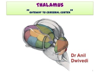
- 1. Thalamus “Gateway to cerebral cortex” 1
- 2. Thalamus • Development • Anatomical & Functional • Functional Roles Divisions • parts • Functional Organization • Relations • Connections of nuclei • Internal Organization • Blood supply • Clinical correlation 2
- 3. BRAIN: • Embryologically derived from 3 primary brain vesicles: – Prosencephalon (Forebrain) • 5th week, • subdivide into : – Telencephalon - Cerebrum – Diencephalon - Thalamus – Mesencephalon (Midbrain) – Rhombencephalon(Hindbrain) • subdivide into: – Metencephalon - Pons, Cerebellum – Myelencephalon - Medulla 3
- 4. Diencephalon • Paired structure • Located between the brain stem and the cerebral hemisphere • Continuous with the mb rostral part of the p C midbrain mo • Forms the lateral wall of the 3rd ventricle 4
- 5. • Almost entirely surrounded by cerebral hemispheres • A little part seen externally on base of brain caudal to optic chiasma • Other parts seen on sagittal & coronal sections 5
- 6. • medial surface of diencephalon- – Subdivided by hypothalamic sulcus (indicated by black line) into: – Dorsal part – Ventral part 6
- 7. Dorsal part Thalamus & Epithalamus Subthalamus & Hypothalamus H Ventral part 7
- 8. Thalamus Functional Roles • Four basic functional roles: – Sensory • All sensory information (except olfaction) is relayed to cortex via the thalamus – Motor • Motor system outputs from basal ganglia and cerebellum are relayed by thalamus – Emotion/memory • The thalamus is part of Papez circuit and helps control some emotional and memory information going to limbic cortex (cingulate gyrus) – Vegetative • The thalamus has some intrinsic nuclei associated with alertness and arousal. Can be associated with disorders of consciousness 8
- 9. Thalamus • Large mass of grey matter • Shape and size, resembles – small hen’s egg • 80 percent of diencephalon • Forms lat wall of 3rd ventricle • Separated from hypothalamus – hypothalamic sulcus • May be connected to opposite thalamus – interthalamic adhesion (massa intermedia) 9
- 10. Thalamus: In horizontal sections of brain Lower Higher level level 10
- 11. • Anterior pole – Narrow – Close to midline – Tubercle of thalamus – form posterior boundary of the interventricular foramen 11
- 12. • posterior pole – Expanded – Pulvinar – Extend beyond 3rd ventricle – Overhang superior Colliculus – Sup quadrigeminal brachium separates from MGB 12
- 13. Relations Dorsal: lateral ventricle Anterior: interventricular foramen Lateral: Medial: 3rd Internal ventricle capsule Ventral: Subthalamus & Hypothalamus Caudal: midbrain 13
- 14. Surfaces • 4 Surfaces: • Superior • Inferior • Medial S • Lateral L M l 14
- 15. Superior Surface caudate nucleus • Stratum zonale stria terminalis • Bounded laterally by – caudate nucleus – thalamostriate vein – stria terminalis LV • Lateral part – lies in the floor of lat ventricle – covered by ependyma • Medial part-related to : – choroid plexus of the 3rd ventricle thalamo- choroid plexus ependyma striate vein 15
- 16. Lateral Surface • Related to the internal capsule Inferior Surface • Rests on the Subthalamus & hypothalamus 16
- 17. Medial Surface Stria medullaris thalami • Stria medullaris thalami (a fascicle of nerve fibers) courses along its dorsomedial margin • hypothalamic sulcus • Interthalamic adhesion • Forms the upper part of the lateral wall of the 3rd ventricle Hypothalamic sulcus 17
- 18. Internal Organization • composed of – grey matter – interrupted by two vertical sheaths of white matter - medullary laminae. • External medullary lamina: – Located laterally – separates reticular nucleus from rest of the thalamic mass – Contains thalamocortical & corticothalamic fibers 18
- 19. Internal medullary lamina • Y- shaped band • divides thalamus into – Anterior – Medial – Lateral nuclear groups • Contains: – Fibers connecting thalamic nuclei with one another – Neuronal collections called intralaminar nuclei 19
- 20. Anatomical Divisions • Anterior Division – Anterior nucleus • Medial Division – Dorsomedial Nucleus (DM) • Lateral Division – Dorsal Tier • Lateral dorsal (LD) • Lateral Posterior (LP) • Pulvinar – Ventral Tier • Ventral Anterior (VA) • Ventral Lateral (VL) • Ventral Posterior (VP) – Ventral posterolateral (VPL) – Ventral posteromedial (VPM) 20
- 21. Anatomical Divisions • Medial Geniculate Nucleus (MGN) • Lateral Geniculate Nucleus (LGN) • Intralaminar Nuclei – Centromedian (CM) – Parafascicular (PF) • Reticular Nucleus 21
- 22. 22
- 23. Functional Divisions • Relay Nuclei – Relay specific information from a particular tract or modality – This is not just sensory information • Relay nuclei are part of several important modulatory loops in the CNS – This is not simple “passing on” of the signal • Relay nuclei engage in some complex condensing and processing of the incoming raw information 23
- 24. Functional Divisions • Association nuclei – Support areas of association cortex • Prefrontal cortex • Parietal-occipital-temporal cortex – Association cortex is involved in higher cognitive function 24
- 25. Other Nuclei • Intralaminar nuclei – Inputs are diverse! • Cortex, basal ganglia, cerebellum, brainstem reticular formation, Spinothalamic tract – Project to • Widespread areas of cortex • Basal ganglia – Produce general changes in cortical function 25
- 26. Other Nuclei • Reticular nucleus – Sheet-like layer of neurons partially covering the thalamus – Receives input from widespread cortical areas – Only thalamic nucleus with no projections to the cortex – Inhibitory projections to specific thalamic nuclei – Regulates the activity of the thalamus in the form of cortical feedback 26
- 27. Functional Organization • Thalamus is major route for- – Subcortical neuronal activity influences the cerebral cortex • All nuclei of thalamus except reticular nucleus, project to ipsilateral cerebral cortex • whole of cerebral cortex receives input from thalamus • All thalamic nuclei receive corticofugal fibers in a reciprocal fashion 27
- 28. • Based on their connection with the cerebral cortex, the thalamic nuclei are divided into: Specific nuclei Nonspecific nuclei 28
- 29. • Specific nuclei: • Non-specific Nuclei: – Have well-defined – Receive less sensory and motor functionally distinct functions afferent input – Have highly organized – Connect with wider point-to-point area of cortex, connection with including associative sensory & motor and limbic regions regions of cerebral – Include nuclei of cortex dorsal tier of lateral – Lie within the ventral group, and whole of group of the lateral ant and med group nuclear group 29
- 30. Anterior Nuclear Groups • Enclosed bn arms of int medullary lamina • 3 parts: – Anteroventral – Anteromedial – Anterodorsal 30
- 31. Mammillary body of Ant limbic area hypothalamus via cingulate gyrus mammillothalamic tract Parahippocampal gyrus •Functionally part of the limbic system •Involved in control of alertness & attention •Acquisition of memory 31
- 32. Medial Nuclear Group Integrates emotion, thought, and judgment Mediodorsal nucleus & Nucleus reuniens Hypothalamus, amygdala, other thalamic nuclei, prefrontal cortex Prefrontal cortex post parietal cortex limbic structures 32
- 33. Lateral Nuclear Group Ventral Tier • Ventral anterior • Ventral lateral • Ventral posterior: • VPL • VPM • Lateral geniculate • Medial geniculate 33
- 34. Ventral Anterior Nucleus Influences motor activity Ipsilateral globus pallidus & substantia nigra premotor cortex Frontal eye field Premotor & supplementary motor cortex 34
- 35. Ventral Lateral Nucleus Planning & modulation of commands Ipsilateral globus pallidus & substantia nigra Contralateral dentate nucleus Spinothalamic tract & vestibular nu Precentral motor cortical area Primary motor cortex Supplementary motor area 35
- 36. Ventral Posterior Nucleus principal thalamic relay for somasensory pathways C/L Gracile &Cuneate nu, C/L Dorsal horn of spinal cord Primary somatosensory cortex C/L trigeminal sensory nuclei 36
- 37. Medial Geniculate Body Part of the Auditory Pathway Inferior Colliculus Primary auditory cortex 37
- 38. Lateral Geniculate Body Part of the Visual Pathway Ipsilateral temporal hemiretina Contralateral nasal hemiretina Primary visual cortex 38
- 39. Lateral Nuclear Group Dorsal Tier • Lateral Dorsal • Lateral Posterior • Pulvinar 39
- 40. Lateral dorsal nucleus Memory, interpretation of visual stimuli Happocampal formation Pretectal area Superior Colliculus Cingulate gyrus Visual association cortex 40
- 41. Lateral posterior nucleus Interpretation of visual & other sensory stimuli Superior Colliculus Parietal, temporal,& occipital association cortex 41
- 42. Pulvinar Visual, perceptive, cognition & memory Pretectal area, superior Colliculus, retinas Association area of parietotemporal cortex Visual areas in occipital &post temporal lobe 42
- 43. Intralaminar Nuclei Cortical activation, Sensorimotor integration Brainstem reticular formation Spinothalamic tract Cerebellar nu Pallidum Frontal & parietal lobes striatum 43
- 44. Midline Nuclei Part of limbic system, memory & arousal Brainstem reticular formation Hypothalamus Spinothalamic tract midbrain Hippocampal formation Amygdala Nucleus accumbens Cingulate gyrus 44
- 45. Reticular Nucleus Inhibitory modulation of thalamocortical transmission Collaterals of Thalamocortical, Corticothalamic , thalamostriatal , pallidothalamic fibers Body of thalamus C/L thalamus 45
- 46. Blood supply of thalamus •Perforating branches of post cerebral a. •Posteromedial group(thalamo- perforating a) supply medial and anterior part. •Posterolateral group ( thalamo- geniculate br) supply posterior and lateral part of thalamus. •Also receives br from- •posterior communicating •anterior choroidal •posterior choroidal •middle cerebral a. 46
- 47. Clinical correlation • Thalamus – important relay and integrative center – lesions will have profound effects • 3 common etiologies: – Invasion of neoplasm – Degeneration following disease of its arterial supply – Damage by Hemorrhage 47
- 48. • Manifestations: 1. Sensory loss • damage to the VPM and VPL nucleus – loss of all forms of sensation » light touch » tactile localization & discrimination » muscle joint sense from opposite side of body 2. Thalamic pain • may be aroused by light touch or by cold – occurs on the opposite side of the body 48
- 49. 3. Abnormal involuntary movements- 4. Thalamic hand- • due to altered muscle tone: – wrist is pronated and flexed – metacarpophalangeal joints are flexed – interphalangeal joints are extended 49
- 50. Thalamic syndrome •Also known as "Dejerine-Roussy disease", after Joseph jules dejerine & Gustave Roussy. •Vascular lesion- •ventral post part of thalamus (PCA) •Disturbance of •somatosensory aspect •C/L impairment of- •Proprioception •Touch •Pain & temp. 50
- 51. Thalamic syndrome •Sensation is- •Exaggerated •Painful &perverted •exceptionally disagreeable •Development of spontaneous pain •Emotional instability •spontaneous or forced •laughing or crying 51
- 52. Fatal familial insomnia •Caused by prions •Mediodorsal & ventral ant nu •Dementia & other neurological symptoms 52
- 53. Thalamic cauterization •Surgical relief of pain in terminal cancer •Intralaminar nu –relay of pain to cortex 53
- 54. 54