1. Riccia – Structure and Reproduction
Systematic position:
Riccia belongs to the family Ricciaceae, order Marchantiales, class Hepaticopsida and division Bryophyta. The
common Indian species are Riccia siliata, R. hitra, R. discolor, R. glauca, R. gangetica, R. melansspora, R. hirta, R.
crystallina.
Habitat or occurrence: The geneus Riccia with about 200 species, is cosmopolitan in its distribution and commonly
grows in moist soils especially during and after rains. Majority of the species are terrestrial, a few are free
free-floating or
submerged aquatics e. g. R. fluitans, R. natans The plant R. crystallina occurs at an altitude of 14,000 ft. in Western
natans.
Himalayas.
External morphology
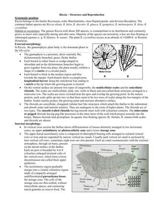
In Riccia, the gametophytic plant body is the dominant phase in
the life cycle.
• The gamtophyte is a prostrate, dorsidorsi-ventrally flat,
dichotomously branched, green, fleshy thallus.
• Each branch is either linear or wedge
r wedge-shaped or
obcordate and as the dichotomous branches begin to
grow together from one place, the plant usually exhibits a
shape of a rosette or a circular patch.
• Each branch is thick in the median region and thin
towards the margin. Each branch shows a conspicuous,
longitudinal furrow along the mid mid-dorsal line ending in
a notch at the tip where the growing point is located.
• On the ventral surface are present two types of outgrowths, the multicellular scales and the unicellular
rhizoids. The scales are multicellular, pink, red, violet or black and one
. one-celled thick structures arranged in a
transverse row. The scales are more crowded near the apex and overlap the growing point. In the mature
portion, each scale splits up into two so that there seem to be two rows of scales along the two margins of the
thallus. Scales mainly protect, the growing point and increase absorptive surface.
• The rhizoids are unicellular, elongated, tubular hair like structures which attach the thallus to the substratum
and absorb water and nutrient solution. They are analogous to the roots of higher plants. The rhizoids are of
roots
two types. The smooth-walled rhizoids having smooth inner wall with colourless contents. The tuberculate
walled
or pegged rhizoids having peg-like processes in the inner layer of the wall which project inwards into the
-like
lumen. Mature rhizoids lack protoplasm. In aquatic free floating species (R. fluitans, R. natans) both scales
men. free-floating
and rhizoids are absent.
Internal morphology:
• In vertical cross section the thallus shows differentiation of tissues distinctly arranged in two horizontal
zones, an upper assimilatory or photosynthetic zone and a lower storage zone.
• The upper dorsal assimilatory zone is composed of chlorophyll bearing cells arranged in isolated vertical
rows or tires and are separated by narrow vertical air canals. Usually each vertical air canal is enclosed by
four vertical cell rows, sometimes eight rows are also present. Each air canal communicates with the external
atmosphere, through air hares, present
on the dorsal surface of the thallus.
Each air pore is bounded by 4 to 8
colourless enlarged terminal cells of
the vertical rows, which form a loos
loose
discontinuous one-celled thick upper
celled
epidermis.
• The assimilatory region gradually
merges into a ventral colourless region
made of compactly arranged
undifferentiated parenchyma tissue
tissue,
the storage zone. The cells of the
storage zone are thin-walled, wit
walled, without
intercellular spaces, and containing
starch granules as reserve food. The
2. lower surface of the tissue containing small cells, compactly arranged, forming a single layer called the lower
epidermis. This layer hears two types of out
out-growths, the
multicellular one-celled thick scales and the unicellular
celled
tubular extension of epidermal cells called rhizoids.
Rhizoids are produced in the midmid-ventral region whereas
the scales arc produced at the margins.
Reproduction: The gametophytic plant body reproduces by
vegetative and sexual methods after attaining a certain stage of
maturity.
Vegetative reproduction:
The vegetative reproduction in Riccia takes place by the following
methods:
• Fragmentation: In this method progressive death and
:
decay of the older part of the thallus from the posterior end
reaches the dichotomy, the two surviving branches become
separate. Then each surviving branch grows independently
by epical growth and finally develops into a new plant.
• Adventitious branches: In some species ( fluitans)
: (R.
special adventitious branches, similar to parent thallus, arise from the mid ventral surface of the thallus.
mid-ventral
These branches get detached and develop into new thalli.
• Tubers: In some species (R. discolor, R. perennis), at the end of growing season, the apex of the thallus
R. perennis),
grows down into the soil and becomes thick forming a thick tuber like body. The tuber which easily survives
tuber-like
a period of drought resumes growth with the commencement of favourable season and develops into a new
thallus.
• Persistent apices: In R. discolor, at the end of growing period, the apices of thalli grow down into the soil.
discolor,
The plant other than the underground apices die. Under favourable condition, these apices come up and
develop into new plants.
• Gemma like body: In R. glauca gemma-like bodies are formed at the tips of rhizoid. These structures
like
ultimately develop into new plants.
Sexual reproduction:
Sexual reproduction in Riccia is oogamous type i.e. union between a motile flagellate male gam and a resting non-
gamete
flagellate female gamete takes place. The gamete bearing organs i.e. sex organs in Riccia are multicellular and are
called antheridium (male) and archegonium (female) respectively.
Both the types of sex organs may develop on the same thallus i.e. the plant is homothallic or monoecious (R.
gangetica, R. glauca) or the sex organs may develop on different thalli i.e. the plants are heterothallic or dioecious
)
(R. discolor, R. personii).
The sex organs develop on the floor of the mid dors longitudinal furrow in an acropetal succession i.e. the first
dorsal
formed (old) sex organ is behind and the last formed (new) sex organ is near the growing apex.
Antheridium:
• A mature antheridium of Riccia is a pear-shaped body within an open antheridial chamber which is formed
ia
by the overarching tissues. The antheridial chamber communicates with the dorsal surface by a pore.
• The antheridium is attached to the base of the antheridial
chamber by means of a few-celled stalk.
celled
• The pear-shaped antheridal bod has got a flat broad base
shaped body
and a conical apex.
• The antheridial body is surrounded by a singlesingle-layered wall
or jacket made of thin-walled cell.
walled
• A central mass of cuboidal cells enclosed by the jacket layer
are the androgonial cells or androcyte mother c cells.
• Each androcyte mother cell, on maturity, divides diagonally
to produce two triangular androcytes.
ngular
• Each androcyte ultimately metamorphoses into a single
biflagellate antherozoid or spermatozoid.
3. • During metamorphosis cell walls of the androgonial ge get
disorganised to form a semifluid mucilaginous content in
which the mature antherozoids float freely.
• Next gelatinization of jacket cells towards the apex marks
it more breakable.
• When water enters into the antheridial chamber the
gelatinized jacket cells absorb it and swell and finally
lls
break open.
• Then the semifluid mucilaginous content of the
antheridium containing the antherozoids, oozes out of the
antheridial chamber to the dorsal surface of the thallus.
Archegonium:
• A mature archcgonium is a flas flask-shaped body embedded
within a chamber called archegonial chamber which
chamber,
communicates with the dorsal surface by a pore.
• The archegonium is attached to the base of the archegonial chamber by means of a short few few-celled stalk.
• The flask-shaped archegonium is differentiated into a basal swollen part the venter and an elongated
um venter,
protruding tubular portion, the neck.
• The venter consists of a single layered wall having more than six cells in perimeter and encloses a lower
large egg or female gamete with an upper small ventral canal cell.
• The wall of upper tubular neck consists of 6 tires of elongated cells
6-9
arranged in 6 vertical rows which encloses a narrow central canal consisting
of 4-6 neck canal cells in a single row.
• The tip of the neck is covered b four specialized cells called cover cells.
by
• When the archegonium is matured, the canal cells (neck and ventral canal
cells) degenerate, leaving a mucilaginous mass.
• Shortly before fertilization, when water enters into the archegonial chamber,
the mucilaginous mass imbibes water, swells and sets up a force which pushes
aginous
the cover cells apart. Thus a free neck canal is formed from the apex of the
archegonium to the egg.
Fertilization:
For fertilization water is necessary. Water helps liberation of antherozoid by the
rupture of the antheridium. Water also acts as a medium for transportation of
antherozoid towards the egg. After rain water is retained as a thin film in the dorsal
furrow of the thallus and acts as a medium for the movement of antherozo
antherozoids. Prior to
fertilization the mucilage that is out of the archegonium attracts the antherozoids towards the archegonium. The
antherozoids thus attracted, arrive near the egg travelling down through the neck canal. Finally a single antherozoid
(n) fuses with the egg (n) and forms a diploid (2n) zygote. The zygote is the first cell of the sporophytic
ith
generation.
Structure of the sporophyte:
• The mature sporophyte of Riccia is a globular
capsule, embedded within the gametophytic plant
,
tissue and is without foot and seta.
oot
• After fertilization, the zygote secretes a wall and
increases in volume until it nearly fills the cavity of
the venter.
• Simultaneously, the cells of the venter divide to
form a two layerd venter enclosing the developing
sporophyte, a structure called calyptra.
• The zygote now divides in both vertical and
transverse planes and produces a more or less
spherical mass of 20-40 cells.
• The spherical mass then differentiates into a
4. peripheral cell layer, the amphithecium and a central mass of cells, the
endothecium.
• The amphithecium forms the jacket or wall of the sporophyte.
• The endothelial cells divide and form a sporogenous tissue, the
archesporium.
• The archesporial cells are finally differentiated into spore mother cells
with dense thick cytoplasm and nurse cells with watery vacuolated
cytoplasm.
• The spore mother cells now undergo meiosis or reduction division
resulting in haploid (n) spore tetrades (four spores).
• In the meantime, the nurse cells, amphithecial layer and also inner
layer of the venter degenerate to form a nutritive viscous fluid. This
r
fluid supplies nourishment to the developing spores.
• The spore tetrad usually remain attached to one another and are finally separated.
• The mature sporophyte, commonly, designated as sporogonium, is a more or less rounded structure
sporogonium,
containing the mature haploid spores, embedded within the gametophytic thallus.
• The mature sporogonium does not contain a single diploid cell the envelope formed from the outer layer of
calyptra, haploid spore, and encircling gametophytic tissue haploid.
• The spores are liberated only by the decay of venter wall and surrounding gametophyte tissue.
Structure of spore:
Spore is the first cell of the gametophytic generation. Each spore is pyramidal or tetrahedral in shape with a clear
triradiate mark at the proximal face. A mature spore shows three layers of wall the outermost thin and cuticularised
wall—the
exosporium, the middle cuticularised mesosporium and the
innermost endosporium.
Germination of spore and formation of the new haploid
gametophyte:
The spore germinates under favourable moist conditions.
During germination the spore takes water and swells up, as a
result the massive black exosporium bust and the thin
endosporium enclosing the spore contents protrudes out in
the form of a tubular outgrowth called the germ tube. The
germ tube elongates and divides to form an eight celled germ
disc. The rhizoid emerges out near the base of the germ tube.
The cells of the germ disc soon divide and re re-divide to form
a multicellular thallus which remains fixed with the soil by
rhizoids.
Life cycle:
In the life cycle of Riccia the haploid gametophytic
generation is independent and is the main vegetative body. It
reproduces both vegetatively and sexually. The asexual
th
reproductive phase i.e. sporophytic generation is dependent
upon the gemetophyte and is embedded within it. It is
represented only by the sporogenous tissues which are
diploid cells. Mature sporophyte or sporogoniu is made up
sporogonium
of haploid cells only, it is a peculiar condition found only in
Riccia.
Dr. Jayakara Bhandary M.
Associate Professor Botany
Professor-
GAS College , Karwar – 581301
Karnataka, India.