
Empfohlen
Empfohlen
Weitere ähnliche Inhalte
Was ist angesagt?
Was ist angesagt? (19)
Ähnlich wie International Journal of Pharmaceutical Science Invention (IJPSI)
Ähnlich wie International Journal of Pharmaceutical Science Invention (IJPSI) (20)
Kürzlich hochgeladen
Kürzlich hochgeladen (20)
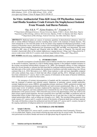
International Journal of Pharmaceutical Science Invention (IJPSI)
- 1. International Journal of Pharmaceutical Science Invention ISSN (Online): 2319 – 6718, ISSN (Print): 2319 – 670X www.ijpsi.org Volume 2 Issue 8‖ August 2013 ‖ PP.09-13 www.ijpsi.org 9 | P a g e In-Vitro Antibacterial Time-Kill Assay Of Phyllanthus Amarus And Diodia Scandens Crude Extracts On Staphylococci Isolated From Wounds And Burns Patients. Ojo, S.K.S. *a1 , Ejims-Enukwe, O. b , Esumeh, F.I. c *a,b Department of Biological Sciences, Novena University, Ogume, P.M.B. 2, Kwale, Delta State, Nigeria c Department of Microbiology, Ambrose Alli University, Ekpoma, P.M.B. 14, Ekpoma, Edo State, Nigeria *a1 Department of Microbiology, Federal University, Oye-Ekiti, Ekiti State, Nigeria ABSTRACT: Medicinal plants are sources of enormous quantities of phytochemical substances which are able to initiate different biological activities including those useful in the treatment of human diseases. This claim initiated the in vitro time-kill studies on the two plants using standard microbiological procedures. Crude extracts of Phyllanthus amarus and Diodia scandens were investigated for the rate of time-kill on staphylococci isolated from clinical samples. Biochemical test, β-lactamase assay, MIC and MBC were determined. The result yielded an MIC value of 128µg/ml and 256µg/ml while the MBCs were at 256µg/ml. The time-kill assay revealed a bactericidal effect on the isolated staphylococci ranging between 3.0log10 and 5.2log10cfu/ml for P. amarus crude extract while for D. scandens crude extract ranges between 3.0log10 and 5.5log10cfu/ml. KEY WORDS: time kill assay, staphylococci, medicinal plants, crude extracts I. INTRODUCTION Scientific investigation of medicinal plants used in folklore remedies have attracted increased attention in the world of medicine, especially in a bid to finding lasting solutions to the problems multiple resistance to the existing conventional antimicrobials (Aiyegoro et al., 2008). The therapeutic efficacy of many indigenous plants for several disorders or ailments has been described by traditional medicine practitioners in India, Africa and other parts of the world, which predates the introduction of antibiotics and other modern drugs into African continent (Ojo et al., 2010; Shrivastava et al., 2009). Green medicine is safer and more dependable compared with costly synthetic base, many of which had side effects (Joseph and Raj, 2011). Although several studies have been conducted on P. amarus, there is still a paucity of information on the nature of bacterial inhibitions, while information on D. scandens has been scanty. The emergence of resistant microorganisms in hospitals and the community is causing problems for both the treatment of patients and infection control. Organisms of particular concern include methicillin- resistant Staphylococcus aureus (MRSA), glycopeptides-resistant enterococci (GRE), gentamicin-resistant and extended-spectrum β-lactamase-producing Klebsiella and multi-resistant pseudomonads (May et al., 2000). A recent major review of antibiotic resistance emphasized the importance of hospital infection control, and the control of these organisms, and many authorities have reiterated the key role of hand-washing with appropriate disinfectants in this process (Larson, 1995).Time-kill assay involves the determination of the rate of kill on the plant extracts against tested organisms. Previous researchers have used MICs and MBCs as prediction tools for antimicrobial action of plant crude extracts, thus limiting the use of such data since it does not consider time- related antimicrobial effects, such as killing rate (Aiyegoro et al., 2009). The microbial killing potential of the crude extracts of P. amarus and D. scandens in terms of the kinetics of bacterial death have not been reported. This study will therefore evaluate the antibacterial and time-kill study of P. amarus and D. scandens, has part of the ongoing exploration of indigenous plants for novel antimicrobial compounds. II. MATERIALS AND METHODS 2.1 Sample Collection The whole plants of P. amarus and D. scandens were harvested from Amai Community, Ukwuani L.G.A in Delta State, Nigeria. It was rinsed in flowing tap water and air-dry for several weeks. The dried plants were pulverized in an electric blender and stored in air-tight plastic bag till required for use. 2.2 Isolation and Species Identification
- 2. In-Vitro Antibacterial Time-Kill Assay Of… www.ijpsi.org 10 | P a g e Fifty (50) clinical samples were obtained from wounds and burns patients at the General Hospital, Kwale, Delta State and inoculated on freshly prepared nutrient agar and mannitol salt agar (Oxoid) slants using sterile swab sticks. Slants were incubated at 37o C for 24hrs. Colonies growing on slants were streaked on freshly prepared mannitol salt agar (MSA) plates and incubated at 35o C. Primary characterization and identification of isolates was based on Gram stain, morphological and cultural characteristics on different media, fermentation on MSA, catalase and coagulase (tube) test. 2.3 Preparation of Extracts The ethanolic extracts of the active ingredients of the plants were carried out using the method described by Harbone (1994). 25g of the pulverized powdered plants were soxhlet extracted using 250ml of the absolute ethanol. The extraction lasted for 6hrs and the volatile oil obtained was purified by filtration through Whatman No.1 filter paper (Atata et al., 2003) and further sterilized through Millipore membrane filter (0.45µm pore size) (Ronald, 1995). This was concentrated by evaporation using water bath at 50o C until molten semi- solid extracts were obtained (Mbata and Saika, 2008). 2.4 Susceptibility Testing MICs for the staphylococcal isolates were determined by the micro dilution method recommended by Clinical Laboratory Standards Institute (CLSI) (2008). Micro dilution plates were inoculated with 100µl of Mueller-Hinton (MH) broth containing the appropriate antimicrobial (plant extracts) concentrations and a final concentration of 105 cfu/ml of the test isolates. After an incubation of 24h at 35o C, plates were examined for turbidity, indicating growth. The MBCs were determined by plating out 0.1µl of the MIC on MH agar incubated at 35o C for 24h. Reference type S. aureus strain (ATCC 29523) was used as positive control. 2.5 Preparation of inocula for time-kill assay Inocula for the time-kill determination were prepared as described by May et al. (2000). Organisms were grown in Brain Heart Infusion (BHI) broth (Oxoid). The overnight broth was adjusted to a 0.5 McFarland standard as described by CLSI (2008). The tubes for all isolates were shaken at 150rpm for 90mins at 37o C to ensure that organisms were out of their phases and into their logarithmic phases. 2.6 Time-kill (Bactericidal kinetic) assay Bactericidal kinetic assay were performed in glass tubes containing 10ml of Mueller Hinton broth. The extracts of P. amarus and D. scandens were used at the MIC and 2xMIC concentrations. An inoculums containing approximately 5x10⁵ cfu/ml was introduced into the Mueller Hinton broth containing various extracts and incubated at 37o C. 500µl sample was removed from culture at 6, 12, 18 and 24h, diluted serially and 100µl of the diluted samples were inoculated on Mueller Hinton agar and incubated at 37o C for 24h. Control include extract free Mueller Hinton broth seeded with the test inoculums, viable counts were calculated to give cfu/ml, and kill curves were plotted with time against logarithm of the viable count. Each experiment was performed in duplicate and mean variance obtained. 2.7 Determining time-kill endpoints A bactericidal effect is defined as 3log10 decrease in the cfu/ml or a 99.9% kill over a specified time. The definition of kill for this study was as described by May et al. (2000) with modification. A constant logarithmic rate of kill has been assumed during a time-kill. A 90% kill at 6h is equivalent to a 99% kill at 24h. In this study, the kill measurement was determined by the actual reduction in viable counts at 6h for each isolate and in comparison with 12, 18, and 24h time-kill period. III RESULTS Fifty (50) wounds and burns samples were analyzed in this study, obtaining 21 staphylococci isolates, with 3(14%) as Coagulase-negative staphylococci (CoNS) and 18(86%) Coagulase positive S. aureus (Table 1). The MICs of P. amarus crude extracts on various isolates ranged between 128µg/ml and 256µg/ml while MIC for D. scandens was 256µg/ml. The MBCs of P. amarus and D. scandens were as observed in MIC (Table 2). Table 3 showed the bactericidal activity of the crude extracts on staphylococci isolates at 6h intervals after incubation. The average logarithm reduction in viable cell count for P. amarus crude extract ranged between 2.6log10cfu/ml and 4.0log10cfu/ml after 6h of incubation, 2.7log10cfu/ml and 3.8log10cfu/ml after 12h, 2.7log10cfu/ml and 4.3log10cfu/ml after 18h and 0log10cfu/ml and 5.2log10cfu/ml after 24h in 1xMIC and 2xMIC. For D. scandens crude extract, it ranges between 2.6log10cfu/ml and 4.0log10cfu/ml after 6h of interaction, 2.6log10cfu/ml and 4.4log10cfu/ml after 12h, 2.7log10cfu/ml and 4.4log10cfu/ml after 18h, and 2.5log10cfu/ml and 5.5log10cfu/ml after 24h of interaction in 1Xmic and 2xMIC. The greatest reduction in cell
- 3. In-Vitro Antibacterial Time-Kill Assay Of… www.ijpsi.org 11 | P a g e viability among the staphylococci isolates tested on P. amarus was 5.2log10cfu/ml while for D. scandens, it was 5.5log10cfu/ml. The S. aureus type ATCC 25923 tested on P. amarus had a higher cell reduction in density of 3.1log10cfu/ml as compared to that tested on D. scandens with 3.0log10cfu/ml. Table1. Identification and biochemical characterization of wounds and burns isolates from hospital patients. Biochemical test No. of staphylococcal isolates (%) Positive Negative/others Gram staining 21(42%) 29(58%) Catalase 21 - Coagulase 18(86%) 3(14%) Mannitol fermentation 16(76%) 5(24%) Table2. Determination of MIC and MBC of the staphylococci isolates on P. amarus and D. scandens crude extracts Isolate P. amarus D. scandens No. MIC (µg/ml) MBC (µg/ml) MIC (µg/ml) MBC (µg/ml) W44 128 NG 256 G W39 128 NG 128 NG W34GY 128 NG 256 G B29 128 NG 128 G B40M 128 NG 256 NG W28 128 NG 256 NG B18 128 NG 256 G B7 128 NG 256 NG B5 256 NG 256 NG W42 256 NG 256 NG W38 256 NG 256 NG W37 256 NG 256 NG W40GY 256 NG 256 NG W25 256 NG 256 NG W41 128 NG 256 G B36 256 NG 256 NG W33 128 NG 256 NG W17 128 NG 256 NG W10 256 NG 256 NG B22 128 NG 256 NG W19 128 NG 256 NG S aureus (ATCC25923) 128 NG 256 NG Key: GY- Golden Yellow, M- Milky, W- wound, B- burns, G- Growth, NG- No Growth Table3. Nature of inhibition of crude extracts of P. amarus and D. scandens on wounds and burns staphylococci Isolate 6h(log10kill) 12h(log10kill) 18h(log10kill) 24h(log10kill) No MIC 2xMIC MIC 2xMIC MIC 2xMIC MIC 2xMIC PA 42 *3.1 *3.3 *3.1 *3.4 2.8 *3.1 *3.0 *4.2 PA 7 *3.4 *3.4 *3.5 *3.6 2.7 *3.0 0 0 PA 39 2.7 *3.2 2.9 *3.5 *3.4 *3.9 *3.3 *4.6 PA29 2.7 *3.2 2.8 *3.2 *4.3 *4.1 *4.4 *4.7 PA41 2.7 *3.1 *3.1 *3.1 *3.1 *3.3 *3.0 *4.1 PA40M 2.6 *3.1 *3.1 *3.4 *3.0 *3.1 2.8 2.8 PA5 2.9 *3.4 *3.1 *3.6 2.8 *3.1 2.7 *3.4 PA28 *3.0 *3.2 *3.2 *3.3 *3.2 *3.5 *3.1 *3.1 PA34GY 2.7 2.8 2.7 2.9 2.8 *3.3 2.9 *3.8 PA38 *3.0 *3.6 *3.4 *3.8 2.8 *3.2 2.9 *3.8 PA44 2.7 *3.0 *3.3 *3.7 *3.1 *4.3 *3.0 *4.4 PA37 2.8 *3.1 *3.0 *3.3 *3.2 *4.0 *3.4 *4.3 PA33 *3.0 *3.5 *3.0 *3.6 2.9 *3.8 2.8 *5.2 PA18 2.6 2.6 2.7 2.7 2.8 2.8 *3.0 *3.8 PA ATCC *3.1 *3.1 2.9 2.9 *3.0 *3.0 2.6 2.6 DS29 *3.0 *3.0 *3.0 *3.0 *3.1 *3.5 2.9 2.9
- 4. In-Vitro Antibacterial Time-Kill Assay Of… www.ijpsi.org 12 | P a g e DS36 2.6 2.6 2.9 2.9 *3.0 *3.3 2.5 2.7 DS5 2.6 2.8 2.9 2.9 2.7 *3.2 *3.0 *3.0 DS19 *3.0 *3.3 *3.0 *3.4 *3.1 *3.7 2.9 *5.1 DS34GY *3.4 *4.0 *3.2 *4.4 *3.2 *3.7 *3.1 *5.5 DS40GY 2.7 *3.0 2.6 *3.0 2.9 2.9 2.8 *3.4 DS28 *3.0 *3.0 *3.0 *3.1 *3.1 *3.1 2.7 *3.7 DS39 2.7 2.7 2.9 *3.3 *3.0 *3.0 *3.0 *3.4 DS25 *3.0 *3.2 *3.1 *3.2 *3.1 *4.2 2.7 *3.5 DS38 2.6 *3.0 2.8 *3.1 2.9 2.9 *3.0 *3.0 DS7 2.9 *3.1 *3.0 *3.4 *3.1 *4.4 *3.0 *4.1 DS37 2.9 *3.0 *3.0 *3.0 2.8 *3.4 2.7 *5.5 DS33 *3.0 *3.6 *3.0 *3.8 2.9 *3.0 2.8 2.9 DS ATCC 2.6 2.6 2.9 2.9 2.7 2.7 *3.0 *3.0 Key: PA: isolates tested with P. amarus; DS: isolates tested with D. scandens; *: Bactericidal effect; PA ATCC: S. aureus ATCC 25923 tested with P. amarus; DS ATCC: S. aureus ATCC 25923 tested with D. scandens Fig.1. Time-kill curve of Staphylococci by P. amarus crude extracts Fig.2. Time-kill curve of Staphylococci by D. scandens crude extracts IV DISCUSSION
- 5. In-Vitro Antibacterial Time-Kill Assay Of… www.ijpsi.org 13 | P a g e The incidence of multi-resistant Staphylococcus spp. to antibiotics of choice is increasing and is a problem of global concern, which needs an urgent attention especially in the development of herbal drugs. The results from this study evaluated the possible claim of P. amarus and D. scandens as having antimicrobial properties used in folkloric medicines for the treatment of various human infections. The rate of killing as demonstrated in this study with fixed concentration of the crude extracts has further proved the in vitro time-kill assay as a reliable method in determining tolerance, thus conforming to earlier studies by Aiyegoro et al. (2008 and 2009), and May et al. (2009). The time-kill study on P. amarus and especially D. scandens was the first to be reported with reference to Aiyegoro et al. (2009), that the conventional bactericidal activity standard is a 3log10cfu/ml or greater reduction in the viable colony number, which was achieved in this study. It was also observed in this study that the rate of killing was high in 2xMIC (Fig.1&2) for the various hours under study indicating a higher antimicrobial efficacy of the crude extracts against clinical staphylococci isolates. It could be ascertained from this study that an increase in the concentration of extracts with time dependence could yield a better result. This was similar to an earlier report by Aiyegoro et al. (2008) who worked on H. pendunculatum and observed a high killing effect on 2xMIC at longer duration of interaction (12h). Although May et al. (2009) and Shrivastava et al. (2009) concluded on 90% kill at 6h is equivalent to a 99.9% kill at 24h. However, this study which was corroborated by Aiyegoro et al. (2008, 2009), revealed a longer duration (time dependent) of kill at 12h of interaction. V CONCLUSION On the basis of the results reported in this study, we conclude that the crude extracts of P. amarus and D. scandens exhibit significant antibacterial activity against tested staphylococci. It also suggests that these plants are a potential candidate in bioprospecting for antimicrobial drugs. However, the rate of kill ascertained from this study should be compared with the rate of kill from conventional antibiotics. ACKNOWLEDGEMENT We sincerely appreciate Prof. Ogi Okwumabua of Wisconsin University, U.S.A for the provision of CLSI standards and Dr. Omumu and his team at the General Hospital, Kwale, Delta State, Nigeria for their unflinching support during sample collection. REFERENCES [1]. Aiyegoro OA, Afolayan AJ and Okoh AI. In vitro antibacterial time kill studies of leaves extracts of Helichrysum longifolium. Journal of Medicinal Plants Research 2009; 3(6): 462-467 [2]. Aiyegoro OA, Afolayan AJ and Okoh AI. Studies on the in vitro time kill assessment of crude acetone and aqueous extracts of Helichrysum pedunculatum leaves. African Journal of Biotechnology 2009; 7(20): 3718-3722. [3]. Shrivastava SM, Kumar S and Chaudhary M. Time-kill curve studies of ampucare against Escherichia coli, Staphylococcus aureus, Klebsiella pnuemoniae and Proteus vulgaris. Research Journal of Medicinal Plant 2009:1-6. [4]. May J, Chan CH, King A, Williams L, and French GL. Time-kill studies of tea tree oils on clinical isolates. Journal of Antimicrobial Chemotherapy 2000; 45:639-643. [5]. Ojo SKS, Idemudia AJ, Alikwe PCN, Awokoya OO. Antimicrobial potency of Diodia scandens and Phyllanthus amarus on some clinical isolates. International Journal of Pharma. Research & Development 2010; 2(2): 1-7. [6]. Joseph B and Raj SB. An overview: Pharmacognostic properties of Phyllanthus amarus. International Journal of Pharmacology 2011; 7:40-45. [7]. Larson EL. APIC guideline for handwashing and hand antisepsis in health care settings. American Journal of Infection Control 1995; 23: 251-69. [8]. Harbone NV. Phytochemical methods. A guide to modern techniques of plant analysis. 2nd ed. Chapman and Hall, London 1994 pp 425. [9]. Atata RF, Sani A and Ajewole SM. Effects of stem bark extracts of Enantia chloranta on some clinical isolates. Nigerian Journal of Microbiology 2003; 20(11): 649-654. [10]. Mbata TI and Saikia A. Antibacterial activity and phytochemical screening of crude ethanolic extract of leaves of Ocimum gratissimum L. on Listeria monocytogenes. The Internet Journal of Microbiology 2008; 4(2). [11]. Ronald MA. Microorganisms in our World. Mosby Year Book Inc., St. Louis 1995 pp 765. [12]. Clinical Laboratory Standards Institute. Performance standards for antimicrobial susceptibility testing: 18th Informational supplement. M100-S18, Wayne, Pennyslvania, USA 2008.