2. 430 H. Y. Al-Matubsi and R. J. Fairclough
inhibiting prostaglandin synthesis using finadyne, which is were placed in heparinized glass tubes and the catheter was
an inhibitor of the cyclo-oxygenase pathway. refilled with heparinized saline (50 iu ml–1).
Ovarian venous blood was collected using the method
described by McCracken et al. (1969). Approximately 5 ml
Materials and Methods
ovarian blood was allowed to drain freely every 30 min
Experimental animals from the open end of the catheter into heparinized 15 ml
graduated centrifuge tubes. The time taken to collect this
Border Leicester Merino ewes (n = 9) were prepared
sample was measured using a stopwatch. Three millilitres of
with ovarian autotransplants as described by Goding et al.
ovarian venous blood were collected at alternate 15 min
(1967). The ewes were housed individually in metabolic
intervals after collection of each 5 ml sample after
cages in a temperature-controlled room (20 C) and were fed
oestradiol injection. Thus, samples were collected every
once a day with 800 g of a pelleted ration consisting of
15 min for determination of hormone concentrations and
hammer milled lucerne (60%) and oats (40%). Water was
every 30 min for determination of blood flow (Lamsa et al.,
available ad libitum. The study was carried out at CSIRO
1989). The blood samples were centrifuged at 1900 g for
Division of Animal Production, Australia. All protocols
15 min. Plasma was collected and stored at –20 C until
were approved by the Animal Experimentation Ethics
assayed for oxytocin and progesterone (ovarian venous
Committees of Victoria University of Technology and the
plasma) or PGFM and oestradiol (jugular venous plasma) by
CSIRO Division of Animal Production.
radioimmunoassay (RIA). Blood flow (ml min–1) was
calculated by measuring the time taken to collect a known
Experimental design
volume of ovarian venous blood. The packed cell volume
As ewes with autotransplanted ovaries do not naturally (PCV) was determined at 1 h intervals and the plasma flow
undergo oestrous cycles, oestrus was induced synchro- (ml min–1) was calculated by multiplying the blood flow by
nously by two injections of 125 µg synthetic PGF2α 100-PCV divided by 100. The secretion rate of oxytocin and
(Estrumate; ICI, Sydney) given 15 days apart. After the progesterone (ng min–1) was obtained by multiplying the
second injection, oestrus was detected by inspection twice plasma flow (ml min–1) by the concentration of hormone in
a day for the presence of crayon marks after mating with a the ovarian venous plasma (ng ml–1).
ram fitted with a sire-o-sine harness (Radford et al., 1960).
The day that the ewes displayed oestrous behaviour Hormone analysis
was designated day 0. On day 15 of the cycle, all ewes
PGFM assay. Plasma PGFM concentrations were
were injected i.m. with 50 µg oestradiol benzoate
measured by RIA (Burgess et al., 1990) with a sensitivity of
(Intervet, Sydney) in peanut oil. In addition, four of
8 pg ml–1. The intra- and interassay coefficients of variation
these ewes were injected i.m. with 2.2 mg kg–1 of the
were 8 and 11%, respectively.
prostaglandin synthetase inhibitor, finadyne (Allhank
Trading Company, Melbourne) at 3 h intervals starting at
Oestradiol assay. The concentration of oestradiol was
the time of oestradiol injection. The remaining five ewes
measured in peripheral blood plasma by RIA (Burgess et al.,
received vehicle only.
1990) with a sensitivity of 7 pg ml–1. The samples were
measured in a single assay and the intra-assay coefficient of
Cannulation of jugular and ovarian veins
variation was 4.7%.
Cannulations were carried out under local anaesthesia
(10% lignocaine hydrochloride spray: xylocaine; Astra Progesterone assay. Progesterone was assayed in 100 µl
Pharmaceuticals, Sydney) as described by McCracken et al. ovarian plasma extracted with 2 ml n-hexane (Crown
(1969) at least 24 h before the start of blood sampling. In Scientific, Victoria) according to the method of Rice et al.
brief, a polyvinyl catheter was inserted into the jugular vein (1986). The sensitivity of the assay was 0.25 ng ml–1. The
exteriorized in the skin loop to cannulate the ovarian vein. samples were measured in a single assay and the intra-assay
The tip of the catheter was positioned at the junction of the coefficient of variation was 7%.
ovarian and jugular veins. An additional polyvinyl catheter
(50 cm) was inserted into the contralateral jugular vein. The Oxytocin assay. Plasma oxytocin concentrations were
catheters were filled with heparinized saline (1000 iu ml–1). measured by RIA as described by Al-Matubsi et al. (1997).
The sensitivity of the assay was 16 pg ml–1, and the intra-
Blood sampling and interassay coefficients of variation were 6 and 11.9%,
respectively.
On day 15 after oestrus, 5 ml and 3 ml samples of blood
were collected from the ovarian and contralateral jugular
Statistical analysis
veins, respectively, at 30 min intervals for 6 h before
oestradiol or finadyne injections and subsequently at Statistically significant pulses of ovarian vein oxytocin
15 min intervals for up to 9 h after injection. The blood and jugular vein PGFM were determined using a Pulsar
samples (3 ml) collected from the contralateral jugular vein program (Merriam and Wachter, 1982). Assay variability
3. Effects of finadyne on oestradiol-induced secretion of oxytocin and PGF2α during late oestrus 431
(a) was estimated by regression analysis of the standard
3000 60 500 deviation for duplicate determinations and the mean at
55 a
2500 50
400
each point. Baseline was calculated representing the
45
2000 40
contribution of long-term trends (15 h) but not fluctuations
35 300 of shorter duration (30 min). The amplitudes of the ovarian
1500 30
25
oxytocin and peripheral PGFM pulses were calculated by
200
1000 20 subtracting baseline values. The resulting values were then
15
500 10 100 rescaled in terms of standard deviation units by dividing the
5 rescaled values by an estimate of assay variability. The
0 0 0
–6 –5 –4 –3 –2 –1 0 1 2 3 4 5 6 7 8 9 amplitude of the rescaled pulses was identified by applying
height and duration criteria specified by user-defined cut-off
(b) points [G(n)] for pulses. These calculations were repeated
3000 60 500
55 until two iterations produced the same values for pulses or
2500 50
400 until the preset limit of six iterations was completed. The
45
2000 40 quadratic (a), linear (b), and constant (c) terms for Pulsar
35 300 were as follows: for oxytocin: a = 0.00, b = 11.91 and
1500 30
25 200 c = 0.00; and for PGFM: a = 0.00, b = 11.17 and c = 0.00.
1000 20
15
The following G(n) values were selected for both oxytocin
500 10 100 and PGFM pulses: G(1) = 6.5, G(2) = 4.45, G(3) = 3.25,
5
0 0 0 G(4) = 2.57 and G(5) = 2.05. Coincident episodes in the
–6 –5 –4 –3 –2 –1 0 1 2 3 4 5 6 7 8 9 secretion of oxytocin and PGFM were defined as those that
(c) showed an increase in the value of the PGFM pulse at the
same time as a defined oxytocin pulse. The plasma
Progesterone (ng min–1)
3000 60 500
55 a secretion rates of oxytocin and concentrations of PGFM
2500 50
PGFM (pg ml–1)
400
Oxytocin (ng min–1)
45 pulses were expressed in ng min–1 and pg ml–1, respectively,
2000 40
35 300 and the duration of that pulse was designated as τ being the
1500 30 number of minutes between the last time point before and
25 200
1000 20 the first time point after a significant increase in hormone
15
100 concentration as detected by the Pulsar program. The area
500 10
5 under the significant ovarian oxytocin and peripheral PGFM
0 0 0 pulses was then calculated for each ewe and was expressed
–6 –5 –4 –3 –2 –1 0 1 2 3 4 5 6 7 8 9
as (ng min–1) τ and (pg ml–1) τ, respectively. The overall
(d) mean concentration, pulse amplitude and duration of the
3000 60 500
55
ab pulse, and the area under the pulse were obtained using the
2500 50
400 Pulsar analysis program. The values were expressed as
45
2000 40 mean SEM. Individual characteristics of these responses
35 300 and the differences in concentrations of oestrogen and
1500 30
25 200 progesterone were compared using a Student’s unpaired t
1000 20 test. The number of ewes that showed pulses of oxytocin
15
500 10
100 and PGFM after oestradiol or oestradiol plus finadyne
5
0 0 0
injections was compared using a chi-squared test.
–6 –5 –4 –3 –2 –1 0 1 2 3 4 5 6 7 8 9
(e) Results
3000 60
a 500
55 The progesterone secretion rate of oestradiol-treated ewes
2500 50
45
400 (679.04 ± 87.98 ng min–1) was not significantly different
2000 40 from that in the oestradiol–finadyne-treated ewes
35 300
1500 30
(762.77 141.76 ng min–1). Progesterone secretion re-
25 200 mained high during the sampling period, indicating the
1000 20
15 presence of a functional corpus luteum in both groups (Figs
100
500 10 1 and 2). In both treated groups, circulating concentrations
5
0 0 0
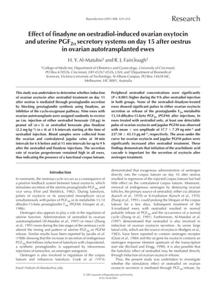
–6 –5 –4 –3 –2 –1 0 1 2 3 4 5 6 7 8 9
treated with oestradiol only on day 15 after oestrus. a and b indicate
Time (h)
significant episodes in secretion of ovarian oxytocin and PGFM,
Fig. 1. Oxytocin ( ) and progesterone ( ) secretion rates into respectively. : Identifies synchronous episodes of secretion of
ovarian venous plasma and concentrations of peripheral 13,14- both compounds. ⇓: Indicates time of oestradiol injection and ↓
dihydro-15-keto PGF2α (PGFM; ) from individual ewes (a–e) indicates times of injection of finadyne vehicle (control).
4. 432 H. Y. Al-Matubsi and R. J. Fairclough
(a) The effect of intramuscular injections of oestradiol only
3000 60
b
500 and oestradiol plus finadyne on peripheral PGFM
55
2500 50 concentrations and ovarian oxytocin secretion are shown
400
45 (Figs 1 and 2, respectively). The mean basal ovarian
2000 40
35 300 oxytocin secretion rate for oestradiol–finadyne-treated ewes
1500 30
(0.47 0.09 ng min–1) was not significantly different from
25 200
1000 20 that in oestradiol-treated ewes (0.50 0.16 ng min–1).
15
500 10
100 During the first 6 h of the sampling period, before the
5 oestrogen and finadyne injections, the mean amplitude
0 0 0
–6 –5 –4 –3 –2 –1 0 1 2 3 4 5 6 7 8 9
(6.57 1.39 ng min–1) and the mean area under the curve
(2.33 0.88 ng min–1) τ for the ovarian oxytocin pulses in
(b) oestradiol–finadyne-treated ewes were not significantly
3000 60 500
55
a different from those in oestradiol-treated ewes
2500 50
400 (10.17 3.23 ng min–1 and 10.68 3.31 ng min–1 τ,
45
2000 40 respectively).
35 300 Administration of oestradiol plus finadyne to ovarian
1500 30
25 200 autotransplanted ewes on day 15 of the oestrous cycle
1000 20 significantly (P < 0.05) reduced the number of ewes
Progesterone (ng min–1)
15
500 10 100 PGFM (pg ml–1) showing pulses of oxytocin (n = 0 versus n = 5) and PGFM
Oxytocin (ng min–1)
5
0 0 0 (n = 0 versus n = 5) when compared with ewes treated with
–6 –5 –4 –3 –2 –1 0 1 2 3 4 5 6 7 8 9 oestradiol only. None of the oestradiol–finadyne-treated
(c)
ewes showed significant pulses in ovarian oxytocin
3000 60 500 secretion after injection. In oestradiol-treated ewes, at least
55 one detectable pulse of ovarian oxytocin was observed after
2500 50
45 400 oestrogen injection. The mean amplitude (17.7 7.29 ng
2000 40
35 300 min–1) of these pulses was not significantly different from
1500 30 those measured before oestrogen injection (10.17
25 200
1000 20
3.23 ng min–1). However, a significant (P < 0.05) increase
15
100 in the area under the curve for ovarian oxytocin secretion
pulses (30.57 7.3 ng min–1) τ was observed after
500 10
5
0 0 0 oestrogen injection when compared with samples collected
–6 –5 –4 –3 –2 –1 0
before injection (10.68 3.31 ng min–1) τ. In these ewes,
1 2 3 4 5 6 7 8 9
(d) the ovarian oxytocin pulses were detected at a mean of
3000 60
a
500 5.05 0.37 h and the mean inter-pulse interval was
55
2500 50
3.36 0.45 h. In oestradiol-treated ewes, administration of
400
45 oestrogen significantly (P < 0.05) increased the duration of
2000 40
35 300 ovarian oxytocin pulses (54 5.5 versus 26.25 7.2 min)
1500 30 compared with corresponding values measured before
25 200
1000 20 oestrogen injection.
15 None of the oestradiol–finadyne-treated ewes and all of
500 10
100
5 the ewes treated with oestradiol only showed significant
0 0 0 pulses of PGFM in peripheral plasma after oestradiol or
–6 –5 –4 –3 –2 –1 0 1 2 3 4 5 6 7 8 9
finadyne injections. Mean basal circulating concentrations
Time (h)
of PGFM were significantly (P < 0.05) different in
Fig. 2. Oxytocin ( ) and progesterone ( ) secretion rates into
oestradiol–finadyne-treated ewes (14.55 3.0 pg ml–1)
ovarian venous plasma and concentrations of peripheral 13,14-
dihydro-15-keto PGF2α (PGFM; ) from individual ewes (a–d)
compared with ewes that received oestrogen only
treated with oestradiol–finadyne on day 15 after oestrus. a and b (28.45 2.10 pg ml–1) over the sampling period.
indicate significant episodes in secretion of ovarian oxytocin and In oestradiol-treated ewes, at least one detectable pulse
PGFM, respectively. : Identifies synchronous episodes of in plasma PGFM concentration was observed after
secretion of both compounds. ⇓ and ↓: Indicate times of oestradiol injection. The mean amplitude of these pulses
and finadyne injections, respectively. (237.18 43.13 pg ml–1) was not significantly different
from those measured before oestrogen injection
of oestradiol were significantly (P < 0.001) higher during (176.16 68.37 pg ml–1). However, there was a significant
the 9 h after oestradiol injection compared with the 6 h (P < 0.05) increase in the area under the curve
period before oestradiol injection (21.48 1.14 versus (1062.11 309.67 versus 302.65 128.91 pg ml–1) τ and
9.99 0.89 and 18.66 1.29 versus 10.66 1.0 pg ml–1, duration of the pulse (109.5 16.65 versus 36 6 min) of
respectively). the PGFM response measured in peripheral plasma
5. Effects of finadyne on oestradiol-induced secretion of oxytocin and PGF2α during late oestrus 433
collected after oestrogen injection compared with samples the absence of luteal oxytocin release. Hooper et al. (1986)
collected before injection. Plasma PGFM pulses were reported that, in ewes, 56% of oxytocin pulses were
observed in all ewes treated with oestradiol only at > 4 h coincident with pulses in uterine PGF2α and 97% of all
after injection. pulses of uterine PGF2α release were accompanied or
During the sampling period, 62.5% of ovarian oxytocin followed by pulses of oxytocin in the ovarian vein. In the
pulses were associated with, or preceded, the increase in present study, the percentage of PGFM pulses that occurred
peripheral PGFM concentrations. In contrast, 46.15% of the immediately before or coincided with a significant increase
plasma PGFM pulses occurred immediately before or in ovarian oxytocin pulses was decreased (46.15%) by
coincided with a significant increase in the ovarian administration of oestradiol. Thus, the present study
oxytocin pulses. reaffirms the findings of Zhang et al. (1991), who reported
similar effects of oestradiol administered to ewes treated
with either sham or X-irradiated ovarian follicles.
Discussion
The mechanism by which oestrogen stimulates ovarian
In this study, ovarian autotransplanted ewes were used as a oxytocin and uterine PGF2α release is not fully understood.
model to determine whether oestrogen acts to stimulate Oestrogen may act indirectly, perhaps via the uterus, to
release of ovarian oxytocin directly or indirectly via release release PGF2α, which, in turn, could stimulate ovarian
of PGF2α, which in turn stimulates ovarian oxytocin. The oxytocin release. Such a mechanism of action is unlikely to
concentrations of oxytocin in ovarian venous plasma were have occurred in the present study as PGF2α would need to
20–1403 pg ml–1 in the present study, which are similar to act through the systemic circulation to stimulate ovarian
those detected by Hooper et al. (1986) in the utero–ovarian oxytocin and it has been shown that 99% of PGF2α is
vein (50–1499 pg ml–1) and were much higher than those in cleared from blood after one passage through the lungs
peripheral plasma (20–220 pg ml–1; Hooper et al., 1986). (Piper et al., 1970). However, using ewes with ovarian
Together, these observations indicate that, in the present autotransplants does not necessarily preclude the possibility
study, oxytocin in ovarian venous plasma represents luteal that the effects of oestrogen and finadyne are mediated
rather than posterior pituitary secretion. through uterine release of PGF2α, as PGFM is known to
As would be expected in ovarian autotransplanted ewes stimulate luteal oxytocin–neurophysin secretion (Watkins
(Goding et al., 1967), the secretion of progesterone and Moore, 1987). Another possibility is that the corpus
remained high in both groups, indicating that the corpus luteum of ewes bearing ovarian autotransplants becomes
luteum of the transplanted ovary is maintained despite hypersensitive to low concentrations of PGF2α in the
intermittent surges of peripheral plasma PGFM in all ewes absence of normal basal concentrations from the adjacent
before oestradiol injection and in ewes treated with uterine horn. Such hypersensitivity may allow the corpus
oestradiol only after injection. Our observation that the luteum to release luteal oxytocin in response to even
administration of oestradiol can induce the simultaneous low concentrations of PGF2α that escape degradation by
release of ovarian oxytocin and uterine PGF2α in ovarian the lungs. An alternative site of oestradiol action may
autotransplanted ewes after a latency period of 4 h in all be directly on the ovary to induce ovarian oxytocin
ewes treated with oestradiol only is in agreement with the release. Oestradiol (Glass et al., 1984) and PGF2α (Fitz
study of Al-Matubsi et al. (1997). et al., 1982) receptors have been reported in large luteal
In the present study, synchronous pulses of ovarian cells, which are the sites of oxytocin synthesis (Rodgers
oxytocin and uterine PGF2α were observed during the first et al., 1983). Infusion of oestrogen into the corpus luteum
6 h of the sampling period before oestradiol treatment. causes luteal regression (Cook et al., 1974) and luteal
However, this did not affect subsequent synchronous cells from sheep (Tsai and Wiltbank, 1997) and cows
secretion of these hormones after oestradiol treatment. (Milvae and Hansel, 1983; Tsai et al., 1996) can produce
Thus, the uterine refractoriness to ovarian oxytocin release prostaglandins, such as PGF2α, PGE2 and PGI2. On the basis
and uterine PGF2α secretion can be eliminated as a reason of these findings it is possible that oestrogen stimulates
for variability in timing of the response. The results from the release of ovarian oxytocin through luteal prostaglandins
present study and other studies (Hooper et al., 1986; Al- (or some other metabolite of arachidonic acid) (Cooke
Matubsi et al., 1998) demonstrate that oxytocin pulses in and Ahmed, 1998). However, the physiological role of
utero–ovarian or ovarian venous plasma frequently occur in luteal prostaglandins during the oestrous cycle and the
the absence of any significant increase in utero–ovarian mechanisms controlling its production remain to be
PGF2α or peripheral PGFM concentrations and indicate that elucidated.
ovarian oxytocin can occur independently of uterine Thus, in intact ewes, the initiation of the arachidonic
PGF2α. In contrast, Lamsa et al. (1989) observed that uterine acid cascade is of importance for the secretion of oxytocin
PGF2α secretion into the utero–ovarian vein begins to after oestrogen treatment.
increase before the discharge of luteal oxytocin. Mann
(1999) demonstrated that normal frequency of episodes of The authors would like to thank J. Downing for helping with
PGF2α release, with lower amplitude and of longer cannulation of the animals and K. Tellbach for assisting with the
duration, can occur at the anticipated time of luteolysis in collection of blood samples.
6. 434 H. Y. Al-Matubsi and R. J. Fairclough
References McCracken JA, Uno A, Goding JR, Ichikawa Y and Baird DT (1969) The in-
vivo effects of sheep pituitary gonadotrophins on the secretion of
Al-Matubsi HY, Downing J, Jenkin G and Fairclough R (1997) Effect of
steroids by the autotransplanted ovary of the ewe Journal of
oestradiol on ovarian oxytocin secretion rate and luteolysis in the ewe
Endocrinology 45 425–440
after ovarian auto-transplantation Reproduction, Fertility and Mann GE (1999) The role of luteal oxytocin in episodic secretion of
Development 9 683–688 prostaglandin F2α at luteolysis in the ewe Animal Reproduction Science
Al-Matubsi HY, Downing J, Jenkin G and Fairclough R (1998) Stimulation 57 167–175
of ovarian oxytocin secretion and uterine prostaglandin release by Merriam GR and Wachter KW (1982) Algorithms for the study of episodic
exogenous progesterone early in the cycle of the ovarian auto- hormone secretion American Journal of Physiology 243 E310–E318
transplanted ewe Journal of Reproduction and Fertility 112 279–288 Milvae RA and Hansel W (1983) Prostacyclin, prostaglandin F2α and
Burgess KM, Ralph MM, Jenkin G and Thorburn GD (1990) Effect of
progesterone production by bovine luteal cells during the estrous cycle
oxytocin and oestradiol on uterine prostaglandin release in non- Biology of Reproduction 29 1063–1068
pregnant and early pregnant ewes Biology of Reproduction 42 822–833 Piper PJ, Vane JR and Wyllie JH (1970) Inactivation of prostaglandins by the
Cook B, Karsch FJ, Foster DL and Nalbandov AV (1974) Estrogen-induced
lungs Nature 225 600–604
luteolysis in the ewe: possible sites of action Endocrinology 94 Radford HH, Watson RH and Wood GF (1960) A crayon and associated
1197–1201
harness for the detection of mating under field conditions Australian
Cooke RG and Ahmed N (1998) Delayed luteolysis after intra-uterine Veterinary Journal 36 57–66
infusion of nordihydroguaiaretic acid in the ewe Animal Reproduction Rice GE, Jenkin G and Thorburn GD (1986) Comparison of particle-
Science 52 113–121 associated progesterone and oxytocin in the ovine corpus luteum
Fitz TA, Mayan MH, Sawyer HR and Niswender GD (1982) Journal of Endocrinology 108 109–116
Characterization of two steroidogenic cell types in the ovine corpus Richard S and Zingg HH (1990) The human oxytocin gene promoter is
luteum Biology of Reproduction 27 703–711 regulated by estrogens Journal of Biological Chemistry 265 6098–6103
Flint APF and Sheldrick EL (1982) Ovarian secretion of oxytocin is Rodgers RJ, O’Shea JD, Frindly JK, Flint APF and Sheldrick EL (1983) Large
stimulated by prostaglandin Nature 297 587–588 luteal cells the source of oxytocin in the sheep Endocrinology 113
Glass JD, Fitz TA and Niswender GD (1984) Cytosolic receptor for 2302–2304
oestradiol in the corpus luteum of the ewe: variation throughout the Tsai SJ and Wiltbank MC (1997) Prostaglandin F2α induces expression of
oestrous cycle and distribution between large and small steroidogenic prostaglandin G/H synthase-2 in the ovine corpus luteum, a potential
cell types Biology of Reproduction 31 967–974 positive feedback loop during luteolysis Biology of Reproduction 57
Goding JR, McCracken JA and Baird DT (1967) The study of ovarian 1016–1022
function in the ewe by means of a vascular autotransplantation Tsai SJ, Wiltbank MC and Bodensteiner KJ (1996) Distinct mechanisms
technique Journal of Endocrinology 39 37–52 regulate induction of messenger ribonucleic acid for prostaglandin G/H
Hooper SB, Watkins WB and Thorburn GD (1986) Oxytocin, oxytocin-
synthase-2, PGE (EP3) receptor, and PGF2α receptor in bovine
associated neurophysin and prostaglandin concentrations in the utero- preovulatory follicles Endocrinology 137 3348–3355
ovarian vein in pregnant and non pregnant sheep Endocrinology 119 Watkins WB and Moore LG (1987) Effect of systemic intravenous infusion
2590–2597
of PGF2α and 13,14-dihydro-15-keto-PGF2α on the release of oxytocin-
Jacobs DSC, Edgerton LA, Silvia WL and Schillo KK (1988) Effect of an associated neurophysin from the ovary in the ewe Journal of
estrogen antagonist (tamoxifen) on cloprostenol-induced luteolysis in Reproduction and Fertility 80 105–112
heifers Journal of Animal Science 66 735–742 Zhang J, Weston PG and Hixon JE (1991) Influence of oestradiol on the
Karsch FJ, Noveroske JW, Roche JF, Norton HW and Nalbandov AV (1970)
secretion of oxytocin and prostaglandin during luteolysis in the ewe
Maintenance of ovine corpora lutea in the absence of ovarian follicles Biology of Reproduction 45 395–403
Endocrinology 87 1228–1236
Lamsa JC, Knot SJ, Eldering JA, Nay MG and McCracken JA (1989)
Prostaglandin F2α stimulated release of ovarian oxytocin in the sheep in
vivo: threshold and dose dependency Biology of Reproduction 40 Received 22 May 2000.
1215–1223 Accepted 16 October 2000.