SlideShare ist ein Scribd-Unternehmen logo
1 von 33
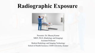
Radiographic Exposure
Presenter: Dr. Dheeraj Kumar
MRIT, Ph.D. (Radiology and Imaging)
Assistant Professor
Medical Radiology and Imaging Technology
School of Health Sciences, CSJM University, Kanpur
Introduction
• Radiographic Exposure in Radiography and Imaging Technology.
• Understanding the fundamentals of radiographic exposure is crucial
for producing high-quality diagnostic images.
• In this presentation, we will delve into the key concepts, factors, and
techniques related to radiographic exposure.
26-09-2023 Radiographic Exposure By- Dr. Dheeraj Kumar 2
Objectives
• Discuss the importance of radiographic exposure in medical imaging.
• Explain the factors affecting radiographic exposure.
• Explore techniques for optimizing exposure settings.
• Provide tips for reducing patient dose.
• Highlight advancements in exposure technology.
26-09-2023 Radiographic Exposure By- Dr. Dheeraj Kumar 3
Importance of Radiographic Exposure
• Radiographic exposure is a critical step in the process of medical
imaging. It involves exposing patients to ionizing radiation for the
specific purpose of creating diagnostic images.
• Understanding its importance is crucial:
26-09-2023 Radiographic Exposure By- Dr. Dheeraj Kumar 4
Image Quality
• Proper radiographic exposure is
essential for obtaining clear and
diagnostically valuable images.
• Inadequate exposure can result in
underexposed images with poor
visibility, while excessive exposure
can lead to overexposed images with
loss of detail.
26-09-2023 Radiographic Exposure By- Dr. Dheeraj Kumar 5
• Diagnostic Accuracy: Radiologists rely on the quality of these images to
make accurate diagnoses. Suboptimal exposure can lead to misdiagnosis,
delayed treatment, or the need for repeat imaging, which increases both
patient radiation dose and healthcare costs.
• Patient Care: Minimizing patient radiation exposure while maintaining
diagnostic image quality is a fundamental principle of radiography.
Achieving this balance requires a thorough understanding of the factors
influencing radiographic exposure.
26-09-2023 Radiographic Exposure By- Dr. Dheeraj Kumar 6
Factors Affecting Radiographic Exposure
• Several factors influence
radiographic exposure
settings, and radiologic
technologists must
consider them when
producing diagnostic
images:
26-09-2023 Radiographic Exposure By- Dr. Dheeraj Kumar 7
kVp (Peak Kilovoltage)
• The peak kilovoltage controls the quality or
penetrating ability of the x-ray beam.
• It affects image contrast, with higher kVp
values resulting in lower contrast and vice
versa.
• Proper kVp selection is vital for different
imaging studies and body parts.
26-09-2023 Radiographic Exposure By- Dr. Dheeraj Kumar 8
mAs (Milliampere-Seconds)
• The milliampere-seconds control the
quantity or number of x-rays produced.
Adjusting mAs affects image brightness
and noise.
• Higher mAs values increase image
brightness but may lead to increased
patient dose.
• Radiographers must balance mAs with
kVp for optimal image quality.
26-09-2023 Radiographic Exposure By- Dr. Dheeraj Kumar 9
Source-to-Image Distance (SID)
• Source-to-Image Distance
(SID) is the distance between
the x-ray tube and the image
receptor (usually the patient or
a detector).
• It plays a significant role in
radiography:
26-09-2023 Radiographic Exposure By- Dr. Dheeraj Kumar 10
• Image Magnification: A
shorter SID results in image
magnification, while a longer
SID reduces magnification. A
longer SID is generally
preferred to minimize
distortion and maintain image
accuracy.
26-09-2023 Radiographic Exposure By- Dr. Dheeraj Kumar 11
• Image Sharpness: Longer SID
settings enhance image sharpness by
reducing the effects of focal spot blur.
• Exposure Intensity: As SID
increases, exposure intensity
decreases. Radiographers must adjust
mAs to compensate for the reduction
in exposure intensity when using a
longer SID.
26-09-2023 Radiographic Exposure By- Dr. Dheeraj Kumar 12
Patient Anatomy and Thickness
Patient anatomy and
tissue thickness vary
widely, and these
differences have a
significant impact
on radiographic
exposure:
26-09-2023 Radiographic Exposure By- Dr. Dheeraj Kumar 13
• Variability in Anatomy: Different body parts and
anatomical structures require varying exposure parameters.
For instance, a chest radiograph requires different settings
than an abdominal one.
• Tissue Thickness: The thickness of the patient's tissues
affects the amount of radiation required to produce a
diagnostic image. Thicker tissues may necessitate higher
mAs settings for adequate penetration.
• Pediatric Imaging: Children have smaller body sizes and
different tissue densities than adults, which requires
specialized techniques and considerations for dose
reduction.
26-09-2023 Radiographic Exposure By- Dr. Dheeraj Kumar 14
Grids
• Grids are used to reduce scatter
radiation, which can degrade image
contrast.
• They are placed between the patient
and the image receptor and consist of
lead strips aligned to allow primary
radiation to pass while absorbing
scattered radiation.
• Grid ratios and focal range play a role
in exposure optimization.
26-09-2023 Radiographic Exposure By- Dr. Dheeraj Kumar 15
Scatter Radiation
• Scatter radiation results from
interactions between x-rays and patient
tissues.
• It can reduce image contrast and
increase patient dose.
• Proper grid use and collimation help
mitigate the effects of scatter radiation.
26-09-2023 Radiographic Exposure By- Dr. Dheeraj Kumar 16
Grid Selection
• Choosing the appropriate
grid ratio depends on factors
like patient thickness and the
body part being imaged.
Radiographers must also
align the grid properly to
avoid grid cutoff.
26-09-2023 Radiographic Exposure By- Dr. Dheeraj Kumar 17
Filtration
• Filtration is the process of removing low-energy x-rays from the
primary beam. This is accomplished by placing a filter, often made of
aluminum, in the path of the x-ray beam.
• The purpose of filtration is twofold: to reduce patient radiation dose
and to improve image quality.
• By removing low-energy x-rays, filtration reduces unnecessary
radiation exposure to the patient's tissues, particularly the skin and
superficial structures.
• It also results in a "harder" x-ray beam, which contributes to better
image contrast.
• Radiographers must ensure that the appropriate filter is selected based
on the clinical context and the energy spectrum of the x-ray tube.
26-09-2023 Radiographic Exposure By- Dr. Dheeraj Kumar 18
Collimation
• Collimation involves restricting the x-ray beam
to the specific area of interest, thereby
minimizing unnecessary exposure to
surrounding tissues.
• The collimator is a device that shapes the x-ray
beam to match the size and shape of the image
receptor and the anatomy being examined.
• Proper collimation reduces patient dose and
helps produce sharper images with less scatter
radiation.
• Radiographers should follow guidelines for
collimation set by regulatory bodies and
healthcare institutions.
26-09-2023 Radiographic Exposure By- Dr. Dheeraj Kumar 19
Beam Restricting Devices
• Beam restricting devices are essential tools for
controlling the x-ray beam and optimizing radiographic
exposure:
• Types of Beam Restricting Devices:
• Aperture Diaphragms: These devices are adjustable
collimators that limit the size of the x-ray field, ensuring that
only the area of clinical interest is irradiated.
• Beam Collimators: These devices help shape the x-ray beam
into a specific size and shape, typically matching the
dimensions of the image receptor.
• Cone or Cylinder Collimators: These devices are used for
specific exams, such as dental radiography, to focus the x-ray
beam accurately.
26-09-2023 Radiographic Exposure By- Dr. Dheeraj Kumar 20
Benefits of Beam Restricting Devices
• They prevent unnecessary radiation exposure to adjacent tissues and
organs.
• They improve image quality by reducing scatter radiation.
• Radiographers must verify that beam restricting devices are
functioning correctly and use them effectively for each patient
examination.
26-09-2023 Radiographic Exposure By- Dr. Dheeraj Kumar 21
Techniques for Optimizing Exposure
• Radiologic technologists employ various
techniques to optimize radiographic
exposure:
• Automatic Exposure Control (AEC)
Systems:
• AEC systems automatically adjust exposure
factors based on the amount of radiation
received by the image receptor. They ensure
consistent image quality while minimizing
patient dose.
26-09-2023 Radiographic Exposure By- Dr. Dheeraj Kumar 22
• Exposure Charts and Technique Charts:
• These charts provide guidelines for selecting
exposure factors (kVp, mAs) based on the body
part, patient size, and imaging modality. They
serve as valuable references for radiographers.
• Exposure Indicator Systems (S-Number):
• Some digital radiography systems display an
exposure indicator (S-number) on the image to
indicate the image receptor's exposure.
Radiographers can use this information to assess
and adjust exposure settings.
26-09-2023 Radiographic Exposure By- Dr. Dheeraj Kumar 23
• Image Receptor Selection (CR vs. DR):
• The choice between computed radiography (CR) and digital radiography (DR)
systems affects exposure techniques. DR systems generally offer faster image
acquisition and may require different exposure settings.
• Repeating Exposures When Necessary:
• If an image is of insufficient quality due to factors such as motion artifacts,
exposure errors, or positioning errors, radiographers may need to repeat the
exposure to ensure diagnostic value.
26-09-2023 Radiographic Exposure By- Dr. Dheeraj Kumar 24
Reducing Patient Dose
• Reducing patient dose is a fundamental
principle in radiography:
• ALARA (As Low As Reasonably
Achievable):
• ALARA is a radiation safety principle aimed at
minimizing radiation exposure while still
achieving the necessary diagnostic information.
• Radiologic technologists should always strive to
keep patient doses as low as reasonably
achievable while maintaining image quality.
26-09-2023 Radiographic Exposure By- Dr. Dheeraj Kumar 25
Shielding and Protective Measures
• Lead aprons and thyroid shields
are used to protect patients from
unnecessary radiation exposure
during x-ray examinations.
• Shielding is especially important
for pregnant patients to protect the
developing fetus.
26-09-2023 Radiographic Exposure By- Dr. Dheeraj Kumar 26
Proper Patient Positioning
• Accurate positioning of the
patient and the x-ray equipment
helps minimize the need for
repeat exposures. Proper
alignment ensures that the x-ray
beam is directed precisely
where needed.
26-09-2023 Radiographic Exposure By- Dr. Dheeraj Kumar 27
Advancements in Exposure Technology
The field of radiography is continuously
evolving, and exposure technology has seen
significant advancements:
Digital Radiography (DR) and Computed
Radiography (CR):
• Transitioning from film-based radiography
to digital technologies has improved image
quality, reduced radiation exposure, and
enhanced workflow efficiency.
26-09-2023 Radiographic Exposure By- Dr. Dheeraj Kumar 28
• Improved Image Processing Algorithms:
• Modern imaging systems incorporate sophisticated image processing algorithms that
enhance image quality and help radiologists make more accurate diagnoses.
• Dose Reduction Technologies (e.g., Pulsed Fluoroscopy):
• Specialized fluoroscopy modes, such as pulsed fluoroscopy, reduce radiation dose
while maintaining real-time imaging capabilities.
• AI-Based Exposure Optimization:
• Artificial intelligence (AI) is being used to assist radiographers in selecting optimal
exposure settings by analyzing patient anatomy and other factors in real time.
26-09-2023 Radiographic Exposure By- Dr. Dheeraj Kumar 29
Conclusion
• Radiographic exposure is a fundamental aspect of medical imaging.
• Understanding the factors affecting exposure and employing proper
techniques are essential for producing high-quality diagnostic images
while minimizing patient dose.
• Staying updated with technological advancements is crucial for a
successful career in Radiography and Imaging Technology.
26-09-2023 Radiographic Exposure By- Dr. Dheeraj Kumar 30
Questions?
Open the floor for questions and discussions.
26-09-2023 Radiographic Exposure By- Dr. Dheeraj Kumar 31
References
• Bushong, S. C. (2016). Radiologic Science for Technologists: Physics, Biology, and Protection.
Elsevier Health Sciences.
• Carlton, R. R., & Adler, A. M. (2017). Principles of Radiographic Imaging: An Art and A Science.
Cengage Learning.
• Fauber, T. L. (2016). Radiographic Imaging and Exposure. Elsevier Health Sciences.
• Fosbinder, A. M. (2018). Radiography PREP Program Review and Exam Preparation (8th ed.).
Lippincott Williams & Wilkins.
• Järvinen, H., & Syväoja, S. (Eds.). (2019). Radiography in Modern Industry (Vol. 1):
Instrumentation and Modern Diagnostic Methods. Springer.
26-09-2023 Radiographic Exposure By- Dr. Dheeraj Kumar 32
Thank You
26-09-2023 Radiographic Exposure By- Dr. Dheeraj Kumar 33

Weitere ähnliche Inhalte

Was ist angesagt?

Tomographic equipment
Tomographic equipmentTomographic equipment
Tomographic equipmentSelf
 
The presentation of radiograph
The presentation of radiographThe presentation of radiograph
The presentation of radiographmr_koky
 
MACRORADIOGRAPHY.pptx
MACRORADIOGRAPHY.pptxMACRORADIOGRAPHY.pptx
MACRORADIOGRAPHY.pptxThejaTej6
 
Radiographic Latent Image .pptx
Radiographic Latent Image .pptxRadiographic Latent Image .pptx
Radiographic Latent Image .pptxDr. Dheeraj Kumar
 
Radiographic image4
Radiographic image4Radiographic image4
Radiographic image4mr_koky
 
xray production.pptx
xray production.pptxxray production.pptx
xray production.pptxAtul Verma
 
Identification of Radiographs
Identification of RadiographsIdentification of Radiographs
Identification of RadiographsDeneicer Guy
 
Intensifying Screen (x-ray)
Intensifying Screen (x-ray)Intensifying Screen (x-ray)
Intensifying Screen (x-ray)Celjhon Ariño
 
Responsibility of radiographer
Responsibility of radiographerResponsibility of radiographer
Responsibility of radiographermohammed althaf
 
Computed Tomography Equipments.pptx
Computed Tomography Equipments.pptxComputed Tomography Equipments.pptx
Computed Tomography Equipments.pptxDr. Dheeraj Kumar
 
Fluoroscopy-Rohit.pptx
Fluoroscopy-Rohit.pptxFluoroscopy-Rohit.pptx
Fluoroscopy-Rohit.pptxRohit Bansal
 
Line focus-principle-and-anode-heel-effect
Line focus-principle-and-anode-heel-effectLine focus-principle-and-anode-heel-effect
Line focus-principle-and-anode-heel-effectJoaquinLuisRinehart1
 
Radiation protection and personnel monitoring devices
Radiation protection and personnel monitoring devicesRadiation protection and personnel monitoring devices
Radiation protection and personnel monitoring devicesRubiSapkota
 
Radiographic grid Swastik
Radiographic grid   SwastikRadiographic grid   Swastik
Radiographic grid SwastikBipul Poudel
 
X ray production & emission
X ray production & emissionX ray production & emission
X ray production & emissionairwave12
 

Was ist angesagt? (20)

Tomographic equipment
Tomographic equipmentTomographic equipment
Tomographic equipment
 
The presentation of radiograph
The presentation of radiographThe presentation of radiograph
The presentation of radiograph
 
MACRORADIOGRAPHY.pptx
MACRORADIOGRAPHY.pptxMACRORADIOGRAPHY.pptx
MACRORADIOGRAPHY.pptx
 
Radiographic Latent Image .pptx
Radiographic Latent Image .pptxRadiographic Latent Image .pptx
Radiographic Latent Image .pptx
 
Radiographic image4
Radiographic image4Radiographic image4
Radiographic image4
 
xray production.pptx
xray production.pptxxray production.pptx
xray production.pptx
 
Automatic exposure control
Automatic exposure controlAutomatic exposure control
Automatic exposure control
 
Identification of Radiographs
Identification of RadiographsIdentification of Radiographs
Identification of Radiographs
 
Intensifying Screen (x-ray)
Intensifying Screen (x-ray)Intensifying Screen (x-ray)
Intensifying Screen (x-ray)
 
Responsibility of radiographer
Responsibility of radiographerResponsibility of radiographer
Responsibility of radiographer
 
Computed Tomography Equipments.pptx
Computed Tomography Equipments.pptxComputed Tomography Equipments.pptx
Computed Tomography Equipments.pptx
 
radiographic Image quality by Astuti Mishra
 radiographic Image quality by Astuti Mishra radiographic Image quality by Astuti Mishra
radiographic Image quality by Astuti Mishra
 
Fluoroscopy-Rohit.pptx
Fluoroscopy-Rohit.pptxFluoroscopy-Rohit.pptx
Fluoroscopy-Rohit.pptx
 
Line focus-principle-and-anode-heel-effect
Line focus-principle-and-anode-heel-effectLine focus-principle-and-anode-heel-effect
Line focus-principle-and-anode-heel-effect
 
Radiation protection and personnel monitoring devices
Radiation protection and personnel monitoring devicesRadiation protection and personnel monitoring devices
Radiation protection and personnel monitoring devices
 
Radiographic grid Swastik
Radiographic grid   SwastikRadiographic grid   Swastik
Radiographic grid Swastik
 
Radiation regulatory bodies
Radiation regulatory bodiesRadiation regulatory bodies
Radiation regulatory bodies
 
Radiographic factor
Radiographic factorRadiographic factor
Radiographic factor
 
Radiographic Grid.pptx
Radiographic Grid.pptxRadiographic Grid.pptx
Radiographic Grid.pptx
 
X ray production & emission
X ray production & emissionX ray production & emission
X ray production & emission
 

Ähnlich wie Radiographic Exposure.pptx

X-Ray Beam Restrictors/Beam Modifying devices.pptx
X-Ray Beam Restrictors/Beam Modifying devices.pptxX-Ray Beam Restrictors/Beam Modifying devices.pptx
X-Ray Beam Restrictors/Beam Modifying devices.pptxDr. Dheeraj Kumar
 
ULTRASOUND COMPONENTS AND THEIR USES.pptx
ULTRASOUND COMPONENTS AND THEIR USES.pptxULTRASOUND COMPONENTS AND THEIR USES.pptx
ULTRASOUND COMPONENTS AND THEIR USES.pptxDr. Dheeraj Kumar
 
SRT AND SRS - Arun.pdf
SRT AND SRS - Arun.pdfSRT AND SRS - Arun.pdf
SRT AND SRS - Arun.pdfBBAdhikari
 
Screen Film Radiographic Technique
Screen Film Radiographic TechniqueScreen Film Radiographic Technique
Screen Film Radiographic TechniqueMuhammad Arif Afridi
 
7-RADIATION PROTECTION IN DENTISTRY===7=2023.pdf
7-RADIATION PROTECTION IN DENTISTRY===7=2023.pdf7-RADIATION PROTECTION IN DENTISTRY===7=2023.pdf
7-RADIATION PROTECTION IN DENTISTRY===7=2023.pdfNASERALHAQ
 
Effects of variation of tube voltage current, filtration..pptx
Effects of variation of tube voltage current, filtration..pptxEffects of variation of tube voltage current, filtration..pptx
Effects of variation of tube voltage current, filtration..pptxDr. Dheeraj Kumar
 
Pre-Clinical-Radiation-Protection_-(002).pptx
Pre-Clinical-Radiation-Protection_-(002).pptxPre-Clinical-Radiation-Protection_-(002).pptx
Pre-Clinical-Radiation-Protection_-(002).pptxEmmanuelOluseyi1
 
Dose reduction in Conventional Radiography and Fluoroscopy
Dose reduction in Conventional Radiography and FluoroscopyDose reduction in Conventional Radiography and Fluoroscopy
Dose reduction in Conventional Radiography and FluoroscopyTarun Goyal
 
QUALITY-ASSURANCE---MAMMOGRAPHY.pdf
QUALITY-ASSURANCE---MAMMOGRAPHY.pdfQUALITY-ASSURANCE---MAMMOGRAPHY.pdf
QUALITY-ASSURANCE---MAMMOGRAPHY.pdfAngelina Protik
 
Getting Ahead of the Expanding Landscape: Radiopharmaceutical Dosimetry
Getting Ahead of the Expanding Landscape: Radiopharmaceutical DosimetryGetting Ahead of the Expanding Landscape: Radiopharmaceutical Dosimetry
Getting Ahead of the Expanding Landscape: Radiopharmaceutical DosimetryMedpace
 
Pre-Clinical-Radiation-Protection_-(002).pptx
Pre-Clinical-Radiation-Protection_-(002).pptxPre-Clinical-Radiation-Protection_-(002).pptx
Pre-Clinical-Radiation-Protection_-(002).pptxChuemmanuelndze
 
Pre-Clinical-Radiation-Protection_-(002).pptx
Pre-Clinical-Radiation-Protection_-(002).pptxPre-Clinical-Radiation-Protection_-(002).pptx
Pre-Clinical-Radiation-Protection_-(002).pptxmedoghoneam
 
Mammographic equipment and its advancement
Mammographic equipment and its advancementMammographic equipment and its advancement
Mammographic equipment and its advancementYashawant Yadav
 
Filters and beam restrictors
Filters  and beam restrictorsFilters  and beam restrictors
Filters and beam restrictorsvishwanath0908
 

Ähnlich wie Radiographic Exposure.pptx (20)

X-Ray Beam Restrictors/Beam Modifying devices.pptx
X-Ray Beam Restrictors/Beam Modifying devices.pptxX-Ray Beam Restrictors/Beam Modifying devices.pptx
X-Ray Beam Restrictors/Beam Modifying devices.pptx
 
ULTRASOUND COMPONENTS AND THEIR USES.pptx
ULTRASOUND COMPONENTS AND THEIR USES.pptxULTRASOUND COMPONENTS AND THEIR USES.pptx
ULTRASOUND COMPONENTS AND THEIR USES.pptx
 
SRT AND SRS - Arun.pdf
SRT AND SRS - Arun.pdfSRT AND SRS - Arun.pdf
SRT AND SRS - Arun.pdf
 
Screen Film Radiographic Technique
Screen Film Radiographic TechniqueScreen Film Radiographic Technique
Screen Film Radiographic Technique
 
MEDICAL IMAGING NOTES
MEDICAL IMAGING NOTESMEDICAL IMAGING NOTES
MEDICAL IMAGING NOTES
 
Mammography.pdf
Mammography.pdfMammography.pdf
Mammography.pdf
 
7-RADIATION PROTECTION IN DENTISTRY===7=2023.pdf
7-RADIATION PROTECTION IN DENTISTRY===7=2023.pdf7-RADIATION PROTECTION IN DENTISTRY===7=2023.pdf
7-RADIATION PROTECTION IN DENTISTRY===7=2023.pdf
 
Effects of variation of tube voltage current, filtration..pptx
Effects of variation of tube voltage current, filtration..pptxEffects of variation of tube voltage current, filtration..pptx
Effects of variation of tube voltage current, filtration..pptx
 
CT dose reduction
CT dose reductionCT dose reduction
CT dose reduction
 
Pre-Clinical-Radiation-Protection_-(002).pptx
Pre-Clinical-Radiation-Protection_-(002).pptxPre-Clinical-Radiation-Protection_-(002).pptx
Pre-Clinical-Radiation-Protection_-(002).pptx
 
Dose reduction in Conventional Radiography and Fluoroscopy
Dose reduction in Conventional Radiography and FluoroscopyDose reduction in Conventional Radiography and Fluoroscopy
Dose reduction in Conventional Radiography and Fluoroscopy
 
QUALITY-ASSURANCE---MAMMOGRAPHY.pdf
QUALITY-ASSURANCE---MAMMOGRAPHY.pdfQUALITY-ASSURANCE---MAMMOGRAPHY.pdf
QUALITY-ASSURANCE---MAMMOGRAPHY.pdf
 
Getting Ahead of the Expanding Landscape: Radiopharmaceutical Dosimetry
Getting Ahead of the Expanding Landscape: Radiopharmaceutical DosimetryGetting Ahead of the Expanding Landscape: Radiopharmaceutical Dosimetry
Getting Ahead of the Expanding Landscape: Radiopharmaceutical Dosimetry
 
Mammography.pptx
Mammography.pptxMammography.pptx
Mammography.pptx
 
Pre-Clinical-Radiation-Protection_-(002).pptx
Pre-Clinical-Radiation-Protection_-(002).pptxPre-Clinical-Radiation-Protection_-(002).pptx
Pre-Clinical-Radiation-Protection_-(002).pptx
 
Pre-Clinical-Radiation-Protection_-(002).pptx
Pre-Clinical-Radiation-Protection_-(002).pptxPre-Clinical-Radiation-Protection_-(002).pptx
Pre-Clinical-Radiation-Protection_-(002).pptx
 
Mammographic equipment and its advancement
Mammographic equipment and its advancementMammographic equipment and its advancement
Mammographic equipment and its advancement
 
Filters and beam restrictors
Filters  and beam restrictorsFilters  and beam restrictors
Filters and beam restrictors
 
gr 8.pdf
gr 8.pdfgr 8.pdf
gr 8.pdf
 
Cbct
CbctCbct
Cbct
 

Mehr von Dr. Dheeraj Kumar

Ultrasound Image Artifacts and Their Remedies.pptx
Ultrasound Image Artifacts and Their Remedies.pptxUltrasound Image Artifacts and Their Remedies.pptx
Ultrasound Image Artifacts and Their Remedies.pptxDr. Dheeraj Kumar
 
MRI Artifacts and Their Remedies/Corrections.pptx
MRI Artifacts and Their Remedies/Corrections.pptxMRI Artifacts and Their Remedies/Corrections.pptx
MRI Artifacts and Their Remedies/Corrections.pptxDr. Dheeraj Kumar
 
Measurement of Radiation and Dosimetric Procedure.pptx
Measurement of Radiation and Dosimetric Procedure.pptxMeasurement of Radiation and Dosimetric Procedure.pptx
Measurement of Radiation and Dosimetric Procedure.pptxDr. Dheeraj Kumar
 
Radiation Dosimetry Parameters and Isodose Curves.pptx
Radiation Dosimetry Parameters and Isodose Curves.pptxRadiation Dosimetry Parameters and Isodose Curves.pptx
Radiation Dosimetry Parameters and Isodose Curves.pptxDr. Dheeraj Kumar
 
Culture and Health Disorders Social change.pptx
Culture and Health Disorders Social change.pptxCulture and Health Disorders Social change.pptx
Culture and Health Disorders Social change.pptxDr. Dheeraj Kumar
 
Units of Radiation Measurements, Quality Specification, Half-Value Thickness,...
Units of Radiation Measurements, Quality Specification, Half-Value Thickness,...Units of Radiation Measurements, Quality Specification, Half-Value Thickness,...
Units of Radiation Measurements, Quality Specification, Half-Value Thickness,...Dr. Dheeraj Kumar
 
Range of Secondary Electrons and Electron Build-Up: Impact on Scatter in Homo...
Range of Secondary Electrons and Electron Build-Up: Impact on Scatter in Homo...Range of Secondary Electrons and Electron Build-Up: Impact on Scatter in Homo...
Range of Secondary Electrons and Electron Build-Up: Impact on Scatter in Homo...Dr. Dheeraj Kumar
 
Transmission of X-ray through body tissues linear energy transfer..pptx
Transmission of X-ray through body tissues linear energy transfer..pptxTransmission of X-ray through body tissues linear energy transfer..pptx
Transmission of X-ray through body tissues linear energy transfer..pptxDr. Dheeraj Kumar
 
X-ray Production A Journey Through History and the X-ray Tube.pptx
X-ray Production A Journey Through History and the X-ray Tube.pptxX-ray Production A Journey Through History and the X-ray Tube.pptx
X-ray Production A Journey Through History and the X-ray Tube.pptxDr. Dheeraj Kumar
 
Population of India and Family welfare programme.pptx
Population of India and Family welfare programme.pptxPopulation of India and Family welfare programme.pptx
Population of India and Family welfare programme.pptxDr. Dheeraj Kumar
 
Artificial Radionuclide Generators in Medicine Applications in Radiotherapy.pptx
Artificial Radionuclide Generators in Medicine Applications in Radiotherapy.pptxArtificial Radionuclide Generators in Medicine Applications in Radiotherapy.pptx
Artificial Radionuclide Generators in Medicine Applications in Radiotherapy.pptxDr. Dheeraj Kumar
 
Radioactivity spectrum of diagnostic imaging and therapy X ray..pptx
Radioactivity spectrum of diagnostic imaging and therapy X ray..pptxRadioactivity spectrum of diagnostic imaging and therapy X ray..pptx
Radioactivity spectrum of diagnostic imaging and therapy X ray..pptxDr. Dheeraj Kumar
 
Atomic structure as applied to generation of X-rays.pptx
Atomic structure as applied to generation of X-rays.pptxAtomic structure as applied to generation of X-rays.pptx
Atomic structure as applied to generation of X-rays.pptxDr. Dheeraj Kumar
 
Guidelines for X-Ray Equipments.pptx
Guidelines for X-Ray Equipments.pptxGuidelines for X-Ray Equipments.pptx
Guidelines for X-Ray Equipments.pptxDr. Dheeraj Kumar
 
Clinical Applications and Procedure of MRI.pptx
Clinical Applications and Procedure of MRI.pptxClinical Applications and Procedure of MRI.pptx
Clinical Applications and Procedure of MRI.pptxDr. Dheeraj Kumar
 
Physical Principle of MRI.pptx
Physical Principle of MRI.pptxPhysical Principle of MRI.pptx
Physical Principle of MRI.pptxDr. Dheeraj Kumar
 
FILM PROCESSING AND DEVELOPMENT.pptx
FILM PROCESSING AND DEVELOPMENT.pptxFILM PROCESSING AND DEVELOPMENT.pptx
FILM PROCESSING AND DEVELOPMENT.pptxDr. Dheeraj Kumar
 
Physical Principle of Ultrasound.pptx
Physical Principle of Ultrasound.pptxPhysical Principle of Ultrasound.pptx
Physical Principle of Ultrasound.pptxDr. Dheeraj Kumar
 

Mehr von Dr. Dheeraj Kumar (20)

Ultrasound Image Artifacts and Their Remedies.pptx
Ultrasound Image Artifacts and Their Remedies.pptxUltrasound Image Artifacts and Their Remedies.pptx
Ultrasound Image Artifacts and Their Remedies.pptx
 
MRI Artifacts and Their Remedies/Corrections.pptx
MRI Artifacts and Their Remedies/Corrections.pptxMRI Artifacts and Their Remedies/Corrections.pptx
MRI Artifacts and Their Remedies/Corrections.pptx
 
Measurement of Radiation and Dosimetric Procedure.pptx
Measurement of Radiation and Dosimetric Procedure.pptxMeasurement of Radiation and Dosimetric Procedure.pptx
Measurement of Radiation and Dosimetric Procedure.pptx
 
Radiation Dosimetry Parameters and Isodose Curves.pptx
Radiation Dosimetry Parameters and Isodose Curves.pptxRadiation Dosimetry Parameters and Isodose Curves.pptx
Radiation Dosimetry Parameters and Isodose Curves.pptx
 
Culture and Health Disorders Social change.pptx
Culture and Health Disorders Social change.pptxCulture and Health Disorders Social change.pptx
Culture and Health Disorders Social change.pptx
 
Units of Radiation Measurements, Quality Specification, Half-Value Thickness,...
Units of Radiation Measurements, Quality Specification, Half-Value Thickness,...Units of Radiation Measurements, Quality Specification, Half-Value Thickness,...
Units of Radiation Measurements, Quality Specification, Half-Value Thickness,...
 
Range of Secondary Electrons and Electron Build-Up: Impact on Scatter in Homo...
Range of Secondary Electrons and Electron Build-Up: Impact on Scatter in Homo...Range of Secondary Electrons and Electron Build-Up: Impact on Scatter in Homo...
Range of Secondary Electrons and Electron Build-Up: Impact on Scatter in Homo...
 
Transmission of X-ray through body tissues linear energy transfer..pptx
Transmission of X-ray through body tissues linear energy transfer..pptxTransmission of X-ray through body tissues linear energy transfer..pptx
Transmission of X-ray through body tissues linear energy transfer..pptx
 
X-ray Production A Journey Through History and the X-ray Tube.pptx
X-ray Production A Journey Through History and the X-ray Tube.pptxX-ray Production A Journey Through History and the X-ray Tube.pptx
X-ray Production A Journey Through History and the X-ray Tube.pptx
 
Population of India and Family welfare programme.pptx
Population of India and Family welfare programme.pptxPopulation of India and Family welfare programme.pptx
Population of India and Family welfare programme.pptx
 
Artificial Radionuclide Generators in Medicine Applications in Radiotherapy.pptx
Artificial Radionuclide Generators in Medicine Applications in Radiotherapy.pptxArtificial Radionuclide Generators in Medicine Applications in Radiotherapy.pptx
Artificial Radionuclide Generators in Medicine Applications in Radiotherapy.pptx
 
Radioactivity spectrum of diagnostic imaging and therapy X ray..pptx
Radioactivity spectrum of diagnostic imaging and therapy X ray..pptxRadioactivity spectrum of diagnostic imaging and therapy X ray..pptx
Radioactivity spectrum of diagnostic imaging and therapy X ray..pptx
 
Atomic structure as applied to generation of X-rays.pptx
Atomic structure as applied to generation of X-rays.pptxAtomic structure as applied to generation of X-rays.pptx
Atomic structure as applied to generation of X-rays.pptx
 
Radiation and Spectra.pptx
Radiation and Spectra.pptxRadiation and Spectra.pptx
Radiation and Spectra.pptx
 
Guidelines for X-Ray Equipments.pptx
Guidelines for X-Ray Equipments.pptxGuidelines for X-Ray Equipments.pptx
Guidelines for X-Ray Equipments.pptx
 
Clinical Applications and Procedure of MRI.pptx
Clinical Applications and Procedure of MRI.pptxClinical Applications and Procedure of MRI.pptx
Clinical Applications and Procedure of MRI.pptx
 
Physical Principle of MRI.pptx
Physical Principle of MRI.pptxPhysical Principle of MRI.pptx
Physical Principle of MRI.pptx
 
Tomography.pptx
Tomography.pptxTomography.pptx
Tomography.pptx
 
FILM PROCESSING AND DEVELOPMENT.pptx
FILM PROCESSING AND DEVELOPMENT.pptxFILM PROCESSING AND DEVELOPMENT.pptx
FILM PROCESSING AND DEVELOPMENT.pptx
 
Physical Principle of Ultrasound.pptx
Physical Principle of Ultrasound.pptxPhysical Principle of Ultrasound.pptx
Physical Principle of Ultrasound.pptx
 

Kürzlich hochgeladen

NDCT Rules, 2019: An Overview | New Drugs and Clinical Trial Rules 2019
NDCT Rules, 2019: An Overview | New Drugs and Clinical Trial Rules 2019NDCT Rules, 2019: An Overview | New Drugs and Clinical Trial Rules 2019
NDCT Rules, 2019: An Overview | New Drugs and Clinical Trial Rules 2019Akash Agnihotri
 
PYODERMA, IMPETIGO, FOLLICULITIS, FURUNCLES, CARBUNCLES.pdf
PYODERMA, IMPETIGO, FOLLICULITIS, FURUNCLES, CARBUNCLES.pdfPYODERMA, IMPETIGO, FOLLICULITIS, FURUNCLES, CARBUNCLES.pdf
PYODERMA, IMPETIGO, FOLLICULITIS, FURUNCLES, CARBUNCLES.pdfDolisha Warbi
 
TEST BANK For Lewis's Medical Surgical Nursing in Canada, 4th Edition by Jane...
TEST BANK For Lewis's Medical Surgical Nursing in Canada, 4th Edition by Jane...TEST BANK For Lewis's Medical Surgical Nursing in Canada, 4th Edition by Jane...
TEST BANK For Lewis's Medical Surgical Nursing in Canada, 4th Edition by Jane...marcuskenyatta275
 
Get the best psychology treatment in Indore at Gokuldas Hospital
Get the best psychology treatment in Indore at Gokuldas HospitalGet the best psychology treatment in Indore at Gokuldas Hospital
Get the best psychology treatment in Indore at Gokuldas HospitalGokuldas Hospital
 
Cytoskeleton and Cell Inclusions - Dr Muhammad Ali Rabbani - Medicose Academics
Cytoskeleton and Cell Inclusions - Dr Muhammad Ali Rabbani - Medicose AcademicsCytoskeleton and Cell Inclusions - Dr Muhammad Ali Rabbani - Medicose Academics
Cytoskeleton and Cell Inclusions - Dr Muhammad Ali Rabbani - Medicose AcademicsMedicoseAcademics
 
Let's Talk About It: Ovarian Cancer (The Emotional Toll of Treatment Decision...
Let's Talk About It: Ovarian Cancer (The Emotional Toll of Treatment Decision...Let's Talk About It: Ovarian Cancer (The Emotional Toll of Treatment Decision...
Let's Talk About It: Ovarian Cancer (The Emotional Toll of Treatment Decision...bkling
 
Integrated Neuromuscular Inhibition Technique (INIT)
Integrated Neuromuscular Inhibition Technique (INIT)Integrated Neuromuscular Inhibition Technique (INIT)
Integrated Neuromuscular Inhibition Technique (INIT)Anjali Parmar
 
SEMESTER-V CHILD HEALTH NURSING-UNIT-1-INTRODUCTION.pdf
SEMESTER-V CHILD HEALTH NURSING-UNIT-1-INTRODUCTION.pdfSEMESTER-V CHILD HEALTH NURSING-UNIT-1-INTRODUCTION.pdf
SEMESTER-V CHILD HEALTH NURSING-UNIT-1-INTRODUCTION.pdfSachin Sharma
 
Tips and tricks to pass the cardiovascular station for PACES exam
Tips and tricks to pass the cardiovascular station for PACES examTips and tricks to pass the cardiovascular station for PACES exam
Tips and tricks to pass the cardiovascular station for PACES examJunhao Koh
 
Hemodialysis: Chapter 1, Physiological Principles of Hemodialysis - Dr.Gawad
Hemodialysis: Chapter 1, Physiological Principles of Hemodialysis - Dr.GawadHemodialysis: Chapter 1, Physiological Principles of Hemodialysis - Dr.Gawad
Hemodialysis: Chapter 1, Physiological Principles of Hemodialysis - Dr.GawadNephroTube - Dr.Gawad
 
CAS 110-63-4 BDO Liquid 1,4-Butanediol 1 4 BDO Warehouse Supply For Excellent...
CAS 110-63-4 BDO Liquid 1,4-Butanediol 1 4 BDO Warehouse Supply For Excellent...CAS 110-63-4 BDO Liquid 1,4-Butanediol 1 4 BDO Warehouse Supply For Excellent...
CAS 110-63-4 BDO Liquid 1,4-Butanediol 1 4 BDO Warehouse Supply For Excellent...ocean4396
 
Mgr university bsc nursing adult health previous question paper with answers
Mgr university  bsc nursing adult health previous question paper with answersMgr university  bsc nursing adult health previous question paper with answers
Mgr university bsc nursing adult health previous question paper with answersShafnaP5
 
Is Rheumatoid Arthritis a Metabolic Disorder.pptx
Is Rheumatoid Arthritis a Metabolic Disorder.pptxIs Rheumatoid Arthritis a Metabolic Disorder.pptx
Is Rheumatoid Arthritis a Metabolic Disorder.pptxSamar Tharwat
 
Overview on the Automatic pill identifier
Overview on the Automatic pill identifierOverview on the Automatic pill identifier
Overview on the Automatic pill identifierNidhi Joshi
 
TEST BANK For Huether and McCance's Understanding Pathophysiology, Canadian 2...
TEST BANK For Huether and McCance's Understanding Pathophysiology, Canadian 2...TEST BANK For Huether and McCance's Understanding Pathophysiology, Canadian 2...
TEST BANK For Huether and McCance's Understanding Pathophysiology, Canadian 2...marcuskenyatta275
 
Unveiling Alcohol Withdrawal Syndrome: exploring it's hidden depths
Unveiling Alcohol Withdrawal Syndrome: exploring it's hidden depthsUnveiling Alcohol Withdrawal Syndrome: exploring it's hidden depths
Unveiling Alcohol Withdrawal Syndrome: exploring it's hidden depthsYash Garg
 
Premium ℂall Girls In Mumbai👉 Dail ℂALL ME: 📞9833325238 📲 ℂall Richa VIP ℂall...
Premium ℂall Girls In Mumbai👉 Dail ℂALL ME: 📞9833325238 📲 ℂall Richa VIP ℂall...Premium ℂall Girls In Mumbai👉 Dail ℂALL ME: 📞9833325238 📲 ℂall Richa VIP ℂall...
Premium ℂall Girls In Mumbai👉 Dail ℂALL ME: 📞9833325238 📲 ℂall Richa VIP ℂall...Avani bhatt
 
Renal Replacement Therapy in Acute Kidney Injury -time modality -Dr Ayman Se...
Renal Replacement Therapy in Acute Kidney Injury -time  modality -Dr Ayman Se...Renal Replacement Therapy in Acute Kidney Injury -time  modality -Dr Ayman Se...
Renal Replacement Therapy in Acute Kidney Injury -time modality -Dr Ayman Se...Ayman Seddik
 
VIP Pune 7877925207 WhatsApp: Me All Time Serviℂe Available Day and Night
VIP Pune 7877925207 WhatsApp: Me All Time Serviℂe Available Day and NightVIP Pune 7877925207 WhatsApp: Me All Time Serviℂe Available Day and Night
VIP Pune 7877925207 WhatsApp: Me All Time Serviℂe Available Day and Nightpatanjali9823#S07
 
Results For Love Spell Is Guaranteed In 1 Day +27834335081 [BACK LOST LOVE SP...
Results For Love Spell Is Guaranteed In 1 Day +27834335081 [BACK LOST LOVE SP...Results For Love Spell Is Guaranteed In 1 Day +27834335081 [BACK LOST LOVE SP...
Results For Love Spell Is Guaranteed In 1 Day +27834335081 [BACK LOST LOVE SP...BabaJohn3
 

Kürzlich hochgeladen (20)

NDCT Rules, 2019: An Overview | New Drugs and Clinical Trial Rules 2019
NDCT Rules, 2019: An Overview | New Drugs and Clinical Trial Rules 2019NDCT Rules, 2019: An Overview | New Drugs and Clinical Trial Rules 2019
NDCT Rules, 2019: An Overview | New Drugs and Clinical Trial Rules 2019
 
PYODERMA, IMPETIGO, FOLLICULITIS, FURUNCLES, CARBUNCLES.pdf
PYODERMA, IMPETIGO, FOLLICULITIS, FURUNCLES, CARBUNCLES.pdfPYODERMA, IMPETIGO, FOLLICULITIS, FURUNCLES, CARBUNCLES.pdf
PYODERMA, IMPETIGO, FOLLICULITIS, FURUNCLES, CARBUNCLES.pdf
 
TEST BANK For Lewis's Medical Surgical Nursing in Canada, 4th Edition by Jane...
TEST BANK For Lewis's Medical Surgical Nursing in Canada, 4th Edition by Jane...TEST BANK For Lewis's Medical Surgical Nursing in Canada, 4th Edition by Jane...
TEST BANK For Lewis's Medical Surgical Nursing in Canada, 4th Edition by Jane...
 
Get the best psychology treatment in Indore at Gokuldas Hospital
Get the best psychology treatment in Indore at Gokuldas HospitalGet the best psychology treatment in Indore at Gokuldas Hospital
Get the best psychology treatment in Indore at Gokuldas Hospital
 
Cytoskeleton and Cell Inclusions - Dr Muhammad Ali Rabbani - Medicose Academics
Cytoskeleton and Cell Inclusions - Dr Muhammad Ali Rabbani - Medicose AcademicsCytoskeleton and Cell Inclusions - Dr Muhammad Ali Rabbani - Medicose Academics
Cytoskeleton and Cell Inclusions - Dr Muhammad Ali Rabbani - Medicose Academics
 
Let's Talk About It: Ovarian Cancer (The Emotional Toll of Treatment Decision...
Let's Talk About It: Ovarian Cancer (The Emotional Toll of Treatment Decision...Let's Talk About It: Ovarian Cancer (The Emotional Toll of Treatment Decision...
Let's Talk About It: Ovarian Cancer (The Emotional Toll of Treatment Decision...
 
Integrated Neuromuscular Inhibition Technique (INIT)
Integrated Neuromuscular Inhibition Technique (INIT)Integrated Neuromuscular Inhibition Technique (INIT)
Integrated Neuromuscular Inhibition Technique (INIT)
 
SEMESTER-V CHILD HEALTH NURSING-UNIT-1-INTRODUCTION.pdf
SEMESTER-V CHILD HEALTH NURSING-UNIT-1-INTRODUCTION.pdfSEMESTER-V CHILD HEALTH NURSING-UNIT-1-INTRODUCTION.pdf
SEMESTER-V CHILD HEALTH NURSING-UNIT-1-INTRODUCTION.pdf
 
Tips and tricks to pass the cardiovascular station for PACES exam
Tips and tricks to pass the cardiovascular station for PACES examTips and tricks to pass the cardiovascular station for PACES exam
Tips and tricks to pass the cardiovascular station for PACES exam
 
Hemodialysis: Chapter 1, Physiological Principles of Hemodialysis - Dr.Gawad
Hemodialysis: Chapter 1, Physiological Principles of Hemodialysis - Dr.GawadHemodialysis: Chapter 1, Physiological Principles of Hemodialysis - Dr.Gawad
Hemodialysis: Chapter 1, Physiological Principles of Hemodialysis - Dr.Gawad
 
CAS 110-63-4 BDO Liquid 1,4-Butanediol 1 4 BDO Warehouse Supply For Excellent...
CAS 110-63-4 BDO Liquid 1,4-Butanediol 1 4 BDO Warehouse Supply For Excellent...CAS 110-63-4 BDO Liquid 1,4-Butanediol 1 4 BDO Warehouse Supply For Excellent...
CAS 110-63-4 BDO Liquid 1,4-Butanediol 1 4 BDO Warehouse Supply For Excellent...
 
Mgr university bsc nursing adult health previous question paper with answers
Mgr university  bsc nursing adult health previous question paper with answersMgr university  bsc nursing adult health previous question paper with answers
Mgr university bsc nursing adult health previous question paper with answers
 
Is Rheumatoid Arthritis a Metabolic Disorder.pptx
Is Rheumatoid Arthritis a Metabolic Disorder.pptxIs Rheumatoid Arthritis a Metabolic Disorder.pptx
Is Rheumatoid Arthritis a Metabolic Disorder.pptx
 
Overview on the Automatic pill identifier
Overview on the Automatic pill identifierOverview on the Automatic pill identifier
Overview on the Automatic pill identifier
 
TEST BANK For Huether and McCance's Understanding Pathophysiology, Canadian 2...
TEST BANK For Huether and McCance's Understanding Pathophysiology, Canadian 2...TEST BANK For Huether and McCance's Understanding Pathophysiology, Canadian 2...
TEST BANK For Huether and McCance's Understanding Pathophysiology, Canadian 2...
 
Unveiling Alcohol Withdrawal Syndrome: exploring it's hidden depths
Unveiling Alcohol Withdrawal Syndrome: exploring it's hidden depthsUnveiling Alcohol Withdrawal Syndrome: exploring it's hidden depths
Unveiling Alcohol Withdrawal Syndrome: exploring it's hidden depths
 
Premium ℂall Girls In Mumbai👉 Dail ℂALL ME: 📞9833325238 📲 ℂall Richa VIP ℂall...
Premium ℂall Girls In Mumbai👉 Dail ℂALL ME: 📞9833325238 📲 ℂall Richa VIP ℂall...Premium ℂall Girls In Mumbai👉 Dail ℂALL ME: 📞9833325238 📲 ℂall Richa VIP ℂall...
Premium ℂall Girls In Mumbai👉 Dail ℂALL ME: 📞9833325238 📲 ℂall Richa VIP ℂall...
 
Renal Replacement Therapy in Acute Kidney Injury -time modality -Dr Ayman Se...
Renal Replacement Therapy in Acute Kidney Injury -time  modality -Dr Ayman Se...Renal Replacement Therapy in Acute Kidney Injury -time  modality -Dr Ayman Se...
Renal Replacement Therapy in Acute Kidney Injury -time modality -Dr Ayman Se...
 
VIP Pune 7877925207 WhatsApp: Me All Time Serviℂe Available Day and Night
VIP Pune 7877925207 WhatsApp: Me All Time Serviℂe Available Day and NightVIP Pune 7877925207 WhatsApp: Me All Time Serviℂe Available Day and Night
VIP Pune 7877925207 WhatsApp: Me All Time Serviℂe Available Day and Night
 
Results For Love Spell Is Guaranteed In 1 Day +27834335081 [BACK LOST LOVE SP...
Results For Love Spell Is Guaranteed In 1 Day +27834335081 [BACK LOST LOVE SP...Results For Love Spell Is Guaranteed In 1 Day +27834335081 [BACK LOST LOVE SP...
Results For Love Spell Is Guaranteed In 1 Day +27834335081 [BACK LOST LOVE SP...
 

Radiographic Exposure.pptx

  • 1. Radiographic Exposure Presenter: Dr. Dheeraj Kumar MRIT, Ph.D. (Radiology and Imaging) Assistant Professor Medical Radiology and Imaging Technology School of Health Sciences, CSJM University, Kanpur
  • 2. Introduction • Radiographic Exposure in Radiography and Imaging Technology. • Understanding the fundamentals of radiographic exposure is crucial for producing high-quality diagnostic images. • In this presentation, we will delve into the key concepts, factors, and techniques related to radiographic exposure. 26-09-2023 Radiographic Exposure By- Dr. Dheeraj Kumar 2
  • 3. Objectives • Discuss the importance of radiographic exposure in medical imaging. • Explain the factors affecting radiographic exposure. • Explore techniques for optimizing exposure settings. • Provide tips for reducing patient dose. • Highlight advancements in exposure technology. 26-09-2023 Radiographic Exposure By- Dr. Dheeraj Kumar 3
  • 4. Importance of Radiographic Exposure • Radiographic exposure is a critical step in the process of medical imaging. It involves exposing patients to ionizing radiation for the specific purpose of creating diagnostic images. • Understanding its importance is crucial: 26-09-2023 Radiographic Exposure By- Dr. Dheeraj Kumar 4
  • 5. Image Quality • Proper radiographic exposure is essential for obtaining clear and diagnostically valuable images. • Inadequate exposure can result in underexposed images with poor visibility, while excessive exposure can lead to overexposed images with loss of detail. 26-09-2023 Radiographic Exposure By- Dr. Dheeraj Kumar 5
  • 6. • Diagnostic Accuracy: Radiologists rely on the quality of these images to make accurate diagnoses. Suboptimal exposure can lead to misdiagnosis, delayed treatment, or the need for repeat imaging, which increases both patient radiation dose and healthcare costs. • Patient Care: Minimizing patient radiation exposure while maintaining diagnostic image quality is a fundamental principle of radiography. Achieving this balance requires a thorough understanding of the factors influencing radiographic exposure. 26-09-2023 Radiographic Exposure By- Dr. Dheeraj Kumar 6
  • 7. Factors Affecting Radiographic Exposure • Several factors influence radiographic exposure settings, and radiologic technologists must consider them when producing diagnostic images: 26-09-2023 Radiographic Exposure By- Dr. Dheeraj Kumar 7
  • 8. kVp (Peak Kilovoltage) • The peak kilovoltage controls the quality or penetrating ability of the x-ray beam. • It affects image contrast, with higher kVp values resulting in lower contrast and vice versa. • Proper kVp selection is vital for different imaging studies and body parts. 26-09-2023 Radiographic Exposure By- Dr. Dheeraj Kumar 8
  • 9. mAs (Milliampere-Seconds) • The milliampere-seconds control the quantity or number of x-rays produced. Adjusting mAs affects image brightness and noise. • Higher mAs values increase image brightness but may lead to increased patient dose. • Radiographers must balance mAs with kVp for optimal image quality. 26-09-2023 Radiographic Exposure By- Dr. Dheeraj Kumar 9
  • 10. Source-to-Image Distance (SID) • Source-to-Image Distance (SID) is the distance between the x-ray tube and the image receptor (usually the patient or a detector). • It plays a significant role in radiography: 26-09-2023 Radiographic Exposure By- Dr. Dheeraj Kumar 10
  • 11. • Image Magnification: A shorter SID results in image magnification, while a longer SID reduces magnification. A longer SID is generally preferred to minimize distortion and maintain image accuracy. 26-09-2023 Radiographic Exposure By- Dr. Dheeraj Kumar 11
  • 12. • Image Sharpness: Longer SID settings enhance image sharpness by reducing the effects of focal spot blur. • Exposure Intensity: As SID increases, exposure intensity decreases. Radiographers must adjust mAs to compensate for the reduction in exposure intensity when using a longer SID. 26-09-2023 Radiographic Exposure By- Dr. Dheeraj Kumar 12
  • 13. Patient Anatomy and Thickness Patient anatomy and tissue thickness vary widely, and these differences have a significant impact on radiographic exposure: 26-09-2023 Radiographic Exposure By- Dr. Dheeraj Kumar 13
  • 14. • Variability in Anatomy: Different body parts and anatomical structures require varying exposure parameters. For instance, a chest radiograph requires different settings than an abdominal one. • Tissue Thickness: The thickness of the patient's tissues affects the amount of radiation required to produce a diagnostic image. Thicker tissues may necessitate higher mAs settings for adequate penetration. • Pediatric Imaging: Children have smaller body sizes and different tissue densities than adults, which requires specialized techniques and considerations for dose reduction. 26-09-2023 Radiographic Exposure By- Dr. Dheeraj Kumar 14
  • 15. Grids • Grids are used to reduce scatter radiation, which can degrade image contrast. • They are placed between the patient and the image receptor and consist of lead strips aligned to allow primary radiation to pass while absorbing scattered radiation. • Grid ratios and focal range play a role in exposure optimization. 26-09-2023 Radiographic Exposure By- Dr. Dheeraj Kumar 15
  • 16. Scatter Radiation • Scatter radiation results from interactions between x-rays and patient tissues. • It can reduce image contrast and increase patient dose. • Proper grid use and collimation help mitigate the effects of scatter radiation. 26-09-2023 Radiographic Exposure By- Dr. Dheeraj Kumar 16
  • 17. Grid Selection • Choosing the appropriate grid ratio depends on factors like patient thickness and the body part being imaged. Radiographers must also align the grid properly to avoid grid cutoff. 26-09-2023 Radiographic Exposure By- Dr. Dheeraj Kumar 17
  • 18. Filtration • Filtration is the process of removing low-energy x-rays from the primary beam. This is accomplished by placing a filter, often made of aluminum, in the path of the x-ray beam. • The purpose of filtration is twofold: to reduce patient radiation dose and to improve image quality. • By removing low-energy x-rays, filtration reduces unnecessary radiation exposure to the patient's tissues, particularly the skin and superficial structures. • It also results in a "harder" x-ray beam, which contributes to better image contrast. • Radiographers must ensure that the appropriate filter is selected based on the clinical context and the energy spectrum of the x-ray tube. 26-09-2023 Radiographic Exposure By- Dr. Dheeraj Kumar 18
  • 19. Collimation • Collimation involves restricting the x-ray beam to the specific area of interest, thereby minimizing unnecessary exposure to surrounding tissues. • The collimator is a device that shapes the x-ray beam to match the size and shape of the image receptor and the anatomy being examined. • Proper collimation reduces patient dose and helps produce sharper images with less scatter radiation. • Radiographers should follow guidelines for collimation set by regulatory bodies and healthcare institutions. 26-09-2023 Radiographic Exposure By- Dr. Dheeraj Kumar 19
  • 20. Beam Restricting Devices • Beam restricting devices are essential tools for controlling the x-ray beam and optimizing radiographic exposure: • Types of Beam Restricting Devices: • Aperture Diaphragms: These devices are adjustable collimators that limit the size of the x-ray field, ensuring that only the area of clinical interest is irradiated. • Beam Collimators: These devices help shape the x-ray beam into a specific size and shape, typically matching the dimensions of the image receptor. • Cone or Cylinder Collimators: These devices are used for specific exams, such as dental radiography, to focus the x-ray beam accurately. 26-09-2023 Radiographic Exposure By- Dr. Dheeraj Kumar 20
  • 21. Benefits of Beam Restricting Devices • They prevent unnecessary radiation exposure to adjacent tissues and organs. • They improve image quality by reducing scatter radiation. • Radiographers must verify that beam restricting devices are functioning correctly and use them effectively for each patient examination. 26-09-2023 Radiographic Exposure By- Dr. Dheeraj Kumar 21
  • 22. Techniques for Optimizing Exposure • Radiologic technologists employ various techniques to optimize radiographic exposure: • Automatic Exposure Control (AEC) Systems: • AEC systems automatically adjust exposure factors based on the amount of radiation received by the image receptor. They ensure consistent image quality while minimizing patient dose. 26-09-2023 Radiographic Exposure By- Dr. Dheeraj Kumar 22
  • 23. • Exposure Charts and Technique Charts: • These charts provide guidelines for selecting exposure factors (kVp, mAs) based on the body part, patient size, and imaging modality. They serve as valuable references for radiographers. • Exposure Indicator Systems (S-Number): • Some digital radiography systems display an exposure indicator (S-number) on the image to indicate the image receptor's exposure. Radiographers can use this information to assess and adjust exposure settings. 26-09-2023 Radiographic Exposure By- Dr. Dheeraj Kumar 23
  • 24. • Image Receptor Selection (CR vs. DR): • The choice between computed radiography (CR) and digital radiography (DR) systems affects exposure techniques. DR systems generally offer faster image acquisition and may require different exposure settings. • Repeating Exposures When Necessary: • If an image is of insufficient quality due to factors such as motion artifacts, exposure errors, or positioning errors, radiographers may need to repeat the exposure to ensure diagnostic value. 26-09-2023 Radiographic Exposure By- Dr. Dheeraj Kumar 24
  • 25. Reducing Patient Dose • Reducing patient dose is a fundamental principle in radiography: • ALARA (As Low As Reasonably Achievable): • ALARA is a radiation safety principle aimed at minimizing radiation exposure while still achieving the necessary diagnostic information. • Radiologic technologists should always strive to keep patient doses as low as reasonably achievable while maintaining image quality. 26-09-2023 Radiographic Exposure By- Dr. Dheeraj Kumar 25
  • 26. Shielding and Protective Measures • Lead aprons and thyroid shields are used to protect patients from unnecessary radiation exposure during x-ray examinations. • Shielding is especially important for pregnant patients to protect the developing fetus. 26-09-2023 Radiographic Exposure By- Dr. Dheeraj Kumar 26
  • 27. Proper Patient Positioning • Accurate positioning of the patient and the x-ray equipment helps minimize the need for repeat exposures. Proper alignment ensures that the x-ray beam is directed precisely where needed. 26-09-2023 Radiographic Exposure By- Dr. Dheeraj Kumar 27
  • 28. Advancements in Exposure Technology The field of radiography is continuously evolving, and exposure technology has seen significant advancements: Digital Radiography (DR) and Computed Radiography (CR): • Transitioning from film-based radiography to digital technologies has improved image quality, reduced radiation exposure, and enhanced workflow efficiency. 26-09-2023 Radiographic Exposure By- Dr. Dheeraj Kumar 28
  • 29. • Improved Image Processing Algorithms: • Modern imaging systems incorporate sophisticated image processing algorithms that enhance image quality and help radiologists make more accurate diagnoses. • Dose Reduction Technologies (e.g., Pulsed Fluoroscopy): • Specialized fluoroscopy modes, such as pulsed fluoroscopy, reduce radiation dose while maintaining real-time imaging capabilities. • AI-Based Exposure Optimization: • Artificial intelligence (AI) is being used to assist radiographers in selecting optimal exposure settings by analyzing patient anatomy and other factors in real time. 26-09-2023 Radiographic Exposure By- Dr. Dheeraj Kumar 29
  • 30. Conclusion • Radiographic exposure is a fundamental aspect of medical imaging. • Understanding the factors affecting exposure and employing proper techniques are essential for producing high-quality diagnostic images while minimizing patient dose. • Staying updated with technological advancements is crucial for a successful career in Radiography and Imaging Technology. 26-09-2023 Radiographic Exposure By- Dr. Dheeraj Kumar 30
  • 31. Questions? Open the floor for questions and discussions. 26-09-2023 Radiographic Exposure By- Dr. Dheeraj Kumar 31
  • 32. References • Bushong, S. C. (2016). Radiologic Science for Technologists: Physics, Biology, and Protection. Elsevier Health Sciences. • Carlton, R. R., & Adler, A. M. (2017). Principles of Radiographic Imaging: An Art and A Science. Cengage Learning. • Fauber, T. L. (2016). Radiographic Imaging and Exposure. Elsevier Health Sciences. • Fosbinder, A. M. (2018). Radiography PREP Program Review and Exam Preparation (8th ed.). Lippincott Williams & Wilkins. • Järvinen, H., & Syväoja, S. (Eds.). (2019). Radiography in Modern Industry (Vol. 1): Instrumentation and Modern Diagnostic Methods. Springer. 26-09-2023 Radiographic Exposure By- Dr. Dheeraj Kumar 32
  • 33. Thank You 26-09-2023 Radiographic Exposure By- Dr. Dheeraj Kumar 33