1. A Novel Hand-held Gamma Camera For Intraoperative Single-photon Emission Computed Tomography (SPECT)
School of Medicine, School of Engineering and Applied Science
Surabhi Nair, 2nd year M.S., Mark B. Williams, Ph.D.
University of Virginia, Charlottesville, VA
Introduction
Sentinel node localization and excision using interstitial
injection of Tc-99m sulphur colloid followed by preoperative
lymphoscintigraphy with a large, fixed gamma camera (FGC)
and intraoperative gamma probe guidance is a standard of
care for several types of soft-tissue malignancies.
Purpose
However, the technique has several limitations, including a
relatively high false negative rate. The availability of
intraoperative imaging could reduce the incidence of missed
nodes, including cases of non-classical sentinel node
drainage in which a node is not apparent until the patient is
in the operating room and positioned for surgery.
Methods & Materials
Our team has designed a novel intraoperative system
incorporating a hand-held gamma camera for rapid 2-
dimensional imaging followed by 3-dimensional image
reconstruction and tracer reconstruction quantification.
System Characterization
Here we describe initial performance evaluation of the system
including spatial resolution, sensitivity, localization accuracy,
and imaging performance using an anthropomorphic torso
phantom.
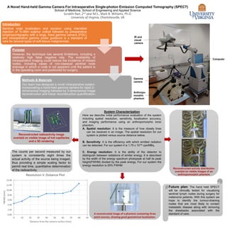
A. Spatial resolution: It is the measure of how closely lines
can be resolved in an image. The spatial resolution for our
system is plotted versus source distance below.
B. Sensitivity: It is the efficiency with which emitted radiation
can be detected. For our system it is 1.75 x 10-04 cps/MBq.
C. Energy resolution: It is the ability of the detector to
distinguish between radiations of similar energy. It is described
by the width of the energy spectrum photopeak at half its peak
height(FWHM) divided by the peak energy. For our system the
energy resolution is 25% FWHM
Reconstructed radioactivity image
overlaid on visible image of hot capillaries
and a 3D rendering
A reconstructed image of a phantom containing three
point sources, showing good geometrical localization.
Reconstructed activity distribution
overlaid on visible images of an
anthropomorphic phantom
Future plan: The hand held SPECT
will be clinically tested for visualizing
sentinel lymph nodes during surgery for
melanoma patients. With this system we
hope to identify the tumour-draining
nodes that are most likely to contain
metastatic disease along with removing
the drawbacks associated with the
standard of care.
Gamma
camera
Anthropo-
morphic
phantom
Computer
IR and
visual
camera
The counts per second measured by our
system is consistently eight times the
actual activity of the source being imaged,
thus providing a simple scaling factor to
permit real time, quantitative determination
of the radioactivity.