Antibacterial and osteo-stimulatory effects of a borate-based glass series doped with strontium ions
1. Original Article
Antibacterial and osteo-stimulatory
effects of a borate-based glass series
doped with strontium ions
Yiming Li1,2
, Wendy Stone3
, Emil H Schemitsch2
, Paul Zalzal4
,
Marcello Papini1
, Stephen D Waldman3,5
and Mark R Towler1,3
Abstract
This work considered the effect of both increasing additions of Strontium (Sr2þ
) and incubation time on solubility and
both antibacterial and osteo-stimulatory effects of a series of glasses based on the B2O3–P2O5–CaCO3–Na2CO3–TiO2–
SrCO3 series. The amorphous nature of all the glasses was confirmed by X-ray diffraction. Discs of each glass were
immersed in de-ionized water for 1, 7 and 30 days, and the water extracts were used for ion release profiles, pH
measurements and cytotoxicity testing. Atomic absorption spectroscopy was employed to detect the release of Naþ
,
Ca2þ
and Sr2þ
ions from the glasses with respect to maturation, which indicated that the addition of Sr2þ
retarded
solubility of the glass series. This effect was also confirmed by weight loss analysis through comparing the initial weight of
glass discs before and after periods of incubation. The incorporation of Sr2þ
in the glasses did not influence the pH of the
water extracts when the glasses were stored for up to 30 days. Cytotoxicity testing with an osteoblastic cell line
(MC3T3-E1) indicated that glasses with the higher (20 mol% and 25 mol%) Sr2þ
incorporation promoted proliferation
of osteoblast cells, while the glasses with lower Sr2þ
contents inhibited cell growth. The glass series, except for Ly-B5
(which contained the highest Sr2þ
incorporation; 25 mol%), were bacteriostatic against S. aureus in the short term (1–7
days) as a result of the dissolution products released.
Keywords
Borate-based glass, strontium, solubility, antibacterial effect, biocompatibility
Introduction
In order to improve the osteointegration of Ti6Al4V
total hip replacement (THR) devices and promote sta-
bility at the implant/bone interface upon implantation,
hydroxyapatite (HA) has been applied as a coating,
because HA is chemically similar to the mineral phase
of human bone.1
Such implants have been employed in
THR for over 20 years, during which 97.1% survival at
a 10-year follow-up clinical study has been recorded.1–3
However, the long-term stability of HA coatings is still
under debate.3,4
Significant loss of the HA coating on
both immobilised and continuously loaded implants
has been demonstrated in vivo.5
Fractures between the
coating and the Ti6Al4V substrate have been observed
after implantation times as short as 12 weeks and as
long as one year;6,7
the primary reason for the failure at
the interface is the residual stress due to the mismatch
of the coefficients of thermal expansion (CTE) of the
ceramic and metal components, which can induce
micro-cracking initiating the de-bonding of the coating
from the substrate.8–10
The micro-cracking of silicate
glass coatings on Ti6Al4V substrates due to mismatch
of CTE has also been recorded.11–13
However, unlike
HA and silicate glasses, borate-based glasses can have
Journal of Biomaterials Applications
0(0) 1–10
! The Author(s) 2016
Reprints and permissions:
sagepub.co.uk/journalsPermissions.nav
DOI: 10.1177/0885328216672088
jba.sagepub.com
1
Department of Mechanical & Industrial Engineering, Ryerson University,
Toronto, ON, Canada
2
Keenan Research Centre, St. Michael’s Hospital, Toronto, ON, Canada
3
Chemistry and Biology, Ryerson University, Toronto, ON, Canada
4
Oakville Memorial Hospital, Oakville, ON, Canada
5
Chemical Engineering, Ryerson University, Toronto, ON, Canada
Corresponding author:
Mark R Towler, Faculty of Engineering and Architectural Science,
Department of Mechanical & Industrial Engineering, Ryerson University,
350 Victoria Street, Toronto M5B 2K3, ON, Canada.
Email: mtowler@ryerson.ca
at RYERSON UNIV on December 5, 2016jba.sagepub.comDownloaded from
2. similar CTEs to Ti6Al4V.14
Borate-based glasses are
also capable of forming chemical bonds between bone
and the implant onto which they are coated,15
and are
reported to offer a favorable substrate for the attach-
ment and proliferation of osteogenic cells,16
and can
contribute to the healing of segmental defects
in vivo.17
Borate-based bioactive glasses, then, can be
considered as coating candidates for THR devices.14
Dissolution products of borate-based glasses have
been reported to facilitate the formation of new
bone.18–20
The rate and extent of ion release will influ-
ence new bone formation and borate-based glasses tend
to have a high dissolution rate.21
Cell damage can be
induced by high concentrations of dissolution products
and the dramatic change in pH of the environment can
occur as a result of this degradation.22
High concentra-
tions of Ca2þ
(>32 mg/L) can decrease osteoblast viabil-
ity,23
and higher than 2.5 mM BO3À
3 can inhibit bone cell
(MC3TC-E1) proliferation.24
In addition, the resorption
and degradability of the glass coating can result in the
loss of the coating–substrate bond strength and subse-
quently retard implant fixation.25
It is therefore critical
to tune the solubility of the borate-based glasses to the
rate of formation of new bone. Addition of Ca2þ
has
been reported to retard the dissolution rate of the glasses
by impeding the migration pathway of Naþ
in the glass
structure.26
Sr2þ
has a similar ionic structure to Ca2þ
,
and therefore, Sr2þ
incorporation is expected to modify
the dissolution of the glasses. Sr2þ
has also been reported
to increase osteoblast proliferation in vivo and stimulate
bone formation in vitro.27
Previous studies have investi-
gated the influence of different Sr2þ
on the solubility and
bioactivity of borate-based glasses by incorporating
strontium by partially substituting it for other modifiers
(like MgO or CaO).18,28–32
Varying amounts of glass
modifiers substituting for B2O3 have also been reported
to influence the physical properties of borate-based
glasses.33–35
However, the influence of different amounts
of SrCO3 (or SrO) at the expense of B2O3 on the solu-
bility and bioactivity of borate-based glasses has not
been researched.
In addition to introducing chemical bonding
between the bone and the implant, the bioactive coating
can be formulated to impart an antibacterial effect to
the surrounding environment as it degrades. Prosthetic
joint infection can occur at the time of implantation.
Staphylococcus aureus (S. aureus) is often the major
pathogen in metallic implant infections;36,37
a study of
isolates from 242 orthopedic patients confirmed that S.
aureus is the most prevalent etiological agent of ortho-
pedic infection.38
Previous studies indicated that stron-
tium-containing bone cements reduce the cell counts of
S. aureus,39
and boron-containing bioactive glasses
(MBG0118 and MBG0123) exert antibacterial effects
against S. aureus.40
The aim of this study is to investigate the influence of
a range of Sr2þ
contents and incubation time on the
solubility and osteo-stimulatory and antibacterial
effects on S. aureus of borate-based glasses designed
for use as coatings on surgical implants.
Materials and methods
Glass sample preparation
Six glasses (Ly-B0 to Ly-B5) were formulated based on
the B2O3–P2O5–CaCO3–Na2CO3–TiO2–SrCO3 glass
series with increasing amounts of SrCO3 (from 5 to
25 mol%) at the expense of B2O3. The control glass,
Ly-B0, was free of SrCO3. The compositions of the
glasses are presented in Table 1. Glasses were prepared
by weighing out appropriate amounts of analytical
grade reagents in powder form, firing the mixtures
(1300
C, 1 h) in a silica crucible, and subsequently
shock quenching them into water. The resulting frits
were dried, ground and sieved to retrieve powders
with a mean particle size of less than 20 mm.
X-ray diffraction
Diffraction patterns were collected using a D2 PHASER
(Bruker AXS Inc., WI, USA). Glass powder samples
were packed into standard stainless steel sample
holders. A generator voltage of 30 kV and a tube current
of 10 mA were employed. Diffractograms were collected
in the range 20
2y 90
, at a scan step size 0.02
and a
count time of 0.3 s.
Atomic absorption spectroscopy
Glass powder discs (2.2 Â 6.4 mm, n ¼ 9) fabricated
for atomic absorption spectroscopy (AAS) were pro-
duced by pressing the glass powders into moulds and
then annealing at 50
C above their Tgs, previously
determined by a combined differential thermal
Table 1. Compositions of the borate glass series, displayed in
mol%.
LY-B0 LY-B1 LY-B2 LY-B3 LY-B4 LY-B5
B2O3 59 54 49 44 39 34
CaCO3 13 13 13 13 13 13
P2O5 3 3 3 3 3 3
Na2CO3 15 15 15 15 15 15
TiO2 10 10 10 10 10 10
SrCO3 0 5 10 15 20 25
Total 100 100 100 100 100 100
2 Journal of Biomaterials Applications 0(0)
at RYERSON UNIV on December 5, 2016jba.sagepub.comDownloaded from
3. analyser–thermal gravimetric analyser (DTA-TGA,
SDT Q600, TA Instruments, New Castle, DE, USA).
The discs were then immersed in 15 mL de-ionized
water for 1, 7 and 30 days (three samples of each
glass for each incubation period). Ionic concentrations
of Naþ
, Ca2þ
and Sr2þ
were evaluated from the water
extracts utilising a Perkin Elmer Analyst 800 Atomic
Absorption Spectrometer (Waltham, MA, USA). The
water extracts were subsequently used for pH analysis
and cell culture testing.
pH analysis
Changes in pH of the water extracts were monitored by
a Corning 430 pH meter (Corning, NY, USA). Prior to
testing, the pH meter was calibrated using pH buffer
solution 4.00 Æ 0.02 and 7.00 Æ 0.02 (Fisher Scientific,
Pittsburgh, PA, USA). Sterile de-ionized water
(pH ¼ 7.0) was used as a control and was measured at
each time period for calibration purposes.
Weight loss
Weight loss measurements were carried out after
removing the glass powder discs from de-ionized
water after the incubation times of 1, 7 and 30 days
and dried for 24 h at 37
C. The equation used to calcu-
late the weight loss (ÁW) is
ÁW ¼
W0 À W
W0
ð1Þ
where W0 is the initial mass of the glass disc, which is
approximately 0.1 g, and W is the mass of the disc after
a certain incubation period.
Agar disk-diffusion test
The antibacterial activity of the borate-based glasses
was evaluated against S. aureus using the agar disk dif-
fusion method. Tryptic Soy Broth (TSB; Sigma Aldrich,
Oakville, ON, Canada) was used for the culture of S.
aureus. All organisms were grown in 100 mL TSB to a
cell concentration of 1 Â 107
cells/mL (20 h, 37
C, aer-
obically, 250 r/min). Preparation of the TSA disk-diffu-
sion plates involved aseptically spreading 100 mL of the
undiluted culture per plate. The pressed glass powder
discs with heat treatment were also used in the antibac-
terial test. Glass powder discs (n ¼ 3) were placed on the
inoculated plates and the plates were cultured for 1, 7
and 30 days at 37
C, sealing the bags to prevent desic-
cation. Three glass disks, of different compositions,
were assessed per plate. Callipers were used to measure
the diameter of glass powder discs and the halo of
inhibition at three different points for each disk, and
then zone sizes were calculated as follows
Inhibition Zone ðmmÞ ¼
Halo À Disc
2
ð2Þ
All glasses were analysed in triplicate and mean zone
sizes standard deviations were calculated.
Cytotoxicity testing
Pre-osteoblastic MC3T3-E1 cells (ATCC CRL-2593,
ATCC, Manassas, VA, USA) from passages 3–5 were
used for this study and were maintained in aMEM
media supplemented with 10% FBS and 1% (2 mM)
L-glutamine (Cambrex, MD, USA) within a cell culture
incubator at 37
C/5% CO2/95% air atmosphere. Cells
were seeded into 24-well plates at a density of 5500
cells/cm2
and incubated for 24 h prior to testing.
Culture media (1 mL) was then further supplemented
with 100 mL of liquid extract (from the solubility sam-
ples at 30 days for all glasses; n ¼ 3 per sample well) and
then incubated for 24 h at 37
/5% CO2. The MTT was
added in an amount equal to 10% of the culture
medium volume/well. The cultures were then re-incu-
bated for a further 2 h (37
C/5% CO2) after which they
were removed from the incubator and the resultant for-
mazan crystals dissolved by adding an amount of MTT
Solubilisation Solution (10% Triton x-100 in Acidic
Isopropanol (0.1 n HCI)) equal to the original culture
medium volume. Once the crystals were fully dissolved,
the absorbance was measured at a wavelength of
570 nm. All results were expressed relative to the meta-
bolic activity of cells seeded (at the same density) on
tissue culture plastic (n ¼ 3) as controls.
Statistical analysis
One-way analysis of variance (ANOVA) was employed
to compare the changes in ion release profiles, pH
values, weight loss, inhibition zone and MTT assay
data of the experimental materials in relation to (1)
different incubation times (e.g. 1, 7 and 30 days), of
each composition and (2) different glass compositions
with the same incubation time. The comparison of rele-
vant means was performed using the post hoc
Bonferroni test. Differences between groups were
deemed significant when p 0.05.
Results and discussion
X-ray diffraction
X-ray diffraction (XRD) patterns of all the materials
are shown in Figure 1. The broad XRD curves without
Li et al. 3
at RYERSON UNIV on December 5, 2016jba.sagepub.comDownloaded from
4. any detectable sharp peaks confirm the glassy nature of
the original glasses and annealed discs.41,42
Crystalline
phases can inhibit the dissolution of bioactive glasses,
influencing glass solubility and biocompatibility.43
Therefore, it is important to retain the amorphous
nature of the samples.
AAS
The concentrations of Naþ
, Ca2þ
and Sr2þ
released,
with respect to incubation time and glass composition,
are shown in Figures 2 to 4, respectively.
Unfortunately, the concentration of BO3À
3 cannot be
accurately determined by AAS because of the insensi-
tivity of this technique for low atomic number elem-
ents.21
For all six glasses, the concentrations of Naþ
and Ca2þ
in the water extracts (Figures 2 to 4) were
found to significantly increase with incubation time
(p 0.05, Table 2), while the concentrations of Naþ
and Ca2þ
decreased along with increased Sr2þ
incorp-
oration (p 0.05, Table 2) in the glasses after 30-day
incubation. For Ly-B0, Ly-B1 and Ly-B2, the concen-
trations of Naþ
measured after seven-day incubation
were very close to those measured after 30-day incuba-
tion, while Naþ
concentrations released after seven-day
incubation for the other three glasses were less than
those after 30-day incubation time. Thus, the release
rate of Naþ
in the first week was retarded by more
than 15 mol% addition of Sr2þ
in the glass series.
The first step in the degradation of a glass in an
aqueous environment is the ion exchange44,45
between
the glass network modifier cations and Hþ
from the
immersing solution. Usually, the Naþ
-water reaction
dominates the process due to the initial enrichment of
Naþ
on the glass surface.26,45
This explains why the
concentrations of Naþ
released from each glass are
higher than those of Ca2þ
and Sr2þ
(Figures 2 to 4).
Additional ions are then transported from the glass
bulk to the surface to complete the dissolution process.
One of the pathways of ion migration is assisted by
correlated forward–backward motion of an ion by
20
Ly-B5
Ly-B4
Ly-B3
Ly-B2
Ly-B1
Ly-B0
30 40 50
Degree° (2 Theta)
60 70 80 90
Figure 1. XRD patterns of the original glasses and glass power
discs.
900
ConcentrationofNa+(mg/L)
821.2
1 Day
7 Days
30 Days754.3
743.3 726.6
603.3
506.5
750
600
450
300
150
0
Ly-B0 Ly-B1 Ly-B2 Ly-B3 Ly-B4 Ly-B5
Na+
Figure 2. Concentration of Naþ from the water extracts of
the glass series versus incubation time.
80
ConcentrationofSr2+(mg/L)
Ly-B0 Ly-B1 Ly-B2 Ly-B3 Ly-B4 Ly-B5
72.9
63.9
57.9
1 Day
7 Days
30 Days
Sr2+
40.35
31.7
60
40
20
0
Figure 4. Concentration of Sr2þ from the water extracts of
the glass series versus incubation time.
35
ConcentrationofCa2+(mg/L)
29.9
1 Day
7 Days
30 Days
18.8
15.7
14.2
10.6
5.3
30
25
20
15
10
5
0
Ly-B0 Ly-B1 Ly-B2 Ly-B3 Ly-B4 Ly-B5
Ca2+
Figure 3. Concentration of Ca2þ from the water extracts of
the glass series versus incubation time.
4 Journal of Biomaterials Applications 0(0)
at RYERSON UNIV on December 5, 2016jba.sagepub.comDownloaded from
5. moving to an intermediate position and then returning
to its initial site after the passage of the migrating ion.26
The movement of a modifier cation in the matrix
involves a change in its coordination, where at least a
fraction of the coordinated oxygen atoms have been
replaced;26
that is, a number of R–O bonds have to
be broken. It is difficult for Sr2þ
to provide such ‘‘tran-
sient sites’’26
due to the high strength of the ionic Sr–O
bonding;46
thus Sr2þ
might block the pathway of other
cations. As a result, it is proposed that Sr2þ
hindered
the movement of other dissolution products, reducing
solubility of the glasses.
As expected, the ion release profile of Sr2þ
(Figure 4)
experienced a significant increase with both incubation
time and Sr2þ
incorporation in the glass series (p 0.05,
Table 2). The highest Sr2þ
concentrations in the water
extracts ranged from 31.7 mg/L to 72.9 mg/L (form Ly-
B0 to Ly-B5) after 30-day incubation. Previous studies
have reported that Sr2þ
concentrations in the range
from 8.76 mg/L to 87.62 mg/L induce stimulatory
effects on osteoblasts and inhibit bone resorption
in vitro.47,48
It has also been presented that the higher
the Sr2þ
concentration, the more pronounced the
inhibitory effect on osteoclasts differentiation up to
Sr2þ
concentrations as high as 2102.8 mg/L.49
Therefore, addition of Sr2þ
to this borate-based glass
series is expected to be beneficial for bone cell prolifer-
ation, and this hypothesis will be investigated in the
cytotoxicity testing.
Weight loss
As ion release from the glass is accompanied by a
decrease in mass, weight loss measurements provide a
useful parameter for monitoring the kinetics of glass
solubility. Weight loss of the glass powder discs with
respect to incubation time is shown in Figure 5. Weight
loss of the glasses after the 30-day incubation period
decreased in line with increased Sr2þ
incorporation
in the glasses (p 0.05, Table 3), which indicated
that lower amount of ions released from the glasses
with more Sr2þ
incorporation. In addition, for Ly-B0,
Ly-B1 and Ly-B2, there was no significant difference
(p ! 0.05, Table 3) between weight loss recorded after
7- and 30-day incubation period, which indicated that
the amount of ions released from the three glasses
reached the limit only after the 7-day incubation
period. In other words, the dissolution rate of the
three glasses was highest in the first week. However,
the weight loss of Ly-B3, Ly-B4 and Ly-B5 increased
significantly (p 0.05, Table 3) from 7-day to 30-day
incubation, which implied that the solubility of
the glasses in the first seven days was retarded due
to more than 15 mol% Sr2þ
incorporation in the
glasses.
Table 2. Comparison of each ion release profile (n ¼ 3) with respect to incubation time, and comparison of each ion concentration
(n ¼ 3) after 30-day incubation with respect to different Sr2þ
incorporation in the glasses, where p 0.05 represents significant
difference.
Incubation time (1 day vs. 30 days) Different Sr2þ
incorporation
Ions Ly-B0 Ly-B1 Ly-B2 Ly-B3 Ly-B4 Ly-B5 Ly-B0 vs.
Ly-B1
Ly-B0 vs.
Ly-B2
Ly-B0 vs.
Ly-B3
Ly-B0 vs.
Ly-B4
Ly-B0 vs.
Ly-B5
Naþ
0.000 0.000 0.000 0.000 0.000 0.000 0.001 0.000 0.000 0.000 0.000
Ca2þ
0.000 0.000 0.000 0.000 0.000 0.000 0.000 0.000 0.000 0.000 0.000
Ly-B1 vs.
Ly-B2
Ly-B1 vs.
Ly-B3
Ly-B1 vs.
Ly-B4
Ly-B1 vs.
Ly-B5
Sr2þ
0.000 0.000 0.000 0.000 0.000 0.000 0.000 0.000 0.000 0.000
60
WeightLoss(%)
50
40
30
20
10
0
1 Day 7 Days
Ly-B0
Ly-B1
Ly-B2
Ly-B3
Ly-B4
Ly-B5
30 Days
Figure 5. Weight Loss of the glass discs versus incubation time.
Table 3. Comparison of weight loss of each glass (n ¼ 3) with
respect to incubation time, where p 0.05 represents significant
difference.
Incubation Time (7 Days vs. 30 Days)
Ly-B0 Ly-B1 Ly-B2 Ly-B3 Ly-B4 Ly-B5
1.000 0.590 0.875 0.001 0.001 0.003
Li et al. 5
at RYERSON UNIV on December 5, 2016jba.sagepub.comDownloaded from
6. pH
pH of the water extracts of the glass series over 1, 7 and
30 days are shown in Figure 6. The pH of de-ionized
water was 7.0. The statistical analysis of pH profiles
demonstrated that the pH of the water extracts
increased significantly with more than 5 mol% addition
of Sr2þ
(p 0.05, Table 4). However, for each glass, pH
values did not change with immersion time (p ! 0.05,
Table 4).
In the reaction between water and glass, Hþ
is
donated to NBO and the remaining OHÀ
from the
water molecule is freed. As a consequence, pH of the
solution increases. Based on previous studies, the pH of
silicate glasses immersed in a neutral aqueous environ-
ment for 30 days are in the range of 11–12,40,41
while
the pH of borate-based glasses are in the range of 9–
10.21,50
It is the acidity of B(OH)3 that causes this
effect.21
However, the pH of the solution still increases
because the strong alkaline NaOH overwhelms the
weak acidic B(OH)3.
The alkaline pH resulting from the degradation of
the glasses has a positive influence on bioactivity.51,52
It
has been reported that bone cells respond to pH change
and higher pHs inhibit the activity of osteoclasts redu-
cing bone resorption.51
Pro-resorptive agents such as
RANKL and parathyroid hormone have little or no
stimulatory activity on osteoclasts at pH of 7.4 or
above.52
The results of pH testing also manifest that,
for each glass, pH of the water extracts remained in a
certain range (p ! 0.05, Table 4). Since the mechanism
of bone cell formation is very sensitive to change of
acidic balance, precise maintenance of pH value in
the blood and extracellular fluid is required.53
Antibacterial effect
The diameters of inhibition zones of the borate glasses
against S. aureus with respect to maturation are shown
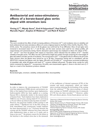
in Figure 7. There is no inhibition zone for Ly-B5
against S. aureus. The mean sizes of the inhibition
zones after one-day incubation are 5.6 mm for Ly-B0,
5.8 mm for Ly-B1, 3.5 mm for Ly-B2, 4.6 mm for Ly-B3
and 4.1 mm for Ly-B4. Based on statistical analysis,
there is no difference (p ! 0.05, Table 5) among the
sizes of the inhibition zones for these five glasses after
Table 4. Comparison of pH of each glass (n ¼ 3) with respect to incubation time, and comparison of pH after 30-day incubation with
respect to different Sr2þ
incorporation in the glasses, where p 0.05 represents significant difference.
Incubation time (1 day vs. 30 days)
Ly-B0 Ly-B1 Ly-B2 Ly-B3 Ly-B4 Ly-B5
0.008 0.293 0.593 0.387 0.105 0.130
Different Sr2þ
incorporation
Ly-B0 vs. Ly-B1 Ly-B0 vs. Ly-B2 Ly-B0 vs. Ly-B3 Ly-B0 vs. Ly-B4 Ly-B0 vs. Ly-B5
0.657 0.000 0.000 0.000 0.000
10
Diameterofinhibitionzone(mm)
1-Day
NOInhibitionZoon
NOInhibitionZoon
NOInhibitionZoon
NOInhibitionZoon
7-Day
30-Day
8
6
4
2
0
Ly-B0 Ly-B1 Ly-B2 Ly-B3 Ly-B4 Ly-B5
Figure 7. Diameters of inhibition zones of the glasses against S.
aureus with different maturation times, where deviations are
presented.
9.8 1 Day
Ly-B0 Ly-B1 Ly-B2 Ly-B3 Ly-B4 Ly-B5
7 Days
30 Days
pHValue
9.6
9.4
9.2
9.0
8.8
8.6
8.81
8.86
9.02
9.26
9.38
9.57
Figure 6. pH values of the water extracts of the glass series
with different incubation times, where the pH values of 30-day
incubation are tagged on the image.
6 Journal of Biomaterials Applications 0(0)
at RYERSON UNIV on December 5, 2016jba.sagepub.comDownloaded from
7. one-day incubation. In addition, there is no difference
(p ! 0.05, Table 5) among the sizes of inhibition zones
after 7-day and 30-day incubation for Ly-B0 and Ly-
B1. However, the inhibition zones after the 7-day incu-
bation period experienced a significant decrease
(p 0.05, Table 5) for the glasses with 15–25 mol%
addition of Sr2þ
.
Based on the previous studies concerning the anti-
bacterial effects of bioactive glasses, some dissolution
products such as zinc or silver ions kill bacteria by
inhibiting multiple activities in the bacterial cell, such
as glycolysis, trans-membrane proton translocation and
acid tolerance.54
Furthermore, the antibacterial effect is
proportional to the concentration of these ions.54
Sr2þ
has been reported to exhibit antibacterial activity
against S. aureus, but at a weak level.55
It is postulated
that Sr2þ
exerts its antibacterial ability by inhibiting
bacterial growth and reproduction and impeding per-
meability of cytoplasmic membrane, cell wall synthesis,
replication of bacterial chromosomes and cell metabol-
ism.39
Based on the AAS data, the increased Sr2þ
released from the glasses with higher Sr2þ
loadings
has no positive effect on inhibition zone size (Figure
7). An inhibition zone also exists for Ly-B0 which
does not contain, or subsequently release, Sr2þ
. In add-
ition, the dissolution mechanism of Sr2þ
in TSB culture
of small volume (100 mL) might be different from that in
de-ionized water. Here, we assume that other dissol-
ution products may contribute to the inhibition zone.
It has been reported that boron-containing bioactive
glass exerts antibacterial effects against S. aureus due
to BO3À
3 release, but the antibacterial mechanism of
BO3À
3 is still unknown.40
In addition, a boron-based
antibacterial (AN3365) was reported to reveal antibac-
terial activity against S. aureus.56
Naþ
and Ca2þ
can
also inhibit the growth of S. aureus.57,58
The weight loss
data (Figure 5) manifests that lower numbers of dissol-
ution products release from the glasses with Sr2þ
incorporation, especially for the glasses with more
than 5 mol% addition of Sr2þ
, which is in agreement
with the fact that the sizes of the inhibition zones
decreased or disappeared after seven-day incubation
with more than 5 mol% Sr2þ
contents in the glasses.
Thus, these glass discs are able to inhibit bacterial pro-
liferation in one week, but not in the long term. In
summary, the concentrations of all ions may have an
influence on the sizes of the inhibition zones. In other
words, a combined or individual effect of some ions
among BO3À
3 , Sr2þ
, Naþ
and Ca2þ
makes the contribu-
tion to the bacteriostatic59
behaviour of the glasses.
Cytotoxicity testing
The cytotoxicity results from glass powder disc extracts
after 30-day incubation are shown in Figure 8. There is
no difference (p ! 0.05, Table 6) among the cell meta-
bolic activity of Ly-B0, Ly-B1, Ly-B2 and Ly-B3
glasses, which all experienced significantly reduced pro-
liferation compared to control (p 0.05, Table 6).
However, the cell proliferation was significantly
enhanced (p 0.05, Table 6) in response to the Ly-B4
(105%) and Ly-B5 (120%) glass formulations.
Compared to the control group, the enhancement of
cell metabolic activity on Ly-B5 was significant
(p ¼ 0.002).
Based on the results of pH measurement and AAS
analysis, concentrations of Sr2þ
ions released increased
with increasing Sr2þ
contents in the glass, while the
concentrations of BO3À
3 decreased after 30-day incuba-
tion, as would be expected. It has been reported that
high concentrations of BO3À
3 (1 mg/L) inhibit prolif-
eration of osteoblasts (MC3T3-E1),24,60
while concen-
trations of Sr2þ
in the range from 8.76 mg/L to
87.62 mg/L promote the proliferation of osteoblastic
cells (MC3T3-E1) in vitro.43
Therefore, this borate-
125
Normalizedcellmetabolicactivity(%)
100
75
50
25
0
Control Ly-B0 Ly-B1 Ly-B2 Ly-B3 Ly-B4 Ly-B5
Figure 8. Cell metabolic activity normalised by the control
group from sintered glass powder disc extracts after 30-day
incubation.
Table 5. Means comparison of the size of inhibition zones
(n ¼ 3) after 1, 7 and 30 days incubation with respect to different
Sr2þ
incorporation in the glasses, where p 0.05 represents
significant difference.
Different Sr2þ
incorporation
Ly-B0 vs.
Ly-B1
Ly-B0 vs.
Ly-B2
Ly-B0 vs.
Ly-B3
Ly-B0 vs.
Ly-B4
1 Day 1.000 0.205 1.000 0.829
7 Days 1.000 0.000 0.004 0.000
30 Days 0.233
Li et al. 7
at RYERSON UNIV on December 5, 2016jba.sagepub.comDownloaded from
8. based glass series promote the proliferation of osteo-
blastic cells with 20 mol% and 25 mol% Sr2þ
incorpo-
rated in the glasses.
Conclusion
This study was conducted to investigate the solubility
and antibacterial and osteo-stimulatory effects of a
novel borate-based glass series with respect to both
increasing additions of Sr2þ
and incubation time. The
concentrations of Naþ
, Ca2þ
and Sr2þ
in the water
extracts experienced significant increases with incuba-
tion time. However, less Naþ
and Ca2þ
released from
the glasses with increasing Sr2þ
incorporation after 30-
day incubation, indicating that the Sr2þ
doping
retarded the dissolution rate of the glasses. Sr2þ
incorp-
oration also made a contribution to the maintenance of
pH values of the water extracts along with incubation
time. In addition, the glass series promoted prolifer-
ation of osteoblastic cells with 20 mol% and 25 mol%
Sr2þ
contents, while the other glasses impeded cell
growth. All members of the glass series, except for
Ly-B5, exhibited bacteriostatic behaviour against S.
aureus in the short term (1–7 days), which might be a
result of a combined or individual effect of some of the
dissolution products.
Declaration of Conflicting Interests
The author(s) declared no potential conflicts of interest with
respect to the research, authorship, and/or publication of this
article.
Funding
The author(s) disclosed receipt of the following financial sup-
port for the research, authorship, and/or publication of this
article: The authors gratefully acknowledge the support of
both the Canadian Institute of Health Research (CIHR)
and the Natural Sciences and Engineering Research Council
of Canada (NSERC) through the Collaborative Health
Research Project (CHRP) program (grant no. 315694-DAN).
References
1. Geesink R, de Groot K and Klein C. Bonding of bone to
apatite-coated implants. J Bone Joint Surg Br 1988; 70:
17–22.
2. Herrera A, Mateo J, Gil-Albarova J, et al. Cementless
hydroxyapatite coated hip prostheses. BioMed Res Int
2015; 2015: 1–13.
3. Mohseni E, Zalnezhad E and Bushroa A. Comparative
investigation on the adhesion of hydroxyapatite coating
on Ti–6Al–4V implant: a review paper. Int J Adhes Adhes
2014; 48: 238–257.
4. Ong J, Appleford M, Oh S, et al. The characterization
and development of bioactive hydroxyapatite coatings.
JOM 2006; 58: 67–69.
5. Overgaard S. Calcium phosphate coatings for fixation
of bone implants. Evaluated mechanically and histologi-
cally by stereological methods. Acta Orthopaed 2001; 71:
1–74.
6. Ong JL, Carnes DL and Bessho K. Evaluation of tita-
nium plasma-sprayed and plasma-sprayed hydroxyapa-
tite implants in vivo. Biomaterials 2004; 25: 4601–4606.
7. Darimont G, Cloots R, Heinen E, et al. In vivo behaviour
of hydroxyapatite coatings on titanium implants: a quan-
titative study in the rabbit. Biomaterials 2002; 23:
2569–2575.
8. Lu Y-P, Li M-S, Li S-T, et al. Plasma-sprayed hydroxy-
apatite titania composite bond coat for hydroxyapatite
coating on titanium substrate. Biomaterials 2004; 25:
4393–4403.
9. Nakamura S, Otsuka R, Aoki H, et al. Thermal expan-
sion of hydroxyapatite-b-tricalcium phosphate ceramics.
Thermochim Acta 1990; 165: 57–72.
10. Yang Y-C and Chang E. Measurements of residual
stresses in plasma-sprayed hydroxyapatite coatings on
titanium alloy. Surf Coat Technol 2005; 190: 122–131.
11. Donald I, Mallinson P, Metcalfe B, et al. Recent devel-
opments in the preparation, characterization and appli-
cations of glass-and glass–ceramic-to-metal seals and
coatings. J Mater Sci 2011; 46: 1975–2000.
12. Bellucci D, Cannillo V and Sola A. Coefficient of thermal
expansion of bioactive glasses: Available literature data
and analytical equation estimates. Ceram Int 2011; 37:
2963–2972.
13. Pavon J, Jimenez-Pique E, Anglada M, et al. Stress–cor-
rosion cracking by indentation techniques of a glass coat-
ing on Ti6Al4V for biomedical applications. J Eur Ceram
Soc 2006; 26: 1159–1169.
14. Peddi L, Brow RK and Brown RF. Bioactive borate glass
coatings for titanium alloys. J Mater Sci Mater Med
2008; 19: 3145–3152.
15. Xiao W, Luo S-H, Wei X-J, et al. Evaluation of Ti
implants coated with Ag-containing borate bioactive
Table 6. Comparison of cell metabolic activity (relative to control) (n ¼ 3) after 30-day incubation with respect to the control group
and different Sr2þ
incorporation in the glasses, where p 0.05 represents significant difference.
Different Sr2þ
incorporation
Ly-B0 vs. Ly-B1 Ly-B0 vs. Ly-B2 Ly-B0 vs. Ly-B3 Ly-B0 vs. Ly-B4 Ly-B0 vs. Ly-B5
1.000 1.000 1.000 0.002 0.000
Control vs. Ly-B0 Control vs. Ly-B1 Control vs. Ly-B2 Control vs. Ly-B3 Control vs. Ly-B4 Control vs. Ly-B5
0.003 0.004 0.006 0.019 1.00 0.002
8 Journal of Biomaterials Applications 0(0)
at RYERSON UNIV on December 5, 2016jba.sagepub.comDownloaded from
9. glass for simultaneous eradication of infection and frac-
ture fixation in a rabbit tibial model. J Mater Res 2012;
27: 3147–3156.
16. Liu X, Pan H, Fu H, et al. Conversion of borate-based
glass scaffold to hydroxyapatite in a dilute phosphate
solution. Biomed Mater 2010; 5: 015005.
17. Bi L, Zobell B, Liu X, et al. Healing of critical-size seg-
mental defects in rat femora using strong porous bio-
active glass scaffolds. Mater Sci Eng C 2014; 42:
816–824.
18. Pan H, Zhao X, Zhang X, et al. Strontium borate glass:
potential biomaterial for bone regeneration. J R Soc
Interface 2009; 7: 1025–1031.
19. Sheng MH-C, Taper LJ, Veit H, et al. Dietary boron
supplementation enhances the effects of estrogen on
bone mineral balance in ovariectomized rats. Biol Trace
Elem Res 2001; 81: 29–45.
20. Uysal T, Ustdal A, Sonmez MF, et al. Stimulation of
bone formation by dietary boron in an orthopedically
expanded suture in rabbits. Angle Orthod 2009; 79:
984–990.
21. Huang W, Day DE, Kittiratanapiboon K, et al. Kinetics
and mechanisms of the conversion of silicate (45S5),
borate, and borosilicate glasses to hydroxyapatite in
dilute phosphate solutions. J Mater Sci Mater Med
2006; 17: 583–596.
22. El-Ghannam A, Ducheyne P and Shapiro IM. Bioactive
material template for in vitro, synthesis of bone. J Biomed
Mater Res 1995; 29: 359–370.
23. Maeno S, Niki Y, Matsumoto H, et al. The effect of cal-
cium ion concentration on osteoblast viability, prolifer-
ation and differentiation in monolayer and 3D culture.
Biomaterials 2005; 26: 4847–4855.
24. Brown RF, Rahaman MN, Dwilewicz AB, et al. Effect of
borate glass composition on its conversion to hydroxy-
apatite and on the proliferation of MC3T3-E1 cells. J
Biomed Mater Res A 2009; 88: 392–400.
25. Sun L, Berndt CC, Gross KA, et al. Material fundamen-
tals and clinical performance of plasma-sprayed hydroxy-
apatite coatings: a review. J Biomed Mater Res 2001; 58:
570–592.
26. Tilocca A. Sodium migration pathways in multicompo-
nent silicate glasses: Car–Parrinello molecular dynamics
simulations. J Chem Phys 2010; 133: 014701.
27. O’Donnell M, Candarlioglu P, Miller C, et al. Materials
characterisation and cytotoxic assessment of strontium-
substituted bioactive glasses for bone regeneration. J
Mater Chem 2010; 20: 8934–8941.
28. Thind K, Singh K, Kumar V, et al. Compositional
dependence of in-vitro bioactivity in sodium calcium
borate glasses. J Phys Chem Solid 2009; 70: 1137–1141.
29. O’Connell K, Hanson M, O’Shea H, et al. Linear release
of strontium ions from high borate glasses via lanthanide/
alkali substitutions. J Non-cryst Solid 2015; 430: 1–8.
30. Jung SB. Borate based bioactive glass scaffolds for hard
and soft tissue engineering. Doctoral Dissertations, Paper
2075, 2010.
31. Shen L, Coughlan A, Towler M, et al. Degradable borate
glass polyalkenoate cements. J Mater Sci Mater Med
2014; 25: 965–973.
32. Marzouk MA and ElBatal HA. In vitro bioactivity of
soda lime borate glasses with substituted SrO in sodium
phosphate solution. Process Appl Ceram 2014; 8:
167–177.
33. Yiannopoulos Y, Chryssikos GD and Kamitsos E.
Structure and properties of alkaline earth borate glasses.
Phys Chem Glasses-Eur J Glass Sci Technol Part B 2001;
42: 164–172.
34. Pye LD, Fre´ chette VD and Kreidl NJ. Borate glasses:
structure, properties, applications. New York: Springer
Science Business Media, 2012.
35. Lower NP, McRae JL, Feller HA, et al. Physical proper-
ties of alkaline-earth and alkali borate glasses prepared
over an extended range of compositions. J Non-cryst
Solid 2001; 293: 669–675.
36. Murdoch DR, Roberts SA, Fowler VG, et al. Infection of
orthopedic prostheses after Staphylococcus aureus bac-
teremia. Clin Infect Dis 2001; 32: 647–649.
37. An YH and Friedman RJ. Concise review of mechanisms
of bacterial adhesion to biomaterial surfaces. J Biomed
Mater Res 1998; 43: 338–348.
38. Montanaro L, Speziale P, Campoccia D, et al. Scenery of
Staphylococcus implant infections in orthopedics. Future
Microbiol 2011; 6: 1329–1349.
39. Brauer DS, Karpukhina N, Kedia G, et al. Bactericidal
strontium-releasing injectable bone cements based on
bioactive glasses. J R Soc Interf 2012; 10: 20120647–
20120654.
40. Munukka E, Leppa¨ ranta O, Korkeama¨ ki M, et al.
Bactericidal effects of bioactive glasses on clinically
important aerobic bacteria. J Mater Sci Mater Med
2008; 19: 27–32.
41. Petkov V, Ohta T, Hou Y, et al. Atomic-scale structure of
nanocrystals by high-energy X-ray diffraction and atomic
pair distribution function analysis: study of FexPd100-x
(x¼ 0, 26, 28, 48) nanoparticles. J Phys Chem C 2007;
111: 714–720.
42. Warren BE and Biscce J. The structure of silica glass by
X-ray diffraction studies. J Am Ceram Soc 1938; 21:
49–54.
43. Li Y, Placek L, Coughlan A, et al. Investigating the influ-
ence of Naþ and Sr2þ on the structure and solubility of
SiO2–TiO2–CaO–Na2O/SrO bioactive glass. J Mater Sci
Mater Med 2015; 26: 1–12.
44. Tilocca A and Cormack AN. Surface signatures of bio-
activity: MD simulations of 45S and 65S silicate glasses.
Langmuir 2009; 26: 545–551.
45. Tilocca A and Cormack AN. Modeling the water–bio-
glass interface by ab initio molecular dynamics simula-
tions. ACS Appl Mater Interf 2009; 1: 1324–1333.
46. Lao J, Nedelec J-M and Jallot E. New strontium-based
bioactive glasses: physicochemical reactivity and deliver-
ing capability of biologically active dissolution products.
J Mater Chem 2009; 19: 2940–2949.
47. Marie P, Ammann P, Boivin G, et al. Mechanisms of
action and therapeutic potential of strontium in bone.
Calcified Tissue Int 2001; 69: 121–129.
48. Murphy S, Boyd D, Moane S, et al. The effect of com-
position on ion release from Ca–Sr–Na–Zn–Si glass bone
grafts. J Mater Sci Mater Med 2009; 20: 2207–2214.
Li et al. 9
at RYERSON UNIV on December 5, 2016jba.sagepub.comDownloaded from
10. 49. Goel A, Rajagopal RR and Ferreira JM. Influence of
strontium on structure, sintering and biodegradation
behaviour of CaO–MgO–SrO–SiO2–P2O5–CaF2 glasses.
Acta Biomater 2011; 7: 4071–4080.
50. Zhao D, Huang W, Rahaman MN, et al. Mechanism for
converting Al 2O3-containing borate glass to hydroxy-
apatite in aqueous phosphate solution. Acta Biomater
2009; 5: 1265–1273.
51. Arentt TR and Dempster DW. Effect of pH on bone
resorption by rat osteoclasts in vitro. Endocrinology
1986; 119: 119–124.
52. Arnett TR. Extracellular pH regulates bone cell function.
J Nutr 2008; 138: 415S–418S.
53. Arnett TR (ed.) Acid–base regulation of bone metabol-
ism. Int Congr Ser 2007; 1297: 255–267.
54. Boyd D, Li H, Tanner D, et al. The antibacterial effects
of zinc ion migration from zinc-based glass polyalkenoate
cements. J Mater Sci Mater Med 2006; 17: 489–494.
55. Ravi ND, Balu R and Sampath Kumar T. Strontium-
substituted calcium deficient hydroxyapatite nanoparti-
cles: synthesis, characterization, and antibacterial proper-
ties. J Am Ceram Soc 2012; 95: 2700–2708.
56. Hernandez V, Cre´ pin T, Palencia A, et al. Discovery of a
novel class of boron-based antibacterials with activity
against Gram-negative bacteria. Antimicrob Agent
Chemother 2013; 57: 1394–1403.
57. Zhang D, Leppa¨ ranta O, Munukka E, et al. Antibacterial
effects and dissolution behavior of six bioactive glasses. J
Biomed Mater Res A 2010; 93: 475–483.
58. Petersen PJ, Bradford PA, Weiss WJ, et al. In vitro and
in vivo activities of tigecycline (GAR-936), daptomycin,
and comparative antimicrobial agents against glycopep-
tide-intermediate Staphylococcus aureus and other resist-
ant Gram-positive pathogens. Antimicrob Agent
Chemother 2002; 46: 2595–2601.
59. Vitale-Brovarone C, Miola M, Balagna C, et al. 3D-
glass–ceramic scaffolds with antibacterial properties for
bone grafting. Chem Eng J 2008; 137: 129–136.
60. Hakki SS, Bozkurt BS and Hakki EE. Boron regulates
mineralized tissue-associated proteins in osteoblasts
(MC3T3-E1). J Trace Elem Med Biol 2010; 24: 243–250.
10 Journal of Biomaterials Applications 0(0)
at RYERSON UNIV on December 5, 2016jba.sagepub.comDownloaded from