Short case...Arterial embolic hemorrhagic infarction
•
2 likes•339 views

Short case...Arterial embolic hemorrhagic infarction http://yassermetwally.com http://yassermetwally.net

Recommended
More Related Content
Viewers also liked
Viewers also liked (12)
Similar to Short case...Arterial embolic hemorrhagic infarction
Similar to Short case...Arterial embolic hemorrhagic infarction (20)
More from Professor Yasser Metwally
More from Professor Yasser Metwally (20)
Recently uploaded
Recently uploaded (20)
Short case...Arterial embolic hemorrhagic infarction
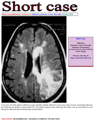
- 1. Short case publication... Version 4.1 | Edited by professor Yasser Metwally | October 2010 Short case Edited by Professor Yasser Metwally Professor of neurology Ain Shams university school of medicine Cairo, Egypt Visit my web site at: http://yassermetwally.com A 65 years old male patient suffered an acute ischemic embolic infarction in the acute stage of acute myocardial infarction and following an episode of paroxysmal AF. The patient clinical course following the stroke was an uneventful on even though the infarction showed hemorragic transformation. DIAGNOSIS: HEMORRHAGIC ARTERIAL EMBOLIC INFARCTION
- 2. Figure 1. Precontrast CT scan showing right posterior parietal wedge-shaped, well defined, cortical/subcortical and predominantly hypodense- embolic infarction. Notice the hemorrhagic transformation of the infarction manifested in the peripheral hyperdensities. The infarction is in the vascular territory of the posterior parietal artery. Figure 2. Precontrast MRI T1 images showing the posterior parietal /occipital infarction shown in figure 1. Notice the peripheral precontrast MRI T1 hyperintensities (Methemoglobin) surrounding the infarction. Mass effect is evident. Punctate hyperintensities are also seen within the infarcted brain tissues.
- 3. Figure 3. MRI T2 images showing the heterogeneous nature of the infarction. The hyperintense areas most probably represent edema and the hypointense areas most probably represent blood products (deoxyhemoglobin in the acute stage and hemosiderin in the chronic stage) Figure 4. MRI FLAIR images showing the heterogeneous nature of the parietal/occipital infarction. The hyperintense areas most probably represent edema and the hypointense areas most probably represent blood products (deoxyhemoglobin in the acute stage and hemosiderin in the chronic stage). Notice the multiple small hyperintense infarctions scattered in the periventricular and cortical/subcortical zones of both hemisphere representing multiplicity of emboli which is quite evident in FLAIR images. References 1. Metwally, MYM: Textbook of neurimaging, A CD-ROM publication, (Metwally, MYM editor) WEB-CD agency for electronic publishing, version 11.4a October 2010
- 4. Addendum A new version of short case is uploaded in my web site every week (every Saturday and remains available till Friday.) To download the current version follow the link "http://pdf.yassermetwally.com/short.pdf". You can download the long case version of this short case during the same week from: http://pdf.yassermetwally.com/case.pdf or visit web site: http://pdf.yassermetwally.com To download the software version of the publication (crow.exe) follow the link: http://neurology.yassermetwally.com/crow.zip At the end of each year, all the publications are compiled on a single CD-ROM, please contact the author to know more details. Also to view a list of the previously published case records follow the following link: (http://wordpress.com/tag/case-record/) or click on it if it appears as a link in your PDF reader To inspect the patient's full radiological study, click on the attachment icon (the paper clip icon in the left pane) of the acrobat reader then double click on the attached file Click here to download the long case version of this short case in PDF format