Short case... Arterial ectasia of the vertebrobasilar system
•
0 gefällt mir•604 views

Short case... Arterial ectasia of the vertebrobasilar system

Empfohlen
Empfohlen
Weitere ähnliche Inhalte
Andere mochten auch
Andere mochten auch (20)
Ähnlich wie Short case... Arterial ectasia of the vertebrobasilar system
Ähnlich wie Short case... Arterial ectasia of the vertebrobasilar system (20)
Mehr von Professor Yasser Metwally
Mehr von Professor Yasser Metwally (20)
Kürzlich hochgeladen
Kürzlich hochgeladen (20)
Short case... Arterial ectasia of the vertebrobasilar system
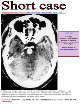
- 1. Short case publication... version 1.5 | Edited by professor Yasser Metwally | January 2008 Short case Edited by Professor Yasser Metwally Professor of neurology Ain Shams university school of medicine Cairo, Egypt Visit my web site at: http://yassermetwally.com A 60 years old male patient presented clinically with a clinical picture simulating the cerebellopontine angle syndrome with 7th, 5th, bulbar cranial nerve manifestations, cerebellar deficits and tinnitus in the right ear. Symptoms are gradual and progressive. The patient is hypertensive, with evidence of concentric left ventricular hypertrophy and type IV hyperlipidemia. The patient was not diabetic. DIAGNOSIS: FUSIFORM ANEURYSM OF THE VERTEBROBASILAR SYSTEM WITH MURAL THROMBOSIS
- 2. Figure 1. Postcontrast CT scan showing fusiform aneurysm affecting the vertebral arteries, the arteries are asymmetrically dilated, oval in shape, laterally placed and encroaching upon the cerebellopontine angle, with significant indentation of the brain stem. Also notice the mural calcification. Figure 2. Postcontrast CT scan showing fusiform aneurysm affecting the vertebral arteries, the arteries are asymmetrically dilated, oval in shape, laterally placed and encroaching upon the cerebellopontine angle, with significant indentation of the brain stem. Also notice the mural calcification.
- 3. Figure 3. Precontrast CT scans showing basilar artery fusiform aneurysm, notice the rounded configuration, the midline location in the prepontine cistern, and the precontrast hyperdensity that could be due to thrombosis ( density below that of calcification). Figure 4. Conventional angiography showing basilar artery fusiform aneurysm. Mural thrombosis is probably present.
- 4. SUMMARY Intracranial fusiform aneurysms Commonly involve the vertebrobasilar system and might extend to involve other arteries around the circle of Willis Involved arteries are diffusely dilated, tortuous, kinked, abnormally prolonged with frequent mural thrombosis and occasional wall calcification. Fusiform aneurysms rarely rupture or produce subarachnoid haemorrhage Fusiform aneurysms are commonly associated with microvascular brain disease The clinical presentation of fusiform aneurysms includes Ischemic manifestations Pressure due to the mass effect of greatly dilated fusiform aneurysms Addendum A new version of this software is uploaded in my web site every week (every Saturday and remains available till Friday.) To download the current version follow the link quot;http://pdf.yassermetwally.com/short.pdfquot;. You can download the long case version of this short case during the same week from: http://pdf.yassermetwally.com/case.pdf or visit web site: http://pdf.yassermetwally.com To download the software version of the publication (crow.exe) follow the link: http://neurology.yassermetwally.com/crow.zip At the end of each year, all the publications are compiled on a single CD-ROM, please contact the author to know more details. Screen resolution is better set at 1024*768 pixel screen area for optimum display References 1. Metwally, MYM: Textbook of neurimaging, A CD-ROM publication, (Metwally, MYM editor) WEB-CD agency for electronic publishing, version 9.1a January 2008