
Empfohlen
Weitere ähnliche Inhalte
Was ist angesagt?
Was ist angesagt? (20)
Ähnlich wie Osteosarcoma ppt
Ähnlich wie Osteosarcoma ppt (20)
Kürzlich hochgeladen
Kürzlich hochgeladen (20)
Osteosarcoma ppt
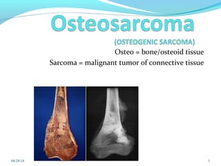
- 1. Osteo = bone/osteoid tissue Sarcoma = malignant tumor of connective tissue 04/28/14 1
- 3. What is osteosarcoma ? Highly malignant tumor arising from primitive mesenchymal bone-forming cells. Histologic hallmark is production of malignant osteoid. Mc histological form of primary bone cancer . 2nd most common primary malignant bone tumor after MM. 3
- 4. • Any bone can be involved but the Mc sites are – distal femur, proximal tibia, proximal humerus. • Mc metastasis to the lungs through blood stream. INCIDENCE: More prevalent in males than female. Involves any age but highest occurrence in adolescence i.e. 12-25yrs. 4
- 5. Distal femur Proximal tibia Proximal humerus (sites of rapid bone growth) others Metaphyseal(89%)>diaphyseal(10%)>epiphyseal(1%) 5 Skeletal distribution
- 6. Etiology Rapid bone growth – adolescence growth spurt in the metaphyseal area near the growth plate. Radiation exposure – mostly causes secondary forms. Genetic predisposition : - hereditary form of retinoblastoma( RB gene mutation) - Li-Fraumeni syndrome (p53 gene mutation) - Rothmund – Thomson syndrome ( autosomal recessive) Paget's disease of bone – mostly secondary forms. 6
- 7. Classification PRIMARY or SECONDARY PRIMARY OSTEOSARCOMAS (15 – 25 yrs) Conventional /classic osteosarcoma (high grade, intra medullar y) Low-grade intramedullary osteosarcoma Paraosteal osteosarcoma Periosteal osteosarcoma High-grade surface osteosarcoma Telangiectatic osteosarcoma, and Small cell osteosarcoma. 7
- 8. Classification SECONDARY OSTEOSARCOMAS Osteosarcoma occurring at the site of another disease process. more common in >50 years of age most commonly a/w premalignant condition like - Paget disease - Previous radiation treatment - endochondromatosis - Fibrous dysplasia - Osteochondromas - Osteogenesis imperfecta 8
- 9. 1. Conventional type: osteoblastic, chondroblastic & fibroblastic OS 2. Telangiectatic or osteolytic type OS 3. Small cell OS 4. Low grade central OS 5. Periosteal OS 6. Paraosteal OS 7. Secondary OS 8. High grade surface OS 9. Extra skeletal OS 9 HISTOPATHOLOGIC VARIENTS:
- 10. Gross pathology 10 Osteoblastic tumor – grayish white hard & gritty feeling when cut. Chondroid type – opalescent & bluish grey. Fibroblastic – typical fish flesh sarcomatous appearance. Telangiectatic – areas of tumor necrosis & blood filled spaces.
- 11. Histologic appearance Stroma - Malignant connective tissue with anaplastic spindle cells in mesenchymal parenchyma. Tumor cells surrounded by osteoid matrix c/f Osteoblastic – lot of new bone fibrous – fibroblasts Chondroid - cartilaginous tissue 11
- 12. Clinical Presentation Pain– progressive pain particularly with activity. Swelling - Palpable mass in the region of metaphysis. - skin over the swelling shiny with prominent veins. - swelling may be warm & tender. Decreased range of motion of the involved joint. Lymphadenopathy – unusual focal & regional lymph node involvement. Respiratory finding – late stage with lung metastasis. Fever & night sweats are rare. 12
- 13. Investigations: Radiological examination - plain X-rays - MRI - CT scan - bone scan • Laboratory studies – serum alkaline phosphatase. • Biopsy – core biopsy, FNAC • Angiogram 13
- 15. Plain X-ray Irregular destruction in metaphysis. New bone formation. Periosteal reaction. Sunray appearance or hair on end - tumor grows into the overlying soft tissue. Codman’s triangle – area off sub periosteal new bone formation seen at the area of tumor -host cortex junction. 15
- 16. 16 Codman triangle is a term used to describe the triangular area of subperiosteal bone that is created when a lesion, often a tumor, raises the periosteum away from the bone.
- 17. MRI : • used to know the soft tissue extant. CT: • Not of much use in osteosarcoma. CT chest to detect lung metastasis 17
- 18. Angiogram • Determine vascularity of the tumour • Detect vascular displacement • Relationship of vessels to the tumour 18
- 19. Bone scan A bone scan to look for skeletal metastases or multi focal disease Thallium scan - Monitor effects of chemotherapy & Detect local recurrence of tumor 19
- 20. laboratory studies Full blood count, ESR, CRP. LDH (elevated level is associated with poor prognosis) ALP (highly osteogenic) Platelet count Electrolyte levels Liver function tests Renal function tests Urinalysis 20
- 21. Biopsy to conform the diagnosis. Types Fine needle aspiration Core needle biopsy Open incisional biopsy 21
- 22. Enneking staging system The staging system is typically depicted as follows Stage I: Low grade tumors I-A intra compartmental I-B extra compartmental Stage II: High grade tumors II-A intra compartmental II-B extra compartmental Stage III: Any tumors with evidence of metastasis 22
- 23. Differential Dx Giant Cell Tumor Chondrosarcoma Fibrosarcoma Aneursymal Bone Cyst Ewings sarcoma Osteoblastoma Metastasis Lymphoma Osteomyelitis Chondroblastoma Post traumatic callus Other variants 23
- 24. Telangiectatic OS Painful radiologically lytic mass lesion in the metaphyseal portion of long bones. Mc site – distal femur. Histology - Blood filled vascular spaces lined by osteoblasts. Presents with local pain, soft tissue mass & fractures. d/d aneurysmal bone cyst. Mets through hematogenous route to bones & lung. 24
- 25. Parosteal OS Arise from the periosteum. Mc occurrence in adults. Mc site Posterior metaphysis of distal femur. slow growing & late to metastasize. Better prognosis. Large ossified mass in centre. 25
- 26. Periosteal OS Arises from diaphysis of long bones. Mc femur & tibia Presents with swelling & tenderness. 25% pathological fractures. X-ray shows sunburst or hair on end periosteal reaction. Histology – lobular & chondroblastic matrix. Intermediate prognosis. 26
- 27. Prognostic Factors Extent of the disease Pts with pulmonary, non pulmonry (bone) or skip metastasis have poor prognosis Grade of the tumor High grade tumor have poor prognosis Size of the primary lesion Large size tumors have worse prognosis then small size tumors Skeletal location proximal tumors do worse than distal tumors. Secondary osteosarcoma: Poor prognosis 27
- 28. Treatment Radiological staging Biopsy to confirm diagnosis Preoperative chemotherapy Repeat radiological staging (access chemo response, finalize surgical treatment plan) Surgical resection with wide margin Reconstruction using one of many techniques Post op chemo based on preop response 28
- 29. Chemotherapy Chemotherapy given preoperatively -Neoadjuvant Given postoperatively - Adjuvant Advantages of neoadjuvant chemotherapy - regression of the primary tumor, making a successful limb salvage operation easier. may decrease the spread of tumor cells at the time of surgery Effectively treating micro metastases at the earliest time possible. It avoid tumor progression, which may occur during any delay before surgery. Given for about 3-4 weeks before definitive procedure 29
- 30. Chemotherapy The drugs used most often to treat osteosarcoma are: Methotrexate with leucovorin (folinic acid) Doxorubicin (Adriamycin) Cisplatin or carboplatin Citrovorum factor. Etoposide Cyclophosphamide Actinomycin D Bleomycin 30
- 31. Surgery The main goal of surgery is to safely and completely remove the tumor. Historically – amputation. Over the past few years - limb-sparing procedures have become the standard, mainly due to advances in chemotherapy and sophisticated imaging techniques Limb salvage procedures now can provide rates of local control and long-term survival equal to amputation. 31
- 32. Limb salvage surgery Removing the tumor with a wide margin of normal tissue surrounding it while preserving vascular and nerve supply to the extremity. 32
- 33. The skeletal defect after limb salvage procedure must be reconstructed by : Autologous bone graft. Allograft Endoprosthesis (most common)– replacing the removed bone with a metal implant. Rotationoplasty- after tumor rresection the distal portion of the leg is rotated 180dgrees & reattached to the thigh at the proximal edge of resection. 33
- 34. Rotationplasty compromise between amputation and limb salvage mc used for osteosarcoma of the distal femur in skeletally immature patients. It is a procedure where the neurovascular structures and distal aspect of the limb (leg) are retained, and re-attached to the proximal portion after the tumor has been removed. For e.g. the distal segment is turned 180 degrees so that the ankle joint functions as a knee joint, thus converting an above-knee to a below-knee amputation in order for prosthetic use to be maximized. 34
- 35. 35
- 36. Radiotherapy Radiation therapy has no major role in osteosarcoma Radiation therapy may be useful in some cases where the tumor cannot be completely removed by surgery. E.g. in pelvic bones or in the bones of the face. Megavoltage (upto 6000-8000 rads) 36
- 37. Immunotherapy Portion of the tumour is implanted in sarcoma survivor & is removed after 14 days. The sensitised lymphocytes from the survivor are infused into the patient. These cells selectively kill the cancer cells. 37
- 38. Follow up and Prognosis Signs of recurrence, metastasis and treatment related complications Physical examination,radiographs of the primary site, serial chest imaging,bone scans and laboratory examinations 50 % cases with high grade osteosarcoma have some type of relapse in 5 months If recurrence is detected, additional surgery (radical amputation)and chemotherapy may be warranted. 5 year survival rate is 5% - 23% 38
- 39. THANK YOU ! 39