Protein gen
•Download as DOCX, PDF•
1 like•290 views

Protein quantification, detection and amplification are necessary and also important for generating data based on the content for critical assays.

Recommended
More Related Content
What's hot
What's hot (20)
Similar to Protein gen
Similar to Protein gen (20)
More from sworna kumari chithiraivelu
More from sworna kumari chithiraivelu (20)
Recently uploaded
Recently uploaded (20)
Protein gen
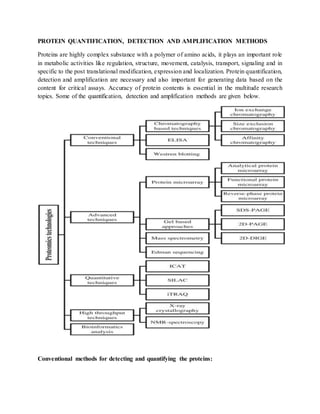
- 1. PROTEIN QUANTIFICATION, DETECTION AND AMPLIFICATION METHODS Proteins are highly complex substance with a polymer of amino acids, it plays an important role in metabolic activities like regulation, structure, movement, catalysis, transport, signaling and in specific to the post translational modification, expression and localization. Protein quantification, detection and amplification are necessary and also important for generating data based on the content for critical assays. Accuracy of protein contents is essential in the multitude research topics. Some of the quantification, detection and amplification methods are given below. Conventional methods for detecting and quantifying the proteins:
- 2. Some of the conventional methods are considerably difficult to identify and quantify the small size of protein macromolecules of particularly unknown protein samples. Several reliable methods are available for quantifying protein and to simplify the process. Quantification methods: Bradford assay method which uses a most common dye coomassie brilliant Blue G-250 to bind to protein. If there is no protein dye is in red but once bound to protein it turns blue at 595nm. This method is sensitive (detects from 1 ug to 60 ug) and do not rely on the tryptophan and tyrosine content. The protein concentration is determined comparing with standard. This assay which allows the rapid quantification and improves sensitivity with addition of low level of non-ionic detergent triton-x 100.(Bradford.,1976) Lowry assay rely on the tryptophan and tyrosine content. It uses folins reagent for accurate quantification and it is stable on acidic conditions and similar to biuret test assays. This method is sensitive at concentration ranges similar to the Bradford method, but requires a minuscule amount more of protein. It determines the level of a protein in a solution in the range of 0.01–1.0 mg/mL result in the blue color and measured by calorimeter. The concentration of the protein is determined by comparing with the known concentration ex. Bovin serum albumin (BSA). Sensitivity and accuracy are the advantage of this assay.(lowry et al., 1951) Warburg-Christian method which quantifies the presence of tryptophan and tyrosine proteins at 280 nanometer, the direct spectrophotometric based on the aromatic acids. But this method is not suitable for determining the mixture of proteins varying with the amounts of the amino acids. Fluorescamine: Quantifies proteins and peptides in solution if primary amine are present in the amino acids, alternative methods are epicocconone, Ellman's reagent (DTNB) and ortho-Phthalaldehyde (OPA) Non-specific methods that detect total protein: Biuret Test Assays: It forms a purple colour complex when the molecules with two or more peptide bonds react with cu2+, after purple color formation, it has been measured at 540nm to determine the protein concentration comparing with the known concentration. Its main disadvantages is low sensitivity and it also requires at least 1mg of protein.
- 3. Bicinchoninic acid assay (BCA assay): also known as the smith assay which Detects proteins down to 0.5 μg/mL results in a color change from green to purple and measured by calorimeter. Amido black: Detection of protein in the range of 1-12 μg/mL and stains the protein in black color, used in western blot Colloidal gold: colloidal gold is a suspension of gold nanoparticle which detects the protein in the range of 20 - 640 ng/mL. Protein quantification in single living cells: Monitoring the protein expression in living cell, linking by genetically, a protein of interest to reporter protein or epitope Ex. Green fluorescent Protein (GFP). Linking of a protein to the reporter with a cis-acting hydrolase element are called as “protein quantitation report”(PQR) allows the reporter and protein which transcribed together but translate separate functional proteins.(Lo et al.,2015) Specific methods which detects a single protein: Spectrometry methods: High-performance liquid chromatography (HPLC): In analytical chemistry, Chromatography method to detect proteins or peptides, quantify and identify each component in a mixture of a compound solution. The basic procedure of the HPLC is to flow sample containing the liquid mobile phase at high pressure driven by a pump through the packed column with suitable matrix. Separation is based on the differential interaction of molecules with the column matrix and passed through the column; the samples are separated by the required time. The column structure provides a possibility of high pressure to drive the mobile phase flowing much faster and complete a separation within short time. The separated molecules are detected in the form of signals, detected by a suitable detector. Further, the data are analyzed on the computer system with processing software.(Gaurav et al., 2016) HPLC
- 4. Liquid chromatography–mass spectrometry (LC/MS): LCMS which detects the proteins at lower concentrations (ng/mL to pg/mL) in blood and body fluids, such as for Pharmacokinetics. Ion- exchange chromatography: IEXC separates proteins on the basis of charge, IEc usually carries the opposite charge. In case of proteins and peptides, ionized histidine, lysine, cysteine provides positive charges and an aspartate and glutamate residue provides the negative charge. At a certain isoelectric point (pI), the total protein positive charge which are equal to the total negative charges and the zero net charge. In IEXC separated protein doesn’t have any net charge at its isoelectric point (pI) and doesn’t interact with the charged medium. (Gaurav et al., 2016) APPLICATIONS:
- 5. HPLC is a highly successful technique for the characterization and separation of proteins and peptides with characteristics of robustness, reproducibility, resolution, and high recoveries. IEXC technique capable of separating two proteins differing by one charged amino acid. The technique is extensively used in protein characterization and purification. IEXC is commonly used to separate alkaline phosphatase, biotinylated proteins and protease. ANTIBODY DEPENDENT METHODS: Enzyme-linked immuno sorbent assay (ELISA): ELISA or solid phase enzyme immunoassay Specifically detects and quantifies protein down to pg/mL in the simple form of specific antigen antibody binding on the microtitre plate, detected using the enzyme- chromogenic substate color formation. Four different types of ELISA, 1. DIRECT ELISA: Detection or quantification of Antibody by Direct ELISA. In this technique, the tested antigen were added in microtiter well (Serum or some other sample). Primary antibody with an attached enzyme or conjugate is added and allowed to react with the antigen. Unattached primary antibodies are washed away and a specific substrate for the enzyme is added. Enzyme which hydrolyzes the substrate to colored products. By spectrophotometric plate readers the amount of colored end product is measured. The higher concentration of the antigens to the primary antibody results in higher color change. 2. INDIRECT ELISA: Detection or quantification of Antibody by indirect ELISA. In this technique, antigens are precoated on the microtiter well. To the microtiter well Serum or some other sample containing primary antibody is added and allowed to react with the coated antigen. Free primary antibody is washed away and the attached antibody to the antigen is detected by an enzyme conjugated secondary antibody that binds to the primary antibody. Unattached secondary antibody washed away and a specific substrate for the enzyme is
- 6. added. Enzyme which hydrolyzes the substrate to colored products. By spectrophotometric plate readers the amount of colored end product is measured. 3. CAPTURE OR SANDWICH ELISA: Detection or quantification of Antibody by Capture ELISA. In this technique, antibody is precoated on the microtiter well. To the microtiter well Serum or some other sample containing antigen is added and allowed to react, resulting in antigen antibody complex. A second monoclonal antibody specific for different epitope attached by an enzyme or conjugate are added. Unattached secondary antibody washed away and a specific substrate for the enzyme is added. Enzyme which hydrolyzes the substrate to colored products. By spectrophotometric plate readers the amount of colored end product is measured. 4. COMPETITIVE ELISA: In this technique, antigen is precoated on the microtiter well. To the microtiter well mixture of antigen antibody solution is added and allowed to react, higher amount of antigen and lesser antibody competitively bind to the antigen coated plate. A secondary antibody attached by an enzyme or conjugate which is specific isotype of the primary antibody are added. Unattached secondary antibody washed away and a specific substrate for the enzyme is added. Enzyme which hydrolyzes the substrate to colored products. By spectrophotometric plate readers the amount of colored end product is measured. The higher concentration of antigen results in the lower absorbance To increase sensitivity in ELISA, minimizing the background noise, lowering the signal, increasing the incubation time, optimizing the blocking and washing steps, which enhance the sensitivity. Factors consideration in developing assay are ELISA plate either polystyrene or polystyrene derivations, plate surface-high(IgG), medium, low binding surfaces for enhancing the binding capacity, solid phase adsorption- direct adsorption of protein, small molecules epitope, covalent linkages of heavy glycosylated protein, proteins with detergent, some most common factors are coating, time, temperature, purity, blocking(BSA, non fat dry milk, casein or caseinate, normal
- 7. serum, fish gelatin), antibody cross reaction, detection molecules(enzymes-horse radish peroxidase, alkaline phosphatase, beta-galactosidase, peroxidase substrates-3,5,3’,5’- tetramethylbenzidine (TMB)2,2-Azino-di (3-ethyl- benzathiazoline) sulphonic acid (ABTS), oNPG and pNPP). Gel based Approaches: Protein immunoprecipitation: It is a technique of precipitating a specific protein out of solution using an specific antibody protein. Ex., protein complex immuno precipitation(protein-protein interaction), individual protein immunoprecipitation. After Immunoprecipitation the proteins were analysed by using SDS-PAGE, western blotting. Immunodiffusion: The agar gel was used for precipitation reaction. The antigen antibody cross linking resulting in the formation of precipitation line. The most common method is agar gel immunodiffusion (AGID) wells are created to load the antigen and antibody. Radial Immunodiffusion (Macini Technique):also called as the Single diffusion technique. It is a Semi-quantitative immunoprecipitin reaction in which the Gel has been impregnated with specific Antibody solution later the well has been created and Antigen is loaded into single well of agarose gel. Antigen will diffuse out from well in radial fashion. The Diameter of immunoprecipitin ring is proportional to the concentration of the Antibody or Antigen. Double immunodiffusion (ouchterlony test): Antibody and Antigen are placed in separate well opposite to each other in the agarose gel. Shape and positioning of precipitation line is proportional to concentration of reactants. A precipitation line is formed at the zone of equivalence where diffusing Antibody and Antigen complex. Staining: Coomassie brilliant blue R- 250,silver staining, fluorescent reagent- flourescein or rhodomine, epicocconone.
- 8. Sodium Dodecyl Sulfate-Polyacrylamide Gel Electrophoresis: SDS-PAGE is a high resolving technique for separating the proteins based on their size, which facilitates separation on molecular weight. Proteins which have moving capability when applied to electric field in a medium with isoelectric point. Mixture proteins or different proteins migrate with different velocities according to the ratio between its mass and charge. Sodium dodecyl sulfate denatures the proteins, therefore separate them absolutely on their molecular weight. Stains which are used in SDS-PAGE Ponceau S, sypro ruby, Epicocconone, coomassie-R-350, amido black, Coomassie brilliant blue R-250,silver staining and cy5. Western blot: Protein separation in the SDS-PAGE, blotted and incubation with antibodies to detect specific proteins in a sample of tissue homogenate or extract. It is a qualitative detection of single proteins and protein modification mainly the post translational modification. Semi quantative estimation of protein based on the size and protein band color intensity in the blot paper. Initially the sample were loaded, electrophoresed with protein ladder.to detect the protein after electrophoresis transferred to the PVDF or nitrocellulose membrane by capillary action. As a result of transfer process, proteins are exposed to the PVDF or nitrocellulose membrane and blocked with any one of the BSA, non fat dry milk, casein or caseinate, normal serum, fish gelatin PBS or PBST (phosphate buffered saline with tween 20) solutions. After blocking the expose the membrane
- 9. to the primary antibody followed with secondary antibody with horseradish peroxidase. Detection and visualization using the substrates-3,5,3’,5’- tetramethylbenzidine (TMB)2,2-Azino-di (3-ethyl-benzathiazoline) sulphonic acid (ABTS), oNPG and pNPP. Some of the stain which are used in the total protein staining before blocking are Ponceau S, sypro ruby, Epicocconone, coomassie-R-350, amido black, Coomassie brilliant blue R-250,silver staining and cy5.(mahmood et al.,2012) Two-dimensional gel electrophoresis: The two-dimensional polyacrylamide gel electrophoreses (2D-PAGE) is a reliable and efficient method for separation of proteins on the based on their charge and mass. 2D-PAGE is capable of resolving more than 5,000 different proteins, depending on the size of gel. The first dimension separates proteins by charge while on second dimension which separates the protein on the basis mass differences. The 2-DE is successfully applied for the evaluation of metabolic pathways characterization of post-translational modifications and mutant proteins. Two-dimensional differential gel electrophoresis 2D-DIGE utilizes the proteins labeled with CyDye by exciting the dye at a specific wavelength that can be easily visualized. Example: The role of apo plastic protein was investiaged in the 10 days old plant using the 2D-DIGE Host cell proteins was screened using the 2D-DIGE in between the null cell line and monoclonal antibody producing cell (Bilal et al.,2017) Advanced techniques:Protein microarray: Protein microarrays or protein chips is a emerging proteomic techniques capable of detecting the small amount of protein sample. Protein microarrays can be classified into three categories; analytical protein microarray, functional protein microarray and reverse-phase protein microarray Analytical protein microarray: Antibody microarray is one of the best representatives for the analytical protein microarray method which detects the proteins by direct protein labeling. These are typically used to measure the expression level and binding affinities of proteins. Examples Highthroughput proteome analysis of cancer cells for profiling the carcinoma cells of oral cavity, bladder cancer.
- 10. Detecting the Staphylococcal enterotoxin B, cholera toxin, Bacillus globigii and B. ricin using the analytical protein micro array. Also developed for identifying the cellular signaling pathways and characterizing Mitogenactivated protein kinases (MAPKs) from Arabidopsis. Functional protein microarray Functional protein microarray for the studying the various interactions including protein–DNA, protein–RNA and protein–protein, protein–drug, protein– lipid, enzyme–substrate relationship. Example: The first use of functional protein microarray was to analyze the substrate specificity of protein kinases in yeast Functional protein microarray characterized the functions of thousands of proteins (Calmodulin- like proteins (CML) and substrates of Calmodulin (CaM) in A. thaliana) Reverse-phase protein microarray: This method is used to determine the dysfunction protein and altered protein in the indicative of a certain diseased cell such as the acute myelogenous leukemia cells and normal hematopoietic stem cell. Cell lysates obtained from different cell states are arrayed on nitrocellulose slide that are probed with antibodies against target proteins. Antibodies are detected with fluorescent, chemiluminescent and colorimetric assays. For protein quantification, reference peptides are printed on slides. Reverse-phase protein microarray approach was evaluated for quantitative analysis of phosphoproteins and other cancer-related proteins in non-small cell lung cancer (NSCLC) cell lines by monitoring the apoptosis, DNA damage, cell-cycle control and signaling pathways.(Bilal et al.,2017) Quantitative techniques: Quantitative proteomic analysis and combination of mass spectrometry led to the emergence of techniques like SILAC (Stable amino acid labeling in cell culture), ICAT (Isotope coded Affinity Tagging) and ITRAQ (Isobaric tagging for Relative and Absolute Quantitation). The ICAT is an isotopic labeling method in which chemical labeling reagents are used for quantification of proteins. The ICAT has also expanded the range of proteins that can be analyzed and permits the accurate quantification and sequence identification of proteins from complex mixtures. The ICAT reagents comprise affinity tag for isolation of labeled peptides, isotopically coded linker and reactive group. PRINCIPLE OF ICAT
- 11. ICAT is an in vitro method of tagging proteins with specific tags. In vitro labeling relies on use of labeling reactions at specific sites in proteins and peptides. In vitro tagging can be classified into various sub-categories based on the chemistry involved. Ideally, there are three broad in vitro labeling methods: a) Specific amino acid tagging: Example: ICAT b) N – Terminal tagging: Example: ITRAQ c) C – Terminal tagging: Example: O18 labeling by proteolysis ICAT REAGENT The ICAT method is based on the chemical reaction between the a chemical tag and cysteine residues of the protein. The tag should have a reactive group that can specifically react with the SH group of cysteine and also with no other cross reactivity. The ICAT reagent consists of three parts a)Iodoacetamide residue (chemically reactive group), which binds to cysteine thiol group. b) Linker region, containing deuterium or hydrogen, which increase in mass. c) Biotin residue, which helps in the enrichment of peptides using Streptavidin assisted affinity chromatography.
- 12. The ICAT reagent comes in two forms: one containing no deuterium or other with eight deuterium atoms. The ICAT reagent binds to the cysteine groups of the protein via the iodoacetamide residue and depending on the number of deuterium ion, there is an increase in the mass of the peptide. WORKING PROTOCOL OF ICAT : ICAT is not only for quantifying proteins but also studies the expression of proteins subject to external stimuli. Ideally, the isolated proteins are first treated with the ICAT reagent in either of the two ways: a) Control with ICAT reagent having no deuterium and treated with ICAT reagent containing eight deuterium b) Tag swapping. The tagged proteins are then trypsinized. ICAT tag also contains a biotin residue, using Streptavidine based affinity chromatography; the peptides are enriched and then subjected to MS analysis and MS/MS analysis for identification and data were analysed. (NPTEL) EX: Using ICAT can compare the dynamic concentration of proteins in two conditions i.e inn normal and diseased conditions. The breast cancer plasma cell sample from the 6 different breast cancer patients as well as with the 6 normal healthy controls were taken and analysed using the ICAT.(kang et al 2010) SILAC-based Proteomics Analysis
- 13. An innovative high throughput technology for quantiting the protein in proteomics is SILAC (SILAQ) which quantitatively analysis the protein-small molecule interactions, protein-protein interactions and large protein complexes. It is a effective method to determine proteins in the cell and also a sensitive method for analysis. SILAC Workflow: SILAC relies on the incorporation of isotope (non radio labelling isotopes) on the heavy or light aminoacid in to a two samples. The labeled amino acids e. g. 12C and 13C labeled L-lysine not interfering in normal cell growth. SILAC approaches are well suited for monitoring the changes in the post-translational modifications. The best Examples for application of SILAC include the measurement of changes in protein phosphorylation and methylation.(NPTEL) Advantage of our SILAC technique:
- 14. Optimized protocols Cutting-edge facilities & In vivo labeling is harmless At one time, Compare up to three samples is possible High sensitivity Suitable for study of protein-protein-interaction Detecting Post-translational modification is possible Isobaric tag for relative and absolute quantitation iTRAQ is based on the tandem mass spectrometryfor multiplex protein labeling. This technique uses the isobaric tags (8-plex and 4-plex) for absolute and relative quantitation. The technique comprises labeling of side chain amine groups of proteins and the N-terminus after labeling it has been fractionated through liquid chromatography and finally analyzed through MassSpectrometry. iTRAQ is an appropriate method that helps to quantify and identify the protein simultaneously and also used in finding the gene regulation to understand the disease mechanism. X-ray crystallography: X-ray crystallography is the most preferred technique for determining the three dimensional structure of proteins. The highly purified crystallized samples are exposed to X-rays and the subsequent diffraction patterns are processed to produce information about the size of the
- 15. repeating unit that forms the crystal and crystal packing symmetry. X-ray crystallography has an range of applications to study the protein–nucleic acid complexes, virus system and immune complexes. The 3-D protein structure provides full information about the drug designing, elucidation of enzyme mechanism, protein– ligand interaction and site-directed mutagenesis. NMR spectroscopy The NMR is a top most tool for the investigating the molecular structure, behavior and folding of proteins. Various phases are involved in the Structure determination through NMR spectroscopy, each using a discrete set of specific techniques. The samples are prepared and measured to confirm the structure. The protein structure is fundamental in different research areas such as functional genomics, homology modeling and structure-based drug design. Protein amplification: Under protein amplification the one only available method is protein misfolding cyclic amplification (PMCA). PMCA which amplifies the misfolded proteins generally the prions. The PMCA technology which are mainly used for understanding the molecular mechanism of prions replication and now this technology which are the targeting the nature of the infectious agent via prions and screens the inhibitors against prion, describing the effect of cellular components, to detect the PrPSc in biological fluids and tissues REFERENCES: 1. Lo C, Kays I, Emran F, Lin T, Cvetkovska V, Chen B. Quantification of Protein Levels in Single Living Cells. Cell Rep. 2015;13:2634-2644. 2. Bradford, M. M. (1976), Anal. Biochem. 72:248 3. Lowry, 0. H., Rosebrough, N. J., Farr, A. L., and Randall, R. J. (1951) J. Biol. Chem. 193,265-275. 4. Gaurav Pratap Singh Jadaun*, Shruti Dixit, Vandana Saklani, Sanjay Mendiratta, Renu Jain, Surinder Singh, HPLC for Peptides and Proteins: Principles, Methods and Applications, Pharm Methods, 2016; 8(1): 139-144 5. Mahmood, Tahrin; Yang, Ping-Chang (September 2012). "Western Blot: Technique, Theory, and Trouble Shooting". N Am J Med Sci. 4 (9): 429–434. doi:10.4103/1947- 2714.100998. PMC 3456489. PMID 23050259. 6. Bilal Aslam, Madiha Basit, Muhammad Atif Nisar, Mohsin Khurshid, and Muhammad Hidayat Rasool Proteomics: Technologies and Their Applications, Journal of Chromatographic Science, 2017, Vol. 55, No. 2, 182–196 doi: 10.1093/chromsci/bmw167
- 16. 7. Kang, U. B. et al., Differential profiling of breast cancer plasma proteome by isotope- coded affinity tagging method reveals biotinidase as a breast cancer biomarker. BMC Cancer 2010, 10:114.