SlideShare ist ein Scribd-Unternehmen logo
1 von 79
THE RESPIRATORY SYSTEM
PRESENTED BY:-
SUCHISMITA SETHI
LECTURER, MSN
INTRODUCTION
• A primary requirement of all body cell
activities & growth is oxygen which is needed
to obtain energy from food.
• The fundamental purpose of respiratory
system is to supply oxygen to the individual
tissue cells & remove there gaseous waste
product , carbon dioxide.
• Breathing or ventilation refers to the
inhalation & exhalation of air.
TYPES OF RESPIRATION
• Pulmonary ventilation:- moving air into & out
of the lungs.
• External respiration:- Gas exchange between
lungs & & blood.
• Internal respiration:- Also called cellular
respiration which involve the uptake of o2
production of co2 within individual cell.
FUNCTION OF THE RESPIRATORY SYSTEM
• Exchanges oxygen and carbon Dioxide between the blood
and external environment
• Exchange of gases takes place within the lungs in
the alveoli.
• NON RESPIRATORY FUNCTIONS:-
• Nose serve as a organ of smell
• Root of water loss & heat elimination
• Defends against inhaled foreign matter
• Enable vocalization ( Speech, singing & other)
• Enhance venous return ( cardiovascular physiology.
• Excretion of volatile substance like ammonia.
• Maintain PH of body.
ORGANS OF THE RESPIRATORY
SYSTEM
1. Nose
2. Pharynx
3. Larynx
4. Trachea
5. Bronchi
6. Lungs – alveoli
7. Organs of the
Respiratory System
1. UPPER AIR WAY:-
Consist of mouth, nose,
nasal cavity, pharynx,
larynx
2. LOWER AIR WAY:-
Consist of trachea,
bronchi, bronchioles,
alveoli.
• RESPIRATORY TRACT:- There are two types :-
• 1. conducting portion:- From nasal cavity to
terminal bronchioles.
• 2. Respiratory Portion:- The respiratory
bronchioles & alveoli.
THE NOSE
• The air from the outside
environment enter the nose
or mouth during inspiration(
inhalation)
• The nose is divided in to two
regions.
• The external nose including
the root, bridge, dorsum
nasi, & the apex.
• The internal nasal cavity is
devided in to two halves by
the nasal septum.
• It contain cilia which is
responsible for filtering out
foreign bodies.
FUNCTION OF NOSE
• Provide an entry & exit for air during
breathing.
• Humidifying & warming the enter air.
• Filtering inspired air & cleaning it of foreign
matter
• Serving as a resonating chamber for speech.
• Housing the olfactory receptors for smell.
ANATOMY OF THE NASAL CAVITY
• Olfactory receptors are located in the mucosa on
the superior surface.
• The rest of the cavity is lined with respiratory
mucosa to moistens air & traps incoming foreign
particles.
• Lateral walls have projections called conchae which
increases surface area & Increases air turbulence
within the nasal cavity.
• The nasal cavity is separated from the oral cavity by
the palate, anterior hard palate (bone)
PARANASAL SINUSES
• Cavities within bones surrounding the nasal cavity
1. Frontal sinus
2. Sphenoid sinus
3. Ethmoid sinus
4. Maxillary sinus
• FUNCTION:-
• Lighten the skull
• Act as resonance chambers for speech
• Produce mucus that drains into the nasal cavity
PARANASAL SINUS
PHARYNX (THROAT)
• Muscular passage from nasal cavity to larynx
• Three regions of the pharynx:-
1. Nasopharynx – superior region behind nasal
cavity
2. Oropharynx – middle region behind mouth
3. Laryngopharynx – inferior region attached to
larynx
• The oropharynx and laryngopharynx are
common passageways for air and food.
• Auditory tubes enter the nasopharynx
• TONSILS OF THE PHARYNX:-
1. Pharyngeal tonsil (adenoids) in the nasopharynx
2. Palatine tonsils in the oropharynx
3. Lingual tonsils at the base of the tongue
4. Tubal tonsil
LARYNX (VOICE BOX)
Function:-
• Routes air and food into proper channels
• Plays a role in speech
• Made of eight rigid hyaline cartilages and a
spoon-shaped flap of elastic cartilage
(epiglottis)
Structures of the Larynx
• It act as a switching mechanism to route air & food
in to the proper channel.
• Prevent food & drink from entering the trachea.
Thyroid cartilage:- Largest hyaline cartilage which
Protrudes anteriorly (Adam’s apple)
Epiglottis:- It is a moveable flap of cartilage that cover
the opening to the larynx( voice box) prevent food
from entering the larynx during swallowing.
Vocal cords :- Vibrate with expelled air to create sound
(speech). Size & thickness determine the pitch of
sound. Short and thin means high pitched sound.
Glottis – opening between vocal cords
TRACHEA (WINDPIPE)
• It is cylindrical shape which connects larynx with
bronchi. Length 11 cm
• Composed of three layers:-
1. mucosa:- made up of goblet cells & cilliated
epithelium which beat continuously in opposite
direction of incoming air & expel mucus loaded
with dust.
2. Submucosa:- connective tissue deep to the
mucosa
3. Adventitia:- Outermost layer made of C- shaped
rings of hyaline cartilage.
BRONCHI
• Formed by division of the trachea
• Enters the lung at the hilus (medial depression)
• Made up of complete ring of cartilage.
• Bronchi divided in to right & left bronchi
• Right bronchus is wider, shorter, and straighter
than left
• Bronchi subdivide into smaller and smaller
branches
Coverings of the Lungs:-
1. Pulmonary (visceral)
pleura covers the lung
surface
2. Parietal pleura lines
the walls of the
thoracic cavity
Pleural fluid fills the area
between layers of
pleura to allow gliding
LUNGS
• The lungs, which is the organ for respiration is a
paired cone shaped organs lying in the thoracic
cavity.
• Each lung has a base resting on the diaphragm
and an apex extending superiorly to a point
approximately 2.5 cm superior to the clavicle.
• It also has a medial surface and with three
borders- anterior, posterior and inferior.
• The broad coastal surface of the lungs is pressed
against the rib cage, while the smaller
mediastinal surface faces medially.
CONT..
• The lungs receives the bronchus, blood vessels,
lymphatic vessels and nerves through a slit in the
mediastinal surface called the helium.
• The right lung is larger and weighs more than the
left lung.
• Since the heart tilts to the left, the left lung is
smaller than the right and has an indentation
called the cardiac impression to accommodate the
heart.
• This indentation shapes the inferior and anterior
parts of the superior lobe into a thin tongue-like
process called the lingual.
Lobes and Fissures of the Lungs
• Each lung is divided into lobes by fissures.
• Both lungs have oblique fissure and the right is
further divided by a transverse fissure.
• The oblique fissure in the left lung separates the
superior and the inferior lobe.
• The oblique and horizontal fissure divides the lungs
into superior, middle and inferior lobes.
• Thus the right lung has three lobes while the left
has two.
• Each lobe is supplied by a lobar bronchus. The
lobes are subdivided by bronchopulmonary
segments which are supplied by the segmental
bronchi
Bronchopulomonary Segment
Trachea
Rt lungs left lungs
Oblique fissure transverse fissure oblique fissure
Upper lobe lower lobe upper lobe lower lobe
a. Apical a. Lateral a. Apical a. apical
b. Posterior b. posterior b. posterior b. anterior
c. Anterior c. anterior c. anterior c. posterior
Middle lobe:- d. Medial d. Superior d. lateral
a. Lateral e. Apical e. inferior
b. medial
ARTERIAL SUPPLY
1) on the right side there is one bronchial artery
which arises from either the third posterior
intercostal artery or from the upper left
bronchial artery
2)On the left side there are two bronchial arteries
both of which arise from the descending
thoracic aorta
3)There are pre capillary anastomoses between
bronchial and pulmonary arteries. these
connections enlarge when any one of them is
obstructed in disease VENOUS DRAIN
VENOUS DRAINAGE
1) Usually there are two bronchial veins on each
side ,the right bronchial vein drain into the
azygous vein
2) The left bronchial vein drains either into the
left superior intercostal vein or into the hemi
azygous vein
3) The greater part of the venous blood from the
lung is drained by the pulmonary veins.
NERVE SUPPLY
1) Para sympathetic nerves are derived from the
vagus .These fibres are:-
a) motor to the bronchial muscles and on
stimulation cause bronchospasm
b) secretomotor to the mucous glands of the
bronchial tree
c) the sensory fibres are responsible for stretch
reflex of the lungs and for the cough reflex
BRONCHI & BRONCHIOLS
• Trachea divided in to two primary bronchi at the
level of 5th thoracic vertebra
• Right bronchus is approximately 2.5 cm long
• This is wider, shorter & more vertical then left
bronchus.
• After entering the right lungs at the hilium it is
divided in to 3 bronchioles each branch is then
subdivided in to smaller branches.
• Left bronchus is about 5 cm long & narrow then
right.
• It is divided in to two branches.
STRUCTURE
• Bronchial wall contain 3 layer & lined with ciliated
columnar epithelium.
• Bronchi sub divided in to bronchioles, terminal
bronchioles, respiratory bronchioles, alveolar
duct and finally alveoli.
• STRUCTURAL CHANGES IN BRONCHIAL
PASSAGE:-
a. Cartilage:- Rigid cartilage interfere with
expansion of lungs tissue & gaseous exchange.
At bronchial level there is no cartilage in airway
wall.
b. Smooth muscle:- cartilage replace by smooth
muscle which allow the diameter of airway to
increase.
It is control by autonomic nervous system which
regulate air flow within the lungs.
c. Epithelial lining:- ciliated epithelium is
gradually replaced with non ciliated
epithelium & goblets cell disappear.
BLOOD SUPPLY
• Right & left bronchial artery supply blood.
• Venous return is through bronchial veins.
• Right side empty in to azygos vein & left side in
to superior intercostal vein.
NERVE SUPPLY:-
• Vagus nerve( parasympathetic) stimulation
contraction of smooth muscle in the bronchial
tree cause bronchoconstriction
• Sympathetic stimulation cause bronchodilation.
FUNCTION:-
• Regulate speed & volume of air flow in to
within the lungs.
• Parasympathetic stimulation cause
constriction & sympathetic stimulation cause
dilation.
• Cough reflex
• Removal of particulate matter
• Warming & humidifying.
RESPIRATORY BRONCHIOLS & ALVEOLI
• In each lobe the lungs tissue is further divided by
connective tissue in to lobule
• Each lobule is supplied with air by terminal
bronchiole which further divided in to respiratory
bronchiole, alveolar duct & large number of
alveoli.
• There are about 150 million alveoli in adult lungs.
• It help in gas exchange.
• Single squamous epithelial cells in the alveolar
duct & alveoli.
• Distal respiratory passage are supported by
loose network of elastic connective tissue in
which macrophages, fibroblast, nerve , lymph
& blood vessel are embedded.
• Alveoli surrounded by dance network of
capillaries
• Alveolar wall & capillary wall fused firmly
together for exchange of o2 & co2.
• In between the squamous cells septal cells
which secrete surfactant .
• Surfactant is a phospholipid fluid which
prevent alveoli from drying out & reduce
surface tension preventing alveolar collapse
during expiration
• NERVE SUPPLY TO BRONCHIOLS:-
Parasympathetic stimulation by vagus nerve
cause bronchoconstriction.
Sympathetic stimulation relax the bronchial
smooth muscle.
EXCHANGE OF GAS
• Exchange of gas occur in two level.
1. Pulmonary exchange( External respiration)
2. Tissue level exchange ( internal respiration)
• This process takes place by partial pressure
of gases & diffusion process.
• Partial Pressure:- Pressure exerted by a gas in
a mixture of gas irrespective of concentration
of other gas.
PROCESS OF GASCIOUS EXCHANGE
• PARTIAL PRESSURE IN INHALED AIR:- (PO2-159
mmhg & PCO2 is 0.3 mmhg
• PARTIAL PRESSURE IN ALVIOLI IS:- PO2-
104mmhg & PCO2 is 40.
• Here PP of inhaled air & PP of alveoli is
different because out of 500 ml(tidal volume)
of inhaled air only 350 ml air riches to the
alveoli.
• Some air remain in the respiratory tract(
Trachae, bronchi & bronchioles) & after we
exhale again there is some gas which is
remaining in the tube.
CONT...
• It means when this fresh air enter it mixes
with the air that is already present in our
respiratory tract & that air has more Co2 &
less of o2.
• So when inhaled air mixed with more Co2 &
less o2 gas partial pressure changes in both o2
& co2 in alveoli
• So in alveoli level the over all partial pressure
of O2 is 104 mmhg & Co2 is 40 mmhg.
CONT...
• The blood which comes to the lungs is
deoxygenated blood by pulmonary artery.
• Because blood comes from the tissue level o2
used & Co2 produced more due to
metabolism.
• So in this blood Po2 is 40 mmhg & Co2 is 46
mmhg.
• The value depends on activity of cell.
CONT...
• Now alveoli has O2 at high PP. Gases diffuse
from their higher PP to their lower PP.
• So here O2 from alveoli which is having higher
pressure diffuse to lower PP o2 which is
present in pulmonary artery.
• Co2 which is present in higher PP pulmonary
artery diffuse with alveoli lower PP Co2.
• After diffusion of PP of O2 increase to 95 mmhg.
• The pulmonary vein carry O2 riched blood to
the left atrium, then to ventricle & then go to
aorta.
CONT.....
• In the tissue level now due to activity of tissue
PP O2 decrease to 20 mmhg & PCo2 is 58
mmhg (aprox)
• Due to o2 partial pressure is more in blood
vessel it diffuse in tissue level & Co2 diffuse
from the tissue to the blood.
• So due to this value now blood vessel again
become deoxygenated & go to the heart
through superior & inferior venacava.
INTRODUCTION TO
RESPIRATION
• The term respiration means exchange of gas
between body cells & their environment.
• It involve two main process:-
1. Breathing (Pulmonary ventilation):- It is the
movement of gas in to & out of the lungs
2. Exchange of gas:- This takes place :-
In lungs:- External Respiration
In tissue :- Internal respiration
1. BREATHING:-
• Breathing supplies oxygen to the alveoli &
eliminate Co2.
MUSCLE OF BREATHING:-
• Expansion of chest during inspiration
occur as a result of muscular activity which is
partly voluntary & partly involuntary.
• The muscle use in normal breathing are
external intercostal muscle & diaphragm.
INTERCOSTAL MUSCLE
• There are 11 pairs of intercostal muscle
occupying the space between the 12 pair of ribs.
• They are arranged in two layers
a. External intercostal muscle:- they extend down
wards & forwards from the lower border of the
ribs above to the upper border of rib below.
b. Internal intercostal muscle:- they extends down
wards and back wards from the lower border of
ribs above to upper border of rib below. This
muscle use when expiration become active
Cont…..
• The first rib is fixed, so when external muscle
contract they pulled all the other ribs to wards
the first rib
• The rib cage move as a unit upward & out
ward enlarge the thoracic cavity.
• Intercostal muscle under the control of
intercostal nerve.
DIAPHRAGM
• The diaphragm is a dome-shaped muscular structure
which separate thoracic and abdominal cavity.
• It consist of a central tendon from which muscle fiber
radiate to attach to the lower ribs & sternum & to
vertebral column by two crura.
• When diaphragm is relaxed the central tendon is at the
level of the 8th thoracic vertebra.
• When it contract its muscle fiber shorten & central
tendon is pulled down to the level of 9th thoracic
vertebra, lengthening the thoracic cavity.
• Thus decrease pressue in the thoracic cavity & increase
pressure in abdominal cavity.
• During inspiration the external intercostal muscle &
diaphragm contract simultaneously to enlarge the
thoracic cavity
ACCESSORY MUSCLE
• When extra respiratory effort is required
additional muscle are required
• Force of inspiration is assisted by the
sternocleidomastoid muscle & scalene muscles
which link to cervical vertebra & first 2 rib. To
increase rib cage expansion.
• Forced expiration is helped by the intercostal
muscle & abdominal muscle. Which increse the
pressure of thorax by squeezing the abdominal
muscle.
CYCLE OF BREATHING
• Average respiratory rate is 12-15 breath/min
• Breathing depends upon change in pressure &
volume in thoracic cavity.
• It follow the principle , “Increase the volume of
container decrease the pressure inside &
decreasing the volume increase the pressure
inside”
• So air flow from area of higher pressure to an
area of low pressure , change the pressure inside
the lungs which determine the direction of air
flow.
Cont....
• Breathing consist of 3 phase
• 1. inspiration
• 2. Expiration
• 3. pause
1. INSPIRATION
• Simultanious contraction of external intercostal muscle &
expand the thorax..
• Parietal pleura is firmly adhered to the diaphragm & inside
the rib cage.
• It is pulled outward along with them.
• This pulls the visceral pleura outward too since the two
pleura are hold together by thin film of pleural fluid.
• Because viseral pluera is firmly adherent to the lung the lungs
tissue also pulled up & out with the ribs & downwards with
the diaphragm.
• This expand the lungs & pressure within the alveoili & in the
air passage fall, drowing air in to the lungs in an attempt to
equalise atmospheric & alveolar pressure.
• The negative pressure created in the thoracic cavity aids
venous return to the heart.
• Inspiration is a active process so it need energy
2. EXPIRATION
• Relaxation of the external intercostal muscle &
the diaphragm result in downward & inward
movement of the rib cage & elastic recoil of the
lungs
• Due to this pressure inside the lungs rise & expel
air from the respiratory tract
• At the end of the expiration the lungs still
contains the air which prevent from complete
collapse by the intact pleura.
• This process is passive which do not need energy.
• After expiration there is pause before next cycle
begin.
PHYSIOLOGICAL VARIABLE AFFECTING
THE BREATHING
1. ELASTICITY:-Elesticity is the ability of the lungs to
return to its normal shape after each breath. Loss of
eleasticity of the connective tissue necessitate forced
expiration & increase effort of inspiration.
2. COMPLIANCE:- this is the stretchebility of the lungs i.e
the effort required to inflate the alveoli. The helathy
lungs is very complient which inlate with very little
effort but compliance is low more effort needed to
inflate.
3. AIRWAY RESISTANCE:- increase resistance cause more
respiratory effort to inflate the lungs.
LUNGS VOLUME & CAPACITY
• There are 15 complete respiratory cycle per
min.
• The lungs and air passage are never empty &
exchange of gases takes place only across the
wall of the alveolar duct & alveoli.
• The remaining capacity of the respiratory
passage is called the anatomical dead space(
150 ml)
CONT....
1. Tidal volume:- this is the amount of air passing in to &
out of the lungs during each cycle of breathing . (500 ml
at rest)
2. Inspiratory reserve volume:- inspiratory reserve
volume (IRV) is the additional amount of air that can be
inhaled after a normal inhalation.
3. Functional residual capacity:- The functional residual
capacity (FRC) is defined as the amount of gas left in the
lungs after normal expiration. This is about 2.5 L in the
average-sized adult or 35 mL/kg. The FRC acts as a buffer
by preventing rapid changes in alveolar gas tensions from
inspired air.
4. Expiratory reserve volume:- expiratory reserve volume is
the amount of extra air — above anormal breath —
exhaled during a forceful breath out. The average ERV
volume is about 1100 mL in males and 800 mL in females
5. Residual volume:- The residual volume (RV) is the amount
of air that is left after expiratory reserve volume is
exhaled.
6. Vital capacity:- Vital capacity (VC) is the maximum
amount of air a person can expel from the lungs after a
maximum inhalation. It is equal to the sum of inspiratory
reserve volume, tidal volume, and expiratory reserve
volume.
7. Total lungs capacity:- Lung capacity or total lung
capacity (TLC) is the volume of air in the lungs upon the
maximum effort of inspiration. Among healthy adults, the
average lung capacity is about 6 liters
8. Alveolar ventilation:- Alveolar ventilation and dead space
A. Alveolar ventilation ( A) is defined as the volume of air
entering and leaving the alveoli per minute. Air ventilating
the anatomic dead space (VD) (Levitzky Fig 3-7), where no
gas exchange occurs, is not included: VT = VD + VA.
COMPOSITION OF INSPIRED &
EXPIRED AIR
• INSPIRED AIR EXPIRED AIR
• OXYGEN:- 21 16
• CARBON DIOXIDE:- 0.04 4
• NITROGEN:- 78 78
• WATER VAPOUR:- VARIABLE SATURATED
CONTROL OF RESPIRATION
• Effective control of respiration enables the
body to regulate blood gas level over a wide
range of physiological environmental &
pathological condition.
• Voluntary control is exerted during activities
such as speaking & singing but is overridden if
blood co2 raise.
The respiratory centre
• Respiratory centre present in medulla which control
rate & depth of breathing.
• Regular discharge of inspiratory neurones within this
centre set the rate & depth of breathing
• Activity of the respiratory rhythmicity centre is
adjusted by nerve in the Pons ( the pneumotaxic
centre & apneustic centre ) in response to input
from other parts of the brain.
• Motor impulses leaving the respiratory centre pass
in Phrenic & intercostal nerves to the diaphragm &
intercostal muscle to stimulate respiration
chemoreceptor's
• These are receptors that respond to changes in
the partial pressure of oxygen & Carbon dioxide
in the blood & CSF.
• Central chemoreceptor's:- located on the surface
of medulla oblongata & are bathed in CSF.
• When arterial PCO2 rise (hypercapnia) the
central chemoreceptor by stimulating the
respiratory centre which increase the ventilation
to reduce the PCO2
Cont..
• Peripheral chemoreceptor's:- these are situated
in the arch of aorta & in the carotid bodies.
• They respond to change in the blood co2 & o2
level.
• It is more sensitivity to CO2 then O2.
• Change in the co2 & o2 level activate these
receptors trigger the nerve impulse to the
respiratory centre via the glossopharyngeal &
vagus nerve.
• This stimulation raise the rate & depth of the
respiration
Respiratory system

Weitere ähnliche Inhalte

Was ist angesagt?

Respiratory system (Structure).pptx
Respiratory system (Structure).pptxRespiratory system (Structure).pptx
Respiratory system (Structure).pptxVipin Chandran
 
Human nervous system
Human nervous systemHuman nervous system
Human nervous systemGotov .kz
 
Respiratory system, physiology of respiratory system and neural control
Respiratory system, physiology of respiratory system and neural control Respiratory system, physiology of respiratory system and neural control
Respiratory system, physiology of respiratory system and neural control sunil JMI
 
Physiology of the respiratory system
Physiology of the respiratory systemPhysiology of the respiratory system
Physiology of the respiratory systemGunJee Gj
 
special sense organs (anatomy and physiology) - a brief discussion
special sense organs (anatomy and physiology) - a brief discussion special sense organs (anatomy and physiology) - a brief discussion
special sense organs (anatomy and physiology) - a brief discussion Pallab Nath
 
Respiratory system
Respiratory systemRespiratory system
Respiratory systemEman Vidallo
 
Anatomy of the respiratory system...
Anatomy of the respiratory system...Anatomy of the respiratory system...
Anatomy of the respiratory system...jagan _jaggi
 
Respiratory system
Respiratory systemRespiratory system
Respiratory systemSoneeshah
 
Respiratory system
Respiratory systemRespiratory system
Respiratory systemcbaker745
 
Glands, Types, classification and functions(Anatomy Topic)
Glands, Types, classification and functions(Anatomy Topic)Glands, Types, classification and functions(Anatomy Topic)
Glands, Types, classification and functions(Anatomy Topic)Swatilekha Das
 
Respiratory system.pdf
Respiratory system.pdfRespiratory system.pdf
Respiratory system.pdfRAMDAS BHAT
 
HUMAN RESPIRATORY SYSTEM ANATOMY & PHYSIOLOGY
HUMAN RESPIRATORY SYSTEM ANATOMY & PHYSIOLOGYHUMAN RESPIRATORY SYSTEM ANATOMY & PHYSIOLOGY
HUMAN RESPIRATORY SYSTEM ANATOMY & PHYSIOLOGYKameshwaran Sugavanam
 
Respiratory System (afreen khan)
 Respiratory System (afreen khan) Respiratory System (afreen khan)
Respiratory System (afreen khan)Shoaib Akhtar
 
Chapter 22: Respiratory System (#1)
Chapter 22: Respiratory System (#1)Chapter 22: Respiratory System (#1)
Chapter 22: Respiratory System (#1)Caroline Tokarski
 

Was ist angesagt? (20)

Respiratory system (Structure).pptx
Respiratory system (Structure).pptxRespiratory system (Structure).pptx
Respiratory system (Structure).pptx
 
Human nervous system
Human nervous systemHuman nervous system
Human nervous system
 
Respiratory System - Anatomy
Respiratory System - AnatomyRespiratory System - Anatomy
Respiratory System - Anatomy
 
Respiratory system, physiology of respiratory system and neural control
Respiratory system, physiology of respiratory system and neural control Respiratory system, physiology of respiratory system and neural control
Respiratory system, physiology of respiratory system and neural control
 
Respiratory System Notes
Respiratory System NotesRespiratory System Notes
Respiratory System Notes
 
Physiology of the respiratory system
Physiology of the respiratory systemPhysiology of the respiratory system
Physiology of the respiratory system
 
special sense organs (anatomy and physiology) - a brief discussion
special sense organs (anatomy and physiology) - a brief discussion special sense organs (anatomy and physiology) - a brief discussion
special sense organs (anatomy and physiology) - a brief discussion
 
Respiratory system
Respiratory systemRespiratory system
Respiratory system
 
Respiratory system
Respiratory systemRespiratory system
Respiratory system
 
Anatomy of the respiratory system...
Anatomy of the respiratory system...Anatomy of the respiratory system...
Anatomy of the respiratory system...
 
Respiratory system
Respiratory systemRespiratory system
Respiratory system
 
Respiratory system
Respiratory systemRespiratory system
Respiratory system
 
Glands, Types, classification and functions(Anatomy Topic)
Glands, Types, classification and functions(Anatomy Topic)Glands, Types, classification and functions(Anatomy Topic)
Glands, Types, classification and functions(Anatomy Topic)
 
Respiratory system
Respiratory systemRespiratory system
Respiratory system
 
Respiratory system.pdf
Respiratory system.pdfRespiratory system.pdf
Respiratory system.pdf
 
HUMAN RESPIRATORY SYSTEM ANATOMY & PHYSIOLOGY
HUMAN RESPIRATORY SYSTEM ANATOMY & PHYSIOLOGYHUMAN RESPIRATORY SYSTEM ANATOMY & PHYSIOLOGY
HUMAN RESPIRATORY SYSTEM ANATOMY & PHYSIOLOGY
 
Respiratory System (afreen khan)
 Respiratory System (afreen khan) Respiratory System (afreen khan)
Respiratory System (afreen khan)
 
Chapter 22: Respiratory System (#1)
Chapter 22: Respiratory System (#1)Chapter 22: Respiratory System (#1)
Chapter 22: Respiratory System (#1)
 
Physiology of respiration
Physiology of respirationPhysiology of respiration
Physiology of respiration
 
Respiratory ststem
Respiratory ststemRespiratory ststem
Respiratory ststem
 

Ähnlich wie Respiratory system

Respiratory_anatomy.pptx
Respiratory_anatomy.pptxRespiratory_anatomy.pptx
Respiratory_anatomy.pptxDesalegnKifle2
 
respiratory system and the related organs
respiratory system and the related organsrespiratory system and the related organs
respiratory system and the related organsCharutaKunjeer1
 
respiratory system and related organs anatomy physiology
respiratory system and related organs anatomy physiologyrespiratory system and related organs anatomy physiology
respiratory system and related organs anatomy physiologyCharutaKunjeer1
 
Respiratory System unit-I cop.pptx
Respiratory System unit-I cop.pptxRespiratory System unit-I cop.pptx
Respiratory System unit-I cop.pptxRaeesShahidBasharat
 
L12__Respiratory_system_anatomy[1].pptx
L12__Respiratory_system_anatomy[1].pptxL12__Respiratory_system_anatomy[1].pptx
L12__Respiratory_system_anatomy[1].pptxssuser31c469
 
Anatomy & mechanics of respiratory system
Anatomy & mechanics of respiratory systemAnatomy & mechanics of respiratory system
Anatomy & mechanics of respiratory systemDharmraj Singh
 
Respiratorysystem 120105050542-phpapp01
Respiratorysystem 120105050542-phpapp01Respiratorysystem 120105050542-phpapp01
Respiratorysystem 120105050542-phpapp01MohdTanveer13
 
Unit-III, Chapter-1- Respiratory System.ppt
Unit-III, Chapter-1- Respiratory System.pptUnit-III, Chapter-1- Respiratory System.ppt
Unit-III, Chapter-1- Respiratory System.pptAudumbar Mali
 
Unit 1; Respiratory System, Educational Platform.pdf
Unit 1; Respiratory System, Educational Platform.pdfUnit 1; Respiratory System, Educational Platform.pdf
Unit 1; Respiratory System, Educational Platform.pdfZiaUddin5613
 
Respiratory system
Respiratory systemRespiratory system
Respiratory systemDAWN V TOMY
 
anatomyandphysiologyoftherespiratorysystem-180129061553.pdf
anatomyandphysiologyoftherespiratorysystem-180129061553.pdfanatomyandphysiologyoftherespiratorysystem-180129061553.pdf
anatomyandphysiologyoftherespiratorysystem-180129061553.pdfsafuraqazi
 
Anatomy and physiology of the respiratory system
Anatomy and physiology of the respiratory systemAnatomy and physiology of the respiratory system
Anatomy and physiology of the respiratory systemDipali Harkhani
 
Respiratory system- Anatomy & Physiology
Respiratory system- Anatomy & PhysiologyRespiratory system- Anatomy & Physiology
Respiratory system- Anatomy & PhysiologyPari Pratik
 
respiratory system.pptx
respiratory system.pptxrespiratory system.pptx
respiratory system.pptxssuser65b0c5
 
Respiratory system Anatomy
Respiratory system AnatomyRespiratory system Anatomy
Respiratory system AnatomyAlok Kumar
 
Respiratory System
Respiratory SystemRespiratory System
Respiratory SystemAsad Kamran
 

Ähnlich wie Respiratory system (20)

Respiratory_anatomy.pptx
Respiratory_anatomy.pptxRespiratory_anatomy.pptx
Respiratory_anatomy.pptx
 
respiratory system and the related organs
respiratory system and the related organsrespiratory system and the related organs
respiratory system and the related organs
 
respiratory system and related organs anatomy physiology
respiratory system and related organs anatomy physiologyrespiratory system and related organs anatomy physiology
respiratory system and related organs anatomy physiology
 
Respiratory System unit-I cop.pptx
Respiratory System unit-I cop.pptxRespiratory System unit-I cop.pptx
Respiratory System unit-I cop.pptx
 
akee ass.pptx
akee ass.pptxakee ass.pptx
akee ass.pptx
 
L12__Respiratory_system_anatomy[1].pptx
L12__Respiratory_system_anatomy[1].pptxL12__Respiratory_system_anatomy[1].pptx
L12__Respiratory_system_anatomy[1].pptx
 
8. respiratory system
8. respiratory system8. respiratory system
8. respiratory system
 
Anatomy & mechanics of respiratory system
Anatomy & mechanics of respiratory systemAnatomy & mechanics of respiratory system
Anatomy & mechanics of respiratory system
 
Respiratory system
Respiratory systemRespiratory system
Respiratory system
 
Respiratorysystem 120105050542-phpapp01
Respiratorysystem 120105050542-phpapp01Respiratorysystem 120105050542-phpapp01
Respiratorysystem 120105050542-phpapp01
 
Unit-III, Chapter-1- Respiratory System.ppt
Unit-III, Chapter-1- Respiratory System.pptUnit-III, Chapter-1- Respiratory System.ppt
Unit-III, Chapter-1- Respiratory System.ppt
 
Unit 1; Respiratory System, Educational Platform.pdf
Unit 1; Respiratory System, Educational Platform.pdfUnit 1; Respiratory System, Educational Platform.pdf
Unit 1; Respiratory System, Educational Platform.pdf
 
Respiratory system
Respiratory systemRespiratory system
Respiratory system
 
anatomyandphysiologyoftherespiratorysystem-180129061553.pdf
anatomyandphysiologyoftherespiratorysystem-180129061553.pdfanatomyandphysiologyoftherespiratorysystem-180129061553.pdf
anatomyandphysiologyoftherespiratorysystem-180129061553.pdf
 
Anatomy and physiology of the respiratory system
Anatomy and physiology of the respiratory systemAnatomy and physiology of the respiratory system
Anatomy and physiology of the respiratory system
 
Respiratory system- Anatomy & Physiology
Respiratory system- Anatomy & PhysiologyRespiratory system- Anatomy & Physiology
Respiratory system- Anatomy & Physiology
 
respiratory system.pptx
respiratory system.pptxrespiratory system.pptx
respiratory system.pptx
 
Respiratory system Anatomy
Respiratory system AnatomyRespiratory system Anatomy
Respiratory system Anatomy
 
08 respiratory system
08 respiratory system08 respiratory system
08 respiratory system
 
Respiratory System
Respiratory SystemRespiratory System
Respiratory System
 

Mehr von suchismita sethi (20)

Burger disease
Burger diseaseBurger disease
Burger disease
 
Rheumatoid heart disease
Rheumatoid heart diseaseRheumatoid heart disease
Rheumatoid heart disease
 
Atherosclerosis
AtherosclerosisAtherosclerosis
Atherosclerosis
 
Anurysm
AnurysmAnurysm
Anurysm
 
Myasthenia gravis
Myasthenia gravisMyasthenia gravis
Myasthenia gravis
 
head injury
 head injury head injury
head injury
 
Stroke
StrokeStroke
Stroke
 
Myocardial infraction
Myocardial infractionMyocardial infraction
Myocardial infraction
 
Heartfailure
HeartfailureHeartfailure
Heartfailure
 
Epilepsy
EpilepsyEpilepsy
Epilepsy
 
Pulmonary edema
Pulmonary edemaPulmonary edema
Pulmonary edema
 
Cor pulmonaly
Cor pulmonalyCor pulmonaly
Cor pulmonaly
 
Cardiogenic shock
Cardiogenic shockCardiogenic shock
Cardiogenic shock
 
Peripheral vasculer disease
Peripheral vasculer diseasePeripheral vasculer disease
Peripheral vasculer disease
 
Aneurysm
AneurysmAneurysm
Aneurysm
 
Urinary system
Urinary systemUrinary system
Urinary system
 
Digestive system
Digestive systemDigestive system
Digestive system
 
Diabetis mellitus
Diabetis mellitusDiabetis mellitus
Diabetis mellitus
 
Cardiovascular ststem
Cardiovascular ststemCardiovascular ststem
Cardiovascular ststem
 
Volume & capacity of lungs
Volume & capacity of lungsVolume & capacity of lungs
Volume & capacity of lungs
 

Kürzlich hochgeladen

Concurrency Control in Database Management system
Concurrency Control in Database Management systemConcurrency Control in Database Management system
Concurrency Control in Database Management systemChristalin Nelson
 
How to do quick user assign in kanban in Odoo 17 ERP
How to do quick user assign in kanban in Odoo 17 ERPHow to do quick user assign in kanban in Odoo 17 ERP
How to do quick user assign in kanban in Odoo 17 ERPCeline George
 
What is Model Inheritance in Odoo 17 ERP
What is Model Inheritance in Odoo 17 ERPWhat is Model Inheritance in Odoo 17 ERP
What is Model Inheritance in Odoo 17 ERPCeline George
 
Student Profile Sample - We help schools to connect the data they have, with ...
Student Profile Sample - We help schools to connect the data they have, with ...Student Profile Sample - We help schools to connect the data they have, with ...
Student Profile Sample - We help schools to connect the data they have, with ...Seán Kennedy
 
INTRODUCTION TO CATHOLIC CHRISTOLOGY.pptx
INTRODUCTION TO CATHOLIC CHRISTOLOGY.pptxINTRODUCTION TO CATHOLIC CHRISTOLOGY.pptx
INTRODUCTION TO CATHOLIC CHRISTOLOGY.pptxHumphrey A Beña
 
ISYU TUNGKOL SA SEKSWLADIDA (ISSUE ABOUT SEXUALITY
ISYU TUNGKOL SA SEKSWLADIDA (ISSUE ABOUT SEXUALITYISYU TUNGKOL SA SEKSWLADIDA (ISSUE ABOUT SEXUALITY
ISYU TUNGKOL SA SEKSWLADIDA (ISSUE ABOUT SEXUALITYKayeClaireEstoconing
 
THEORIES OF ORGANIZATION-PUBLIC ADMINISTRATION
THEORIES OF ORGANIZATION-PUBLIC ADMINISTRATIONTHEORIES OF ORGANIZATION-PUBLIC ADMINISTRATION
THEORIES OF ORGANIZATION-PUBLIC ADMINISTRATIONHumphrey A Beña
 
ACC 2024 Chronicles. Cardiology. Exam.pdf
ACC 2024 Chronicles. Cardiology. Exam.pdfACC 2024 Chronicles. Cardiology. Exam.pdf
ACC 2024 Chronicles. Cardiology. Exam.pdfSpandanaRallapalli
 
Transaction Management in Database Management System
Transaction Management in Database Management SystemTransaction Management in Database Management System
Transaction Management in Database Management SystemChristalin Nelson
 
Difference Between Search & Browse Methods in Odoo 17
Difference Between Search & Browse Methods in Odoo 17Difference Between Search & Browse Methods in Odoo 17
Difference Between Search & Browse Methods in Odoo 17Celine George
 
4.18.24 Movement Legacies, Reflection, and Review.pptx
4.18.24 Movement Legacies, Reflection, and Review.pptx4.18.24 Movement Legacies, Reflection, and Review.pptx
4.18.24 Movement Legacies, Reflection, and Review.pptxmary850239
 
ECONOMIC CONTEXT - PAPER 1 Q3: NEWSPAPERS.pptx
ECONOMIC CONTEXT - PAPER 1 Q3: NEWSPAPERS.pptxECONOMIC CONTEXT - PAPER 1 Q3: NEWSPAPERS.pptx
ECONOMIC CONTEXT - PAPER 1 Q3: NEWSPAPERS.pptxiammrhaywood
 
FILIPINO PSYCHology sikolohiyang pilipino
FILIPINO PSYCHology sikolohiyang pilipinoFILIPINO PSYCHology sikolohiyang pilipino
FILIPINO PSYCHology sikolohiyang pilipinojohnmickonozaleda
 
ANG SEKTOR NG agrikultura.pptx QUARTER 4
ANG SEKTOR NG agrikultura.pptx QUARTER 4ANG SEKTOR NG agrikultura.pptx QUARTER 4
ANG SEKTOR NG agrikultura.pptx QUARTER 4MiaBumagat1
 
Keynote by Prof. Wurzer at Nordex about IP-design
Keynote by Prof. Wurzer at Nordex about IP-designKeynote by Prof. Wurzer at Nordex about IP-design
Keynote by Prof. Wurzer at Nordex about IP-designMIPLM
 
GRADE 4 - SUMMATIVE TEST QUARTER 4 ALL SUBJECTS
GRADE 4 - SUMMATIVE TEST QUARTER 4 ALL SUBJECTSGRADE 4 - SUMMATIVE TEST QUARTER 4 ALL SUBJECTS
GRADE 4 - SUMMATIVE TEST QUARTER 4 ALL SUBJECTSJoshuaGantuangco2
 
4.16.24 21st Century Movements for Black Lives.pptx
4.16.24 21st Century Movements for Black Lives.pptx4.16.24 21st Century Movements for Black Lives.pptx
4.16.24 21st Century Movements for Black Lives.pptxmary850239
 
Influencing policy (training slides from Fast Track Impact)
Influencing policy (training slides from Fast Track Impact)Influencing policy (training slides from Fast Track Impact)
Influencing policy (training slides from Fast Track Impact)Mark Reed
 
Virtual-Orientation-on-the-Administration-of-NATG12-NATG6-and-ELLNA.pdf
Virtual-Orientation-on-the-Administration-of-NATG12-NATG6-and-ELLNA.pdfVirtual-Orientation-on-the-Administration-of-NATG12-NATG6-and-ELLNA.pdf
Virtual-Orientation-on-the-Administration-of-NATG12-NATG6-and-ELLNA.pdfErwinPantujan2
 
Barangay Council for the Protection of Children (BCPC) Orientation.pptx
Barangay Council for the Protection of Children (BCPC) Orientation.pptxBarangay Council for the Protection of Children (BCPC) Orientation.pptx
Barangay Council for the Protection of Children (BCPC) Orientation.pptxCarlos105
 

Kürzlich hochgeladen (20)

Concurrency Control in Database Management system
Concurrency Control in Database Management systemConcurrency Control in Database Management system
Concurrency Control in Database Management system
 
How to do quick user assign in kanban in Odoo 17 ERP
How to do quick user assign in kanban in Odoo 17 ERPHow to do quick user assign in kanban in Odoo 17 ERP
How to do quick user assign in kanban in Odoo 17 ERP
 
What is Model Inheritance in Odoo 17 ERP
What is Model Inheritance in Odoo 17 ERPWhat is Model Inheritance in Odoo 17 ERP
What is Model Inheritance in Odoo 17 ERP
 
Student Profile Sample - We help schools to connect the data they have, with ...
Student Profile Sample - We help schools to connect the data they have, with ...Student Profile Sample - We help schools to connect the data they have, with ...
Student Profile Sample - We help schools to connect the data they have, with ...
 
INTRODUCTION TO CATHOLIC CHRISTOLOGY.pptx
INTRODUCTION TO CATHOLIC CHRISTOLOGY.pptxINTRODUCTION TO CATHOLIC CHRISTOLOGY.pptx
INTRODUCTION TO CATHOLIC CHRISTOLOGY.pptx
 
ISYU TUNGKOL SA SEKSWLADIDA (ISSUE ABOUT SEXUALITY
ISYU TUNGKOL SA SEKSWLADIDA (ISSUE ABOUT SEXUALITYISYU TUNGKOL SA SEKSWLADIDA (ISSUE ABOUT SEXUALITY
ISYU TUNGKOL SA SEKSWLADIDA (ISSUE ABOUT SEXUALITY
 
THEORIES OF ORGANIZATION-PUBLIC ADMINISTRATION
THEORIES OF ORGANIZATION-PUBLIC ADMINISTRATIONTHEORIES OF ORGANIZATION-PUBLIC ADMINISTRATION
THEORIES OF ORGANIZATION-PUBLIC ADMINISTRATION
 
ACC 2024 Chronicles. Cardiology. Exam.pdf
ACC 2024 Chronicles. Cardiology. Exam.pdfACC 2024 Chronicles. Cardiology. Exam.pdf
ACC 2024 Chronicles. Cardiology. Exam.pdf
 
Transaction Management in Database Management System
Transaction Management in Database Management SystemTransaction Management in Database Management System
Transaction Management in Database Management System
 
Difference Between Search & Browse Methods in Odoo 17
Difference Between Search & Browse Methods in Odoo 17Difference Between Search & Browse Methods in Odoo 17
Difference Between Search & Browse Methods in Odoo 17
 
4.18.24 Movement Legacies, Reflection, and Review.pptx
4.18.24 Movement Legacies, Reflection, and Review.pptx4.18.24 Movement Legacies, Reflection, and Review.pptx
4.18.24 Movement Legacies, Reflection, and Review.pptx
 
ECONOMIC CONTEXT - PAPER 1 Q3: NEWSPAPERS.pptx
ECONOMIC CONTEXT - PAPER 1 Q3: NEWSPAPERS.pptxECONOMIC CONTEXT - PAPER 1 Q3: NEWSPAPERS.pptx
ECONOMIC CONTEXT - PAPER 1 Q3: NEWSPAPERS.pptx
 
FILIPINO PSYCHology sikolohiyang pilipino
FILIPINO PSYCHology sikolohiyang pilipinoFILIPINO PSYCHology sikolohiyang pilipino
FILIPINO PSYCHology sikolohiyang pilipino
 
ANG SEKTOR NG agrikultura.pptx QUARTER 4
ANG SEKTOR NG agrikultura.pptx QUARTER 4ANG SEKTOR NG agrikultura.pptx QUARTER 4
ANG SEKTOR NG agrikultura.pptx QUARTER 4
 
Keynote by Prof. Wurzer at Nordex about IP-design
Keynote by Prof. Wurzer at Nordex about IP-designKeynote by Prof. Wurzer at Nordex about IP-design
Keynote by Prof. Wurzer at Nordex about IP-design
 
GRADE 4 - SUMMATIVE TEST QUARTER 4 ALL SUBJECTS
GRADE 4 - SUMMATIVE TEST QUARTER 4 ALL SUBJECTSGRADE 4 - SUMMATIVE TEST QUARTER 4 ALL SUBJECTS
GRADE 4 - SUMMATIVE TEST QUARTER 4 ALL SUBJECTS
 
4.16.24 21st Century Movements for Black Lives.pptx
4.16.24 21st Century Movements for Black Lives.pptx4.16.24 21st Century Movements for Black Lives.pptx
4.16.24 21st Century Movements for Black Lives.pptx
 
Influencing policy (training slides from Fast Track Impact)
Influencing policy (training slides from Fast Track Impact)Influencing policy (training slides from Fast Track Impact)
Influencing policy (training slides from Fast Track Impact)
 
Virtual-Orientation-on-the-Administration-of-NATG12-NATG6-and-ELLNA.pdf
Virtual-Orientation-on-the-Administration-of-NATG12-NATG6-and-ELLNA.pdfVirtual-Orientation-on-the-Administration-of-NATG12-NATG6-and-ELLNA.pdf
Virtual-Orientation-on-the-Administration-of-NATG12-NATG6-and-ELLNA.pdf
 
Barangay Council for the Protection of Children (BCPC) Orientation.pptx
Barangay Council for the Protection of Children (BCPC) Orientation.pptxBarangay Council for the Protection of Children (BCPC) Orientation.pptx
Barangay Council for the Protection of Children (BCPC) Orientation.pptx
 

Respiratory system

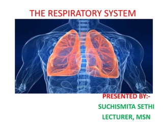
  • 1. THE RESPIRATORY SYSTEM PRESENTED BY:- SUCHISMITA SETHI LECTURER, MSN
  • 2. INTRODUCTION • A primary requirement of all body cell activities & growth is oxygen which is needed to obtain energy from food. • The fundamental purpose of respiratory system is to supply oxygen to the individual tissue cells & remove there gaseous waste product , carbon dioxide. • Breathing or ventilation refers to the inhalation & exhalation of air.
  • 3. TYPES OF RESPIRATION • Pulmonary ventilation:- moving air into & out of the lungs. • External respiration:- Gas exchange between lungs & & blood. • Internal respiration:- Also called cellular respiration which involve the uptake of o2 production of co2 within individual cell.
  • 4. FUNCTION OF THE RESPIRATORY SYSTEM • Exchanges oxygen and carbon Dioxide between the blood and external environment • Exchange of gases takes place within the lungs in the alveoli. • NON RESPIRATORY FUNCTIONS:- • Nose serve as a organ of smell • Root of water loss & heat elimination • Defends against inhaled foreign matter • Enable vocalization ( Speech, singing & other) • Enhance venous return ( cardiovascular physiology. • Excretion of volatile substance like ammonia. • Maintain PH of body.
  • 5. ORGANS OF THE RESPIRATORY SYSTEM 1. Nose 2. Pharynx 3. Larynx 4. Trachea 5. Bronchi 6. Lungs – alveoli 7. Organs of the Respiratory System 1. UPPER AIR WAY:- Consist of mouth, nose, nasal cavity, pharynx, larynx 2. LOWER AIR WAY:- Consist of trachea, bronchi, bronchioles, alveoli.
  • 6. • RESPIRATORY TRACT:- There are two types :- • 1. conducting portion:- From nasal cavity to terminal bronchioles. • 2. Respiratory Portion:- The respiratory bronchioles & alveoli.
  • 7.
  • 8. THE NOSE • The air from the outside environment enter the nose or mouth during inspiration( inhalation) • The nose is divided in to two regions. • The external nose including the root, bridge, dorsum nasi, & the apex. • The internal nasal cavity is devided in to two halves by the nasal septum. • It contain cilia which is responsible for filtering out foreign bodies.
  • 9. FUNCTION OF NOSE • Provide an entry & exit for air during breathing. • Humidifying & warming the enter air. • Filtering inspired air & cleaning it of foreign matter • Serving as a resonating chamber for speech. • Housing the olfactory receptors for smell.
  • 10. ANATOMY OF THE NASAL CAVITY • Olfactory receptors are located in the mucosa on the superior surface. • The rest of the cavity is lined with respiratory mucosa to moistens air & traps incoming foreign particles. • Lateral walls have projections called conchae which increases surface area & Increases air turbulence within the nasal cavity. • The nasal cavity is separated from the oral cavity by the palate, anterior hard palate (bone)
  • 11. PARANASAL SINUSES • Cavities within bones surrounding the nasal cavity 1. Frontal sinus 2. Sphenoid sinus 3. Ethmoid sinus 4. Maxillary sinus • FUNCTION:- • Lighten the skull • Act as resonance chambers for speech • Produce mucus that drains into the nasal cavity
  • 13. PHARYNX (THROAT) • Muscular passage from nasal cavity to larynx • Three regions of the pharynx:- 1. Nasopharynx – superior region behind nasal cavity 2. Oropharynx – middle region behind mouth 3. Laryngopharynx – inferior region attached to larynx • The oropharynx and laryngopharynx are common passageways for air and food. • Auditory tubes enter the nasopharynx
  • 14.
  • 15. • TONSILS OF THE PHARYNX:- 1. Pharyngeal tonsil (adenoids) in the nasopharynx 2. Palatine tonsils in the oropharynx 3. Lingual tonsils at the base of the tongue 4. Tubal tonsil
  • 16. LARYNX (VOICE BOX) Function:- • Routes air and food into proper channels • Plays a role in speech • Made of eight rigid hyaline cartilages and a spoon-shaped flap of elastic cartilage (epiglottis)
  • 17. Structures of the Larynx • It act as a switching mechanism to route air & food in to the proper channel. • Prevent food & drink from entering the trachea. Thyroid cartilage:- Largest hyaline cartilage which Protrudes anteriorly (Adam’s apple) Epiglottis:- It is a moveable flap of cartilage that cover the opening to the larynx( voice box) prevent food from entering the larynx during swallowing. Vocal cords :- Vibrate with expelled air to create sound (speech). Size & thickness determine the pitch of sound. Short and thin means high pitched sound. Glottis – opening between vocal cords
  • 18.
  • 19.
  • 20. TRACHEA (WINDPIPE) • It is cylindrical shape which connects larynx with bronchi. Length 11 cm • Composed of three layers:- 1. mucosa:- made up of goblet cells & cilliated epithelium which beat continuously in opposite direction of incoming air & expel mucus loaded with dust. 2. Submucosa:- connective tissue deep to the mucosa 3. Adventitia:- Outermost layer made of C- shaped rings of hyaline cartilage.
  • 21.
  • 22. BRONCHI • Formed by division of the trachea • Enters the lung at the hilus (medial depression) • Made up of complete ring of cartilage. • Bronchi divided in to right & left bronchi • Right bronchus is wider, shorter, and straighter than left • Bronchi subdivide into smaller and smaller branches
  • 23.
  • 24.
  • 25. Coverings of the Lungs:- 1. Pulmonary (visceral) pleura covers the lung surface 2. Parietal pleura lines the walls of the thoracic cavity Pleural fluid fills the area between layers of pleura to allow gliding
  • 26.
  • 27. LUNGS • The lungs, which is the organ for respiration is a paired cone shaped organs lying in the thoracic cavity. • Each lung has a base resting on the diaphragm and an apex extending superiorly to a point approximately 2.5 cm superior to the clavicle. • It also has a medial surface and with three borders- anterior, posterior and inferior. • The broad coastal surface of the lungs is pressed against the rib cage, while the smaller mediastinal surface faces medially.
  • 28.
  • 29. CONT.. • The lungs receives the bronchus, blood vessels, lymphatic vessels and nerves through a slit in the mediastinal surface called the helium. • The right lung is larger and weighs more than the left lung. • Since the heart tilts to the left, the left lung is smaller than the right and has an indentation called the cardiac impression to accommodate the heart. • This indentation shapes the inferior and anterior parts of the superior lobe into a thin tongue-like process called the lingual.
  • 30. Lobes and Fissures of the Lungs • Each lung is divided into lobes by fissures. • Both lungs have oblique fissure and the right is further divided by a transverse fissure. • The oblique fissure in the left lung separates the superior and the inferior lobe. • The oblique and horizontal fissure divides the lungs into superior, middle and inferior lobes. • Thus the right lung has three lobes while the left has two. • Each lobe is supplied by a lobar bronchus. The lobes are subdivided by bronchopulmonary segments which are supplied by the segmental bronchi
  • 31. Bronchopulomonary Segment Trachea Rt lungs left lungs Oblique fissure transverse fissure oblique fissure Upper lobe lower lobe upper lobe lower lobe a. Apical a. Lateral a. Apical a. apical b. Posterior b. posterior b. posterior b. anterior c. Anterior c. anterior c. anterior c. posterior Middle lobe:- d. Medial d. Superior d. lateral a. Lateral e. Apical e. inferior b. medial
  • 32.
  • 33. ARTERIAL SUPPLY 1) on the right side there is one bronchial artery which arises from either the third posterior intercostal artery or from the upper left bronchial artery 2)On the left side there are two bronchial arteries both of which arise from the descending thoracic aorta 3)There are pre capillary anastomoses between bronchial and pulmonary arteries. these connections enlarge when any one of them is obstructed in disease VENOUS DRAIN
  • 34. VENOUS DRAINAGE 1) Usually there are two bronchial veins on each side ,the right bronchial vein drain into the azygous vein 2) The left bronchial vein drains either into the left superior intercostal vein or into the hemi azygous vein 3) The greater part of the venous blood from the lung is drained by the pulmonary veins.
  • 35. NERVE SUPPLY 1) Para sympathetic nerves are derived from the vagus .These fibres are:- a) motor to the bronchial muscles and on stimulation cause bronchospasm b) secretomotor to the mucous glands of the bronchial tree c) the sensory fibres are responsible for stretch reflex of the lungs and for the cough reflex
  • 36.
  • 37. BRONCHI & BRONCHIOLS • Trachea divided in to two primary bronchi at the level of 5th thoracic vertebra • Right bronchus is approximately 2.5 cm long • This is wider, shorter & more vertical then left bronchus. • After entering the right lungs at the hilium it is divided in to 3 bronchioles each branch is then subdivided in to smaller branches. • Left bronchus is about 5 cm long & narrow then right. • It is divided in to two branches.
  • 38. STRUCTURE • Bronchial wall contain 3 layer & lined with ciliated columnar epithelium. • Bronchi sub divided in to bronchioles, terminal bronchioles, respiratory bronchioles, alveolar duct and finally alveoli. • STRUCTURAL CHANGES IN BRONCHIAL PASSAGE:- a. Cartilage:- Rigid cartilage interfere with expansion of lungs tissue & gaseous exchange. At bronchial level there is no cartilage in airway wall.
  • 39.
  • 40. b. Smooth muscle:- cartilage replace by smooth muscle which allow the diameter of airway to increase. It is control by autonomic nervous system which regulate air flow within the lungs. c. Epithelial lining:- ciliated epithelium is gradually replaced with non ciliated epithelium & goblets cell disappear.
  • 41. BLOOD SUPPLY • Right & left bronchial artery supply blood. • Venous return is through bronchial veins. • Right side empty in to azygos vein & left side in to superior intercostal vein. NERVE SUPPLY:- • Vagus nerve( parasympathetic) stimulation contraction of smooth muscle in the bronchial tree cause bronchoconstriction • Sympathetic stimulation cause bronchodilation.
  • 42. FUNCTION:- • Regulate speed & volume of air flow in to within the lungs. • Parasympathetic stimulation cause constriction & sympathetic stimulation cause dilation. • Cough reflex • Removal of particulate matter • Warming & humidifying.
  • 43. RESPIRATORY BRONCHIOLS & ALVEOLI • In each lobe the lungs tissue is further divided by connective tissue in to lobule • Each lobule is supplied with air by terminal bronchiole which further divided in to respiratory bronchiole, alveolar duct & large number of alveoli. • There are about 150 million alveoli in adult lungs. • It help in gas exchange.
  • 44. • Single squamous epithelial cells in the alveolar duct & alveoli. • Distal respiratory passage are supported by loose network of elastic connective tissue in which macrophages, fibroblast, nerve , lymph & blood vessel are embedded. • Alveoli surrounded by dance network of capillaries • Alveolar wall & capillary wall fused firmly together for exchange of o2 & co2.
  • 45. • In between the squamous cells septal cells which secrete surfactant . • Surfactant is a phospholipid fluid which prevent alveoli from drying out & reduce surface tension preventing alveolar collapse during expiration • NERVE SUPPLY TO BRONCHIOLS:- Parasympathetic stimulation by vagus nerve cause bronchoconstriction. Sympathetic stimulation relax the bronchial smooth muscle.
  • 46. EXCHANGE OF GAS • Exchange of gas occur in two level. 1. Pulmonary exchange( External respiration) 2. Tissue level exchange ( internal respiration) • This process takes place by partial pressure of gases & diffusion process. • Partial Pressure:- Pressure exerted by a gas in a mixture of gas irrespective of concentration of other gas.
  • 47. PROCESS OF GASCIOUS EXCHANGE • PARTIAL PRESSURE IN INHALED AIR:- (PO2-159 mmhg & PCO2 is 0.3 mmhg • PARTIAL PRESSURE IN ALVIOLI IS:- PO2- 104mmhg & PCO2 is 40. • Here PP of inhaled air & PP of alveoli is different because out of 500 ml(tidal volume) of inhaled air only 350 ml air riches to the alveoli. • Some air remain in the respiratory tract( Trachae, bronchi & bronchioles) & after we exhale again there is some gas which is remaining in the tube.
  • 48. CONT... • It means when this fresh air enter it mixes with the air that is already present in our respiratory tract & that air has more Co2 & less of o2. • So when inhaled air mixed with more Co2 & less o2 gas partial pressure changes in both o2 & co2 in alveoli • So in alveoli level the over all partial pressure of O2 is 104 mmhg & Co2 is 40 mmhg.
  • 49. CONT... • The blood which comes to the lungs is deoxygenated blood by pulmonary artery. • Because blood comes from the tissue level o2 used & Co2 produced more due to metabolism. • So in this blood Po2 is 40 mmhg & Co2 is 46 mmhg. • The value depends on activity of cell.
  • 50. CONT... • Now alveoli has O2 at high PP. Gases diffuse from their higher PP to their lower PP. • So here O2 from alveoli which is having higher pressure diffuse to lower PP o2 which is present in pulmonary artery. • Co2 which is present in higher PP pulmonary artery diffuse with alveoli lower PP Co2. • After diffusion of PP of O2 increase to 95 mmhg. • The pulmonary vein carry O2 riched blood to the left atrium, then to ventricle & then go to aorta.
  • 51. CONT..... • In the tissue level now due to activity of tissue PP O2 decrease to 20 mmhg & PCo2 is 58 mmhg (aprox) • Due to o2 partial pressure is more in blood vessel it diffuse in tissue level & Co2 diffuse from the tissue to the blood. • So due to this value now blood vessel again become deoxygenated & go to the heart through superior & inferior venacava.
  • 52.
  • 53.
  • 54. INTRODUCTION TO RESPIRATION • The term respiration means exchange of gas between body cells & their environment. • It involve two main process:- 1. Breathing (Pulmonary ventilation):- It is the movement of gas in to & out of the lungs 2. Exchange of gas:- This takes place :- In lungs:- External Respiration In tissue :- Internal respiration
  • 55. 1. BREATHING:- • Breathing supplies oxygen to the alveoli & eliminate Co2. MUSCLE OF BREATHING:- • Expansion of chest during inspiration occur as a result of muscular activity which is partly voluntary & partly involuntary. • The muscle use in normal breathing are external intercostal muscle & diaphragm.
  • 56. INTERCOSTAL MUSCLE • There are 11 pairs of intercostal muscle occupying the space between the 12 pair of ribs. • They are arranged in two layers a. External intercostal muscle:- they extend down wards & forwards from the lower border of the ribs above to the upper border of rib below. b. Internal intercostal muscle:- they extends down wards and back wards from the lower border of ribs above to upper border of rib below. This muscle use when expiration become active
  • 57.
  • 58. Cont….. • The first rib is fixed, so when external muscle contract they pulled all the other ribs to wards the first rib • The rib cage move as a unit upward & out ward enlarge the thoracic cavity. • Intercostal muscle under the control of intercostal nerve.
  • 59.
  • 60. DIAPHRAGM • The diaphragm is a dome-shaped muscular structure which separate thoracic and abdominal cavity. • It consist of a central tendon from which muscle fiber radiate to attach to the lower ribs & sternum & to vertebral column by two crura. • When diaphragm is relaxed the central tendon is at the level of the 8th thoracic vertebra. • When it contract its muscle fiber shorten & central tendon is pulled down to the level of 9th thoracic vertebra, lengthening the thoracic cavity. • Thus decrease pressue in the thoracic cavity & increase pressure in abdominal cavity. • During inspiration the external intercostal muscle & diaphragm contract simultaneously to enlarge the thoracic cavity
  • 61. ACCESSORY MUSCLE • When extra respiratory effort is required additional muscle are required • Force of inspiration is assisted by the sternocleidomastoid muscle & scalene muscles which link to cervical vertebra & first 2 rib. To increase rib cage expansion. • Forced expiration is helped by the intercostal muscle & abdominal muscle. Which increse the pressure of thorax by squeezing the abdominal muscle.
  • 62. CYCLE OF BREATHING • Average respiratory rate is 12-15 breath/min • Breathing depends upon change in pressure & volume in thoracic cavity. • It follow the principle , “Increase the volume of container decrease the pressure inside & decreasing the volume increase the pressure inside” • So air flow from area of higher pressure to an area of low pressure , change the pressure inside the lungs which determine the direction of air flow.
  • 63.
  • 64.
  • 65. Cont.... • Breathing consist of 3 phase • 1. inspiration • 2. Expiration • 3. pause
  • 66. 1. INSPIRATION • Simultanious contraction of external intercostal muscle & expand the thorax.. • Parietal pleura is firmly adhered to the diaphragm & inside the rib cage. • It is pulled outward along with them. • This pulls the visceral pleura outward too since the two pleura are hold together by thin film of pleural fluid. • Because viseral pluera is firmly adherent to the lung the lungs tissue also pulled up & out with the ribs & downwards with the diaphragm. • This expand the lungs & pressure within the alveoili & in the air passage fall, drowing air in to the lungs in an attempt to equalise atmospheric & alveolar pressure. • The negative pressure created in the thoracic cavity aids venous return to the heart. • Inspiration is a active process so it need energy
  • 67.
  • 68. 2. EXPIRATION • Relaxation of the external intercostal muscle & the diaphragm result in downward & inward movement of the rib cage & elastic recoil of the lungs • Due to this pressure inside the lungs rise & expel air from the respiratory tract • At the end of the expiration the lungs still contains the air which prevent from complete collapse by the intact pleura. • This process is passive which do not need energy. • After expiration there is pause before next cycle begin.
  • 69.
  • 70. PHYSIOLOGICAL VARIABLE AFFECTING THE BREATHING 1. ELASTICITY:-Elesticity is the ability of the lungs to return to its normal shape after each breath. Loss of eleasticity of the connective tissue necessitate forced expiration & increase effort of inspiration. 2. COMPLIANCE:- this is the stretchebility of the lungs i.e the effort required to inflate the alveoli. The helathy lungs is very complient which inlate with very little effort but compliance is low more effort needed to inflate. 3. AIRWAY RESISTANCE:- increase resistance cause more respiratory effort to inflate the lungs.
  • 71. LUNGS VOLUME & CAPACITY • There are 15 complete respiratory cycle per min. • The lungs and air passage are never empty & exchange of gases takes place only across the wall of the alveolar duct & alveoli. • The remaining capacity of the respiratory passage is called the anatomical dead space( 150 ml)
  • 72. CONT.... 1. Tidal volume:- this is the amount of air passing in to & out of the lungs during each cycle of breathing . (500 ml at rest) 2. Inspiratory reserve volume:- inspiratory reserve volume (IRV) is the additional amount of air that can be inhaled after a normal inhalation. 3. Functional residual capacity:- The functional residual capacity (FRC) is defined as the amount of gas left in the lungs after normal expiration. This is about 2.5 L in the average-sized adult or 35 mL/kg. The FRC acts as a buffer by preventing rapid changes in alveolar gas tensions from inspired air. 4. Expiratory reserve volume:- expiratory reserve volume is the amount of extra air — above anormal breath — exhaled during a forceful breath out. The average ERV volume is about 1100 mL in males and 800 mL in females
  • 73. 5. Residual volume:- The residual volume (RV) is the amount of air that is left after expiratory reserve volume is exhaled. 6. Vital capacity:- Vital capacity (VC) is the maximum amount of air a person can expel from the lungs after a maximum inhalation. It is equal to the sum of inspiratory reserve volume, tidal volume, and expiratory reserve volume. 7. Total lungs capacity:- Lung capacity or total lung capacity (TLC) is the volume of air in the lungs upon the maximum effort of inspiration. Among healthy adults, the average lung capacity is about 6 liters 8. Alveolar ventilation:- Alveolar ventilation and dead space A. Alveolar ventilation ( A) is defined as the volume of air entering and leaving the alveoli per minute. Air ventilating the anatomic dead space (VD) (Levitzky Fig 3-7), where no gas exchange occurs, is not included: VT = VD + VA.
  • 74. COMPOSITION OF INSPIRED & EXPIRED AIR • INSPIRED AIR EXPIRED AIR • OXYGEN:- 21 16 • CARBON DIOXIDE:- 0.04 4 • NITROGEN:- 78 78 • WATER VAPOUR:- VARIABLE SATURATED
  • 75. CONTROL OF RESPIRATION • Effective control of respiration enables the body to regulate blood gas level over a wide range of physiological environmental & pathological condition. • Voluntary control is exerted during activities such as speaking & singing but is overridden if blood co2 raise.
  • 76. The respiratory centre • Respiratory centre present in medulla which control rate & depth of breathing. • Regular discharge of inspiratory neurones within this centre set the rate & depth of breathing • Activity of the respiratory rhythmicity centre is adjusted by nerve in the Pons ( the pneumotaxic centre & apneustic centre ) in response to input from other parts of the brain. • Motor impulses leaving the respiratory centre pass in Phrenic & intercostal nerves to the diaphragm & intercostal muscle to stimulate respiration
  • 77. chemoreceptor's • These are receptors that respond to changes in the partial pressure of oxygen & Carbon dioxide in the blood & CSF. • Central chemoreceptor's:- located on the surface of medulla oblongata & are bathed in CSF. • When arterial PCO2 rise (hypercapnia) the central chemoreceptor by stimulating the respiratory centre which increase the ventilation to reduce the PCO2
  • 78. Cont.. • Peripheral chemoreceptor's:- these are situated in the arch of aorta & in the carotid bodies. • They respond to change in the blood co2 & o2 level. • It is more sensitivity to CO2 then O2. • Change in the co2 & o2 level activate these receptors trigger the nerve impulse to the respiratory centre via the glossopharyngeal & vagus nerve. • This stimulation raise the rate & depth of the respiration