
Empfohlen
Weitere ähnliche Inhalte
Ähnlich wie cell biology class 17th N ovember mitochondria 2015.ppt
Ähnlich wie cell biology class 17th N ovember mitochondria 2015.ppt (20)
Kürzlich hochgeladen
Kürzlich hochgeladen (20)
cell biology class 17th N ovember mitochondria 2015.ppt
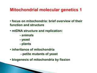
- 1. Mitochondrial molecular genetics 1 • focus on mitochondria: brief overview of their function and structure • mtDNA structure and replication: - animals - yeast - plants • inheritance of mitochondria - petite mutants of yeast • biogenesis of mitochondria by fission
- 2. MITOCHONDRIA • essential for cell life - ATP synthesis - many metabolic intermediates • essential for cell death - unprogrammed death: necrosis ( eg, due to loss of energy status) - programmed cell death (apoptosis - controlled cell destruction)
- 3. • Two membranes • Inner membrane invaginated • Numbers of mitochondria per cell vary but usually 100s/cell Matrix contains the TCA cycle (and other) soluble enzymes Inner membrane contains metabolite transporters and the electron transport chain Mitochondrial structure
- 4. The ribosomes can actually be visualized in some mitochondria. In these figures, they are seen in the matrix as small dark bodies. DNA can also be visualized in mitochondria. The DNA is circular and resembles that of a bacterium in its basic structure. Mitochondria also have their own ribosomes and tRNA: • 22 tRNAs • rRNAs (16S and 12S)
- 5. Mitochondria have their own DNA and Ribosomes Mitochondria have some of their own DNA, ribosomes, and can make many of their own proteins. The DNA is circular and lies in the matrix in structures called "nucleoids". Each nucleoid may contain 4-5 copies of the mitochondrial DNA (mtDNA). mitochondrial DNA
- 6. To visualize the structure of mitochondrial DNA, we have to extract the DNA and float it on a water surface. Then, it can be picked up by a plastic coated grid, and examined in the electron microscope. Mitochondrial circular DNA is shown in the figure.
- 7. Human mtDNA • small, double stranded circular chromosome • 16,569 bp in total • no non-coding DNA • no introns • polycistronic replication which is initiated from the D (displacement)- loop region • followed by splicing of transcript to form messages. Organisation of the mitochondrial chromosome
- 8. Human mtDNA. The human mitochondrial DNA consists of 16,568 base pairs and was the first entire genome sequenced in 1981. It encodes 22 tRNA, the 12 S and the 16 S rRNA, and 13 peptides forming with the 74 nuclear-encoded polypeptides the five respiratory chain complexes. The regulatory region is a non coding sequence, or three stranded D-loop of nearly 600 base pairs. It contains the promoters of the heavy and light strands (HSP and LSP respectively), and the origin of replication of Purine-rich Heavy strand (OH). Transcription is polycistronic, and translation uses a specific code different from the universal. Replication is asymmetric, starting with the synthesis of the Heavy strand until the origin of the Light strand (OL) located about 2/3 from OH from which synthesis proceeds counter-clockwise. Thus, the Heavy strand is single stranded during about half of the replication cycle which lasts about 2 hours. ND1-6: NADH dehydrogenase subunits, COX1-3: Cytochrome c oxidase subunits, ATP6 and ATP8: subunits of ATP synthase, Cyt b: Cytochrome b.
- 11. Human DNA • 16,569 bp; • no non-coding DNA • no introns • polycistronic replication followed by splicing to form messages. Yeast mtDNA • 68-75 kb, similar in structure to bacterial genome • contains introns and non-regions between genes. • Same proteins made as in animals • genes transcribed separately
- 12. Plant mtDNA • chromosome size is much bigger but varies dramatically between species (200-2000 kb) • arranged as different size circles, sometimes with plasmids. • The plant mtDNA contains chloroplast sequences, indicating exchange of genetic information between organelles in plants. • Much of the plant mtDNA is non-coding, but coding regions are larger than animals and fungi. • Number of proteins synthesised not known definitely but more than in animals and yeast (probably about 50) Plant mitochondria have specialised functions • in leaves they participate in photorespiration • sites of vitamin synthesis (vit C, folic acid, biotin)
- 14. Mitochondrial Inheritance Yeast has been used extensively to study mitochondrial inheritance. There is a Yeast strain, called "Petite" that have structurally abnormal mitochondria that are incapable of oxidative phosphorylation. These mitochondria have lost some or all of their DNA. Genetic crosses between petite and wt strains showed that inheritance of this trait did not segregate with any of the nuclear chromosomes.
- 15. Mitochondrial Inheritance Mitochondrial inheritance from yeast is biparental, and both parent cells contribute to the daughter cells when the haploid cells fuse. After meiosis and mitosis, there is random distribution of mitochondria to daughter cells. If the fusion is with yeast that are petite and yeast that are not, a certain percentage of the daughter cells will be "petite".
- 16. Mitochondrial Inheritance in Yeast
- 17. This led to the suggestion that some genetic element existed in the cytoplasm and was inherited in a different manner from nuclear genes. This is called “non- Mendelian inheritance” or “cytoplasmic inheritance”. In yeast and animals, this indicated inheritance of mitochondrial genes: in plants it also includes inheritance of chloroplast genes Mitochondrial Inheritance
- 18. Cytoplasmic Male Sterility • Definition: Cytoplasmic male sterility is total or partial male sterility in plants as the result of specific nuclear and mitochondrial interactions.Male sterility is the failure of plants to produce functional anthers, pollen, or male gametes. • Function • example of non mendelian inheritance • Governed by cytoplasmic factors • Interplay of nuclear and mitochondrial genomes. • Restorer of fertility gene
- 19. Biogenesis of mitochondria • Non de novo synthesis • Molecular basis of biogenesis : it involves transcriptional co activators (peroxisome proliferator activator gamma C family)PGC- 1α, 1β and PRC, co factors NRF 1 and NRF 2
- 24. Schematic representation of the human mitochondrial genome. Richard C. Scarpulla Physiol Rev 2008;88:611-638 ©2008 by American Physiological Society
- 25. Schematic representation of the human mitochondrial genome. Genomic organization and structural features of human mtDNA are depicted in a circular genomic map showing heavy (blue) and light (black) strands assigned as such based on their buoyant densities. Protein coding and rRNA genes are interspersed with 22 tRNA genes (red bars denoted by the single-letter amino acid code). Duplicate tRNA genes for leucine (L) and serine (S) are distinguished by their codon recognition (parentheses). The D-loop regulatory region contains the L- and H-strand promoters (LSP, HSP1, and HSP2), with arrows showing the direction of transcription. The origin of H-strand replication (OH) is within the D-loop, whereas the origin of L-strand replication (OL) is displaced by approximately two-thirds of the genome within a cluster of five tRNA genes (W, A, N, C, Y). Protein coding genes include the following: cytochrome oxidase (COX) subunits 1, 2, and 3; NADH dehydrogenase (ND) subinits 1, 2, 3, 4, 4L, 5, and 6; ATP synthase (ATPS) subunits 6 and 8; cytochrome b (Cyt b). ND6 and the 8 tRNA genes transcribed from the L-strand as template are labeled on the inside of the genomic map, whereas the remaining protein coding and RNA genes transcribed from the H-strand as template are labeled on the outside.
- 26. Mitochondria replicate much like bacterial cells. When they get too large, they undergo fission. This involves a furrowing of the inner and then the outer membrane as if someone was pinching the mitochondrion. Then the two daughter mitochondria split. Of course, the mitochondria must first replicate their DNA. An electron micrograph depicting the furrowing process is shown in these figures. Mitochondrial replication cell division: random distribution of mitos between daughter cells mitochondrial replication
- 27. Sometimes new mitochondria are synthesized in centres that are rich in proteins and polyribosomes needed for their synthesis. The electron micrograph in the following figure shows such a centre. It appears that the cluster of mitochondria are sitting in a matrix of proteins and other materials needed for their production.
- 28. Certain mitochondrial proteins are needed before the mitochondria can divide. This has been shown in a study by Sorgo and Yaffe, J Cell Bio. 126: 1361-1373, 1994. They showed the result of the removal of an outer membrane protein from mitochondria called MDM10. This figure shows the results. The mitochondria are able to take in components and produce membranes and matrix enzymes. However, fission is not allowed and the result is a giant mitochondrion. giant mitochondrion
- 29. Despite having their own genome, most mitochondrial proteins are encoded in the nucleus, made in the cytosol and imported into the mitochondria
- 30. In all organisms, only a few of the proteins of the mitochondrion are encoded by mtDNA, but the precise number varies between organisms • Subunits 1, 2, and 3 of cytochrome oxidase • Subunits 6, 8, 9 of the Fo ATPase • Apocytochrome b subunit of complexIII • Seven NADH-CoQ reductase subunits (except in yeast) The nucleus encodes the remaining proteins which are made in the cytosol and imported into the mitochondrion. Most of the lipid is imported. Synthesis of mitochondrial proteins
- 31. Rice mitochondrial and chloroplast genomes Plant mitochondria contain chloroplast genes - suggesting that genetic transfer occurs between the two organelles
- 32. Mitochondrial DNA of animals and fungi uses a different genetic code than the “universal” code
- 33. RNA processing in mitochondria Plant mitochondria “edit” their RNA transcripts. This was first noticed when comparing cDNA sequences with genomic DNA sequences. The most common change is to replace C with U, although in some instances other changes can occur. Matrix enzymes are thought to be responsible for this, but the reason for the editing is not known. Most of the DNA in plant mitochondria is non-coding, only some of which is transcribed. RNA editing occurs even in non-coding regions such as introns.
- 34. RNA editing is a molecular process through which some cells can make discrete changes to specific nucleotide sequences within a RNA molecule after it has been generated by RNA polymerase. RNA editing is relatively rare, and common forms of RNA processing (e.g. splicing, 5'- capping and 3'-polyadenylation) are not usually included as editing. Editing events may include the insertion, deletion, and base substitution of nucleotides within the edited RNA molecule.
- 35. Evolution of mitochondria Mitochondria are generally thought to have evolved endosymbiotically when an anaerobic prokaryotic cell engulfed an aerobic bacterium and formed a stable symbiosis. Loss of most of the aerobe’s genome to the nucleus of the host allowed the latter to control the former. This is supported by gene sequence analysis which shows remarkable homology between bacteria and mitochondrial genes.