Addition of propranolol and isosorbide mononitrate to endoscopic variceal ligation does not reduce varicealrebleeding incidence.
•
0 gefällt mir•153 views

Addition of propranolol and isosorbide mononitrate to endoscopic variceal ligation does not reduce varicealrebleeding incidence.

Empfohlen
Empfohlen
Weitere ähnliche Inhalte
Was ist angesagt?
Was ist angesagt? (19)
Andere mochten auch
Andere mochten auch (15)
Ähnlich wie Addition of propranolol and isosorbide mononitrate to endoscopic variceal ligation does not reduce varicealrebleeding incidence.
Ähnlich wie Addition of propranolol and isosorbide mononitrate to endoscopic variceal ligation does not reduce varicealrebleeding incidence. (20)
Kürzlich hochgeladen
Kürzlich hochgeladen (20)
Addition of propranolol and isosorbide mononitrate to endoscopic variceal ligation does not reduce varicealrebleeding incidence.
- 1. Addition of Propranolol and Isosorbide Mononitrate to Endoscopic Variceal Ligation Does Not Reduce Variceal Rebleeding Incidence ASHISH KUMAR,* SANJEEV KUMAR JHA,† PRAVEEN SHARMA,* SAROJ DUBEY,† PANKAJ TYAGI,† BARJESH CHANDER SHARMA,† and SHIV KUMAR SARIN*,† *Department of Medical Hepatology, Institute of Liver and Biliary Sciences, New Delhi, India; and † Department of Gastroenterology, G B Pant Hospital, University of Delhi, New Delhi, India See related article, Rocha ECV et al, on page 988 in CGH. BACKGROUND & AIMS: Endoscopic variceal ligation (EVL) and propranolol are standard secondary prophylaxis therapies for variceal bleeding. Addition of isosorbide mono- nitrate (ISMN) to propranolol improves its hemodynamic ef- ficacy; we investigated whether a combination of EVL and propranolol/ISMN was more effective than EVL alone for secondary prophylaxis. METHODS: Patients with a prior variceal bleed were randomly assigned to groups given a com- bination (n ϭ 88) of EVL, propranolol (dose titrated to reduce heart rate to 55 beats per minute), and ISMN (40 mg/day) or EVL alone (n ϭ 89). Primary end points were rebleeding or death; secondary end points were new complications of portal hypertension or serious adverse effects. RESULTS: The actu- arial probabilities of rebleeding 2 years after therapy were 27% in the combination group and 31% in the EVL alone group (P ϭ .822). Two patients in the combination group and 3 patients in the EVL alone group died during the study period (P ϭ .682); no deaths were caused by variceal hemorrhage. In cir- rhotic patients, the actuarial probabilities of rebleeding were 24% and 30%, respectively (P ϭ .720). Secondary end points were comparable between groups. In multivariate analyses, presence of ascites (P ϭ .003), serum albumin Ͻ 3.3 g/dL (P ϭ .008), and hepatic venous pressure gradients Ն 18 mm Hg (P ϭ .009) were independent risk factors for variceal rebleeding. CONCLUSIONS: EVL alone is sufficient to prevent variceal rebleeding in cirrhotic and noncirrhotic patients with history of variceal bleeding. Addition of propranolol and ISMN to EVL does not reduce the incidence of variceal rebleeding but increases severe adverse effects. Risk factors for rebleeding include ascites, low serum al- bumin, and high hepatic venous pressure gradients. The lifetime prevalence of varices in cirrhotics has been reported to be as high as 80%–90%. Variceal bleeding is a serious complication, and it occurs in 30%– 40% of the patients with varices. The mortality of acute variceal hemorrhage has decreased significantly but still ranges between 15% and 20% even with optimal manage- ment.1–3 Patients with portal hypertension, who have bled from esophageal varices, have a 60%–80% risk of variceal rebleeding at 1 year, if no therapy for prevention of variceal rebleeding is instituted.4,5 Rebleeding from esophageal varices is associated with a high morbidity and mortality. Therefore, patients with variceal bleeding should be given effective long-term therapy to prevent recurrence of variceal bleeding. The current recommen- dation, according to the Baveno IV consensus confer- ence,6 for prevention of rebleeding in patients of cirrho- sis, is the use of -blockers or endoscopic variceal ligation (EVL) or both. The combination therapy (-blocker plus EVL) was thought to be better than either therapy alone; however, definite recommendation for its use could not be made because of lack of a sufficient number of trials showing its superiority.6 Conceptually, reduction of portal pressure by combi- nation pharmacotherapy and local obliteration of varices should be more effective and synergistic than either ther- apy administered alone. The hemodynamic response to -blockers is achieved in only approximately one third of the patients, and addition of nitrates has been found to work synergistically to reduce further the portal pressure and improve the overall response rates to pharmacother- apy.7 We conducted a randomized controlled trial of a combination of EVL and -blocker plus nitrate vs EVL alone in the secondary prophylaxis of variceal rebleed from esophageal varices. The main objectives were to evaluate, in patients who had bled from varices in the past, whether endoscopic and combined drug therapy regimen reduces the incidence of variceal rebleeds and improves the survival compared with ligation alone in this group of patients. The adverse effects of the drug therapy were also carefully monitored. Patients and Methods Patients Patients presenting to the Liver Disease Clinic at the G B Pant Hospital, New Delhi, with a history of Abbreviations used in this paper: EVL, endoscopic variceal ligation; HVPG, hepatic venous pressure gradient; ISMN, isosorbide mononi- trate. © 2009 by the AGA Institute 0016-5085/09/$36.00 doi:10.1053/j.gastro.2009.05.049 CLINICAL–LIVER, PANCREAS,AND BILIARYTRACT GASTROENTEROLOGY 2009;137:892–901
- 2. hematemesis and/or melena and proven to have esopha- geal varices as the bleeding source on upper gastrointes- tinal (GI) endoscopy were included in the study. The exclusion criteria were as follows: (1) a history of under- going endoscopic sclerotherapy, EVL, or cyanoacrylate injection; (2) a history of surgery for portal hypertension; (3) coexisting malignancy; (4) severe cardiopulmonary or renal disease; (5) a history of severe adverse effects or contraindications to -blockers like bronchial asthma, uncontrolled diabetes mellitus, heart failure, peripheral vascular disease, prostatic hypertrophy, arterial hypoten- sion (systolic blood pressure Ͻ100 mm Hg), bradycardia (basal heart rate Ͻ55 beats per minute), or complete heart block; and (6) refusal to give consent to participate in the trial. A written informed consent was taken from all the included patients. The Institutional Ethics Com- mittee approved the study protocol. The trial was regis- tered with ClincalTrials.gov vide NCT00766805. Baseline Evaluation The included patients were broadly categorized into cirrhotic patients or non-cirrhotic patients. Cirrho- sis was diagnosed on the basis of clinical, biochemical, histologic, or ultrasonographic evidence. An effort was made to determine the etiology of cirrhosis. Hepatitis B and C virus markers, autoimmune markers, serum ceru- loplasmin, urinary copper, and serum iron and ferritin assays were performed wherever needed. History of alco- hol abuse was sought from all the patients. The severity of liver disease in cirrhotics was assessed by Child–Tur- cotte–Pugh (CPT) score. Non-cirrhotic patients were fur- ther categorized into non-cirrhotic portal fibrosis and extrahepatic portal vein obstruction. Non-cirrhotic por- tal fibrosis was diagnosed when varices were present, and there was no evidence of thrombosis in the splenoportal axis on ultrasonography and no evidence of cirrhosis on liver biopsy.8 Extrahepatic portal vein obstruction was diagnosed when a portal cavernoma was detected by ultrasonography, and there were no signs of cirrhosis.8 The size of esophageal varices was assessed according to Conn’s classification9: grade 1, visible only during 1 phase of respiration or on performance of valsalva ma- neuver; grade 2, visible during both phases of respiration; grade 3, 3–6 mm; and grade 4, Ͼ6 mm. The size of the largest varix was assessed by comparison with the shaft of the biopsy forceps (3 mm) or with the distance between the open jaws of the biopsy forceps (6 mm) in the lumen of the lower 2 to 3 cm of the esophagus. Hepatic Venous Pressure Gradient Measurement Hepatic venous pressure gradient (HVPG) was measured before randomization. HVPG measurement was done after overnight fast and under antibiotic cover. Under local anesthesia, a 7-French central venous cathe- ter (Arrow Medical, Athens, TX) was placed in the right femoral vein or internal jugular vein under fluoroscopic guidance, using the Seldinger technique. HVPG was mea- sured by the standard technique10 in which a balloon catheter was introduced into the right hepatic vein under fluoroscopic guidance. The zero reference point was set at the midaxillary point. The free hepatic venous pressure was obtained by keeping the catheter free into the lumen of the hepatic vein. The balloon of the catheter was then inflated to wedge the lumen of the hepatic vein. Presence of wedging was confirmed by absence of reflux into the inferior vena cava, after the injection of 2 mL intravenous contrast, and appearance of a sinusoidogram. The pres- sure tracing at this juncture showed absence of wave- forms, and the pressure was labeled as wedged hepatic venous pressure. HVPG was determined by subtracting free from wedged hepatic venous pressures (HVPG ϭ wedged hepatic venous pressure Ϫ free hepatic venous pressure). All measurements were performed in triplicate. If the difference between the 2 readings was more than 1 mm Hg, all the readings were discarded, and a fresh set of measurements were taken. The normal value of the HVPG in our hemodynamic laboratory is between 1 and 4 mm Hg. Randomization This was an open-label, randomized controlled trial. Patients were randomized after HVPG, using com- puter generated random numbers, to receive either of the 2 therapies (EVL or EVL plus drugs). The randomization sequence remained with the statistician, and the se- quence remained concealed from the investigators until the intervention was assigned. Those presenting with an acute bleed underwent an emergency variceal band liga- tion and 5-day therapy with terlipressin or somatostatin prior to HVPG and randomization. Endoscopic Variceal Ligation Patients assigned to the EVL group underwent variceal band ligation at the first endoscopy session within the next 24 hours. Ligation was done using a multiband ligator. In each session, as many bands as possible were placed on the varices in the lower 5–7 cm of the esophagus, the number varying from 2 to 10. EVL was done at intervals of 3–4 weeks until the varices were completely obliterated or reduced to grade 1 size and could not be banded any further. Once varices were eradicated, repeat endoscopy was done at monthly inter- vals for 3 months and then at 3-month intervals to check for recurrent varices. Further EVL sessions were under- taken for recurrent varices. Patients were routinely ad- vised to have liquids on the day of EVL and subsequently placed on semisolids and solids. They were prescribed 40 mg of pantoprazole twice a day and a suspension of sucralfate 4 times per day. Patients with ascites were prescribed oral antibiotics. In general, attempts were made to correct the coagulation disorders by giving fresh CLINICAL–LIVER, PANCREAS,AND BILIARYTRACT September 2009 EVL VS EVL؉ DRUGS FOR SECONDARY PROPHYLAXIS DRUGS 893
- 3. frozen plasma. All adverse effects including chest pain, dysphagia, fever, and GI bleeding were recorded. EVL Plus Drug Therapy Patients randomized to the EVL plus drugs ther- apy received EVL plus -blocker (propranolol) and ni- trate (isosorbide mononitrate [ISMN]). Within 24 hours, these patients were started on propranolol after a base- line electrocardiogram and cardiac evaluation. Treatment was started with propranolol at a dose of 40 mg twice a day. The heart rate and blood pressure were checked after 12 to 24 hours. The dose of propranolol was increased at increments of 20 to 40 mg per day until the patient achieved a heart rate of 55 beats per minute (bpm) or a maximum dose of 320 mg/day was achieved. The dose was reduced if any of the following occurred: systolic blood pressure (BP) Ͻ90 mm Hg, heart rate Ͻ55 bpm, or other serious adverse effects. After attaining a stable dose of propranolol, ISMN was added at a dose of 10 mg twice a day. The dose was escalated at increments of 10–20 mg/day until a maximum dose of 40 mg/day was reached or the patient experienced adverse effects such as head- ache, dizziness, or hypotension. Full dosage of -blocker and ISMN was achieved within 2–3 weeks of randomiza- tion, before the second session of EVL. Further EVL sessions and follow-ups were done as in the EVL arm. Patients receiving propranolol were monitored daily until -blockade was adequate then monthly for the first 3 months and every 3 months subsequently. Drug compli- ance was ascertained by interviewing the patient and by measuring the resting heart rate. End Points Primary end points of the study were bleeding and death. Secondary end points included complications (be- cause of bleeding, causes related to the underlying liver disease or unrelated causes), upper GI tract bleeding because of causes not related to portal hypertension, and the development of serious adverse effects that required the discontinuation of therapy. Bleeding Patients presenting with active upper GI bleeding during the study were admitted and subjected to upper GI endoscopy within 12 hours to determine the source of bleeding. Upper GI bleeding was diagnosed and classified using Baveno IV consensus conference criteria.6 Bleeding from esophageal varices was diagnosed if active bleeding, a “white nipple,” or a clot was seen at endoscopy or if there was blood in the stomach in a patient with esoph- ageal varices and no other potential bleeding source. Gastric variceal bleed was diagnosed if active bleeding or a clot was seen on gastric varices on endoscopy or if there was evidence of recent bleeding in a patient with a gastric varix and the bleeding had no other possible cause. Bleed- ing was attributed to portal hypertensive gastropathy (PHG) if distinct lesions of the gastric mucosa were present, and there was endoscopic evidence of an active bleeding lesion, assessed after washing or removal of clots, and there was no evidence of bleeding from esoph- ageal, gastric, or ectopic varices. Esophageal ulcer bleed as a result of band ligation was diagnosed if there was active bleeding from the ulcer at the site of banding or if there was an adherent clot on the esophageal ulcer with ab- sence of any other potentially bleeding lesions in the upper GI endoscopy. Bleeding was categorized as clini- cally significant when the heart rate was Ͼ100 bpm, the systolic blood pressure was Ͻ100 mm Hg, or there was a postural drop of Ͼ20 mm Hg and transfusion require- ment of more than 2 U of blood in 24 hours.6 All patients with bleeding were started on vasoactive drugs (terlipres- sin or somatostatin) immediately. An emergency session of EVL was performed in these patients to stop the bleeding. The vasoactive drugs were continued for 5 days. Sample Size Calculation and Statistical Analysis This study was designed to compare the risks of variceal rebleed as calculated by Kaplan–Meier curves for EVL plus drug therapy vs EVL alone. The probability of rebleed on EVL alone therapy was assumed to be 40% at 1 year. It was hypothesized that EVL plus drug therapy would halve the risk to 20%. Using a 1-tailed test with an ␣ value of .05 and power (1 Ϫ ) of 0.80, the required sample size would be 148, ie, 74 in each group. Quantitative data were expressed as mean (ϮSD) or median (range) and analyzed using Student 2-tailed t test or Mann–Whitney U test. Qualitative data were analyzed by Fisher exact test or Pearson 2 test. Kaplan–Meier plot and log-rank test was used to compare cumulative prob- abilities of rebleeding and death. Subgroup analysis was done for cirrhotic and non-cirrhotic patients separately. Univariate and multivariate analyses were performed to assess the variables predicting rebleed. Statistical analysis was done using the SPSS 15.0 statistical package (SPSS Inc, Chicago, IL). The statistical analysis of the entire data sets pertaining to efficacy (specifically primary and major secondary efficacy end points) and safety (specifi- cally, serious adverse events as defined in federal guide- lines) have been independently confirmed by a biostatis- tician who is not employed by the corporate entity. Results Patients One thousand nine hundred twenty-one new pa- tients with portal hypertension were admitted in our hospital from October 2002 through December 2006 (Supplementary Figure 1). Of these, 653 had history of variceal bleed in past. Four hundred seventy-six patients were excluded for the following reasons: history of un- dergoing endoscopic sclerotherapy, EVL, or cyanoacrylate CLINICAL–LIVER, PANCREAS,AND BILIARYTRACT 894 KUMAR ET AL GASTROENTEROLOGY Vol. 137, No. 3
- 4. injection before presenting to our hospital, 375; history of surgery for portal hypertension, 12; coexisting malig- nancy, 27; severe cardiopulmonary or renal disease, 7; history of severe adverse effects or contraindications to -blockers, 42; and refusal to give consent to participate in the trial, 13. Hence, a remaining 177 patients were included in the trial. One hundred fifty-one patients (85%) were cirrhotic patients, whereas the remaining 26 (15%) were non-cir- rhotic patients (extrahepatic portal vein obstruction, 12, and non-cirrhotic portal fibrosis, 14). Eighteen (10%) patients had presented with acute variceal bleed (ie, within 5 days), and the rest had presented with past history of variceal bleed (Table 1). Randomization and Follow-Up Eighty-eight patients were randomized to the EVLϩdrugs group, and 89 patients were randomized to the EVL alone group. The baseline characteristics of pa- tients were similar in the 2 groups (Table 1). Ten patients were lost to follow-up before the second session of EVL (4 in EVLϩdrugs group and 6 in EVL alone group, P ϭ .536). These patients were excluded, and, thus, subsequent analysis was done on 167 patients. The mean follow-up was 15 Ϯ 12 months (range, 0.3–52 months) in the EVLϩdrugs group and 15 Ϯ 11 months (range, 1–55 months) in the EVL group (P ϭ .662). EVL was done at an average of 4-weeks in both the groups. The median dose of propranolol used was 120 (range, 40–320) mg/day and of ISMN was 40 (range, 20–40) mg/day in the EVLϩdrugs group. Primary End Points During a mean follow-up of 15 Ϯ 12 months, 30 patients developed rebleed: 14 of 84 (17%) in the EVLϩdrugs group and 16 of 83 (19%) in the EVL group Table 1. Baseline Characteristics of Study Groups Parameters EVLϩdrugs EVL alone P value All patients (n ϭ 177) (n ϭ 88) (n ϭ 89) Mean age (ϮSD), y 42 (14) 41 (14) .716 Sex, n (%) .667 Males 75 (85) 78 (88) Females 13 (15) 11 (12) Cause of portal hypertension, n (%) .832 Cirrhosis 76 (86) 75 (84) Non-cirrhotic portal hypertension 12 (14) 14 (16) EHPVO 6 6 NCPF 6 8 Last bleed, n (%) .590 Յ5 days ago 11 (12) 7 (8) 6Ϫ42 days ago 43 (49) 45 (50) Ͼ42 days ago 34 (39) 37 (42) Mean (ϮSD) grade of esophageal varices 3.1 (0.8) 3.2 (0.7) .292 Patients with gastric varices, n (%) 51 (58) 46 (52) .451 Patients with PHG, n (%) 40 (45) 48 (54) .294 Mean (ϮSD) platelet count, 103/mm3 155 (52) 160 (59) .755 Cirrhotic patients (n ϭ 151) (n ϭ 76) (n ϭ 75) Etiology of cirrhosis, n (%) .354 Alcohol 33 (43) 30 (40) Viral 13 (17) 20 (27) Others 30 (40) 25 (33) CTP class, n (%) .292 A 35 (46) 26 (35) B 31 (41) 34 (45) C 10 (13) 15 (20) Mean (ϮSD) CTP score 7.3 (2.0) 7.8 (2.1) .138 Ascites, n (%) 24 (32) 33 (44) .133 Encephalopathy, n (%) 1 (1) 4 (5) .209 Median (range) serum bilirubin, mg/dL 1.5 (0.5Ϫ16.9) 1.6 (0.2Ϫ14.4) .527 Mean (ϮSD) serum albumin, g/dL 3.2 (0.5) 3.2 (0.6) .707 Prothrombin time prolongation, n (%) .559 0–3 s 42 (56) 35 (47) 4–6 s 17 (22) 21 (28) Ͼ6 s 17 (22) 19 (25) Mean (ϮSD) serum creatinine, mg/dL 0.9 (0.5) 0.9 (0.4) .659 Mean (ϮSD) HVPG, mm Hg 18.0 (5.7) 17.7 (5.3) .772 EVL, endoscopic variceal ligation; SD, standard deviation; EHPVO, extrahepatic portal vein obstruction; NCPF, non-cirrhotic portal fibrosis; PHG, portal hypertensive gastropathy; CTP, Child–Turcotte–Pugh; HVPG, hepatic venous pressure gradient. CLINICAL–LIVER, PANCREAS,AND BILIARYTRACT September 2009 EVL VS EVL؉ DRUGS FOR SECONDARY PROPHYLAXIS DRUGS 895
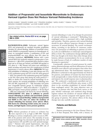
- 5. (P ϭ .842). The actuarial probability of rebleed at 2 years was 27% in the EVLϩdrugs group and 31% in the EVL group (P ϭ .822, log-rank test) (Figure 1). Of the 14 bleeds in the EVLϩdrugs arm, 6 (43%) were from esophageal varices prior to eradication (including post-EVL variceal ulcers), 5 (36%) were from recurrent esophageal varices, 3 (21%) were from gastric varices, and none were from portal hypertensive gastropathy. In the EVL group, of the 16 bleeds, 11 (69%) patients bled from esophageal varices (including post-EVL variceal ulcers), 4 (25%) from gastric varices, 1 (6%) from portal hyperten- sive gastropathy, and none from recurrent esophageal varices. Five patients died during the study period (2 [2%] in the EVLϩdrugs group and 3 [4%] in the EVL group, P ϭ .682) (Figure 2). None of the patients died of variceal hemorrhage. In the subgroup analysis of only cirrhotic patients (Table 2), 11 of 72 (15%) rebled in the EVLϩdrugs group, and 13 of 69 (19%) rebled in the EVL group (P ϭ .656). The actuarial probability of rebleed at 2 years was 24% in the EVLϩdrugs group and 30% in the EVL group (P ϭ .720, log-rank test) (Figure 3). One (1%) patient died in the EVLϩdrugs group, whereas 3 (4%) died in the EVL group (P ϭ .359). In another subgroup analysis of only those patients who had their last bleed within 42 days (ie, patients who had the highest chance of rebleed), no significant differ- ence was found in the rate of rebleed in the 2 groups: 8 of 52 (15%) rebled in the EVLϩdrugs group, and 10 of 48 (21%) rebled in the EVL group (P ϭ .604). Secondary End Points Nine patients developed new ascites (4/84 [5%] in EVLϩdrugs group vs 5/83 [6%] in EVL group; P ϭ .746), 15 patients had episode(s) of hepatic encephalopathy (8/84 [10%] in EVLϩdrugs group vs 7/83 [8%] in EVL group; P ϭ 1), 4 patients had episode(s) of spontaneous bacterial peritonitis (2/84 [2%] in EVLϩdrugs group vs 2/83 [2%] in EVL group; P ϭ 1), and 6 patients had episode(s) of hepatorenal syndrome (3/84 [4%] in EVLϩ drugs group vs 3/83 [4%] in EVL group; P ϭ 1). Fifty-six patients were hospitalized (26/84 [31%] in EVLϩdrugs group vs 30/83 [36%] in EVL group; P ϭ .515) for bleed, ascites, spontaneous bacterial peritonitis, hepatorenal syndrome, encephalopathy, fever, and others (Table 2). Adverse Effects of Therapy Ten patients developed transient chest pain fol- lowing EVL, which resolved in 1 day (3/84 [4%] in EVLϩdrugs group and 7/83 [8%] in EVL alone group; P ϭ .211). Nine (11%) patients developed adverse effects to drugs in the EVLϩdrugs groups. Adverse effects of pro- pranolol were as follows: dizziness because of bradycardia (n ϭ 2, improved with dose reduction), hypotension (n ϭ 2, improved with dose reduction), and dyspnea (n ϭ 1, improved with stoppage of propranolol). Adverse effect of ISMN was headache (n ϭ 4, required dose reduction in 2 and stoppage in 2). Recurrence of Varices Variceal eradication could be achieved in 77 of 167 patients (44/84 [52%] in the EVLϩdrugs group and 33/83 [40%] in the EVL group, P ϭ .121). The mean sessions of EVL required to eradicate esophageal varices were similar in both the groups (4.6 Ϯ 1.9 EVLϩdrugs vs 4.6 Ϯ 1.6 EVL sessions, P ϭ .970). In 28 of 77 (36%) patients, there was variceal recurrence, defined as appear- ance or an increase in the grade of varices after achieving successful eradication. The recurrence rate was similar in both the groups (16/44 [36%] in the EVLϩdrugs group and 12 of 33 [36%] in the EVL group, P ϭ 1). Recurrences of varices were further treated by repeat sessions of EVL. Effect on Gastric Varices and PHG At baseline, 97 of 177 (55%) patients had gastric varices. On follow-up, 91 of 167 (54%) patients had gas- tric varices that were similar to baseline (P ϭ 1). There was no change in the frequency of gastric varices in either of the groups (EVLϩdrugs: baseline 51/88 [58%] vs fol- Figure 2. Kaplan–Meier graph showing cumulative probability of death in the 2 groups, in all patients. Figure 1. Kaplan–Meier graph showing cumulative probability of re- bleed in the 2 groups, in all patients. CLINICAL–LIVER, PANCREAS,AND BILIARYTRACT 896 KUMAR ET AL GASTROENTEROLOGY Vol. 137, No. 3
- 6. low-up 50/84 [60%], P ϭ .878; EVL alone: baseline 46/89 [52%] vs follow-up 41/83 [49%], P ϭ .879). At baseline, 88 of 177 (50%) patients had PHG. There was significant increase in the frequency of PHG on follow-up: 109 of 167 (65%, P ϭ .004). The increase in frequency of PHG was similar in both the groups (EVLϩdrugs: baseline 40/88 [45%] vs follow-up 51/84 [61%], P ϭ .049; EVL alone: baseline 48/89 [54%] vs follow-up 58/83 [70%], P ϭ .041). Predictors of Rebleed On univariate analysis, the baseline predictors of rebleed were as follows: presence of ascites, encephalop- athy, high bilirubin, low albumin, deranged prothrombin time, high HVPG, alcohol as etiology, and high CTP score (Table 3). CTP class of B or C as compared with A was the strongest risk factor of rebleed (relative risk, 5.04 [95% CI: 1.57–16.11]). All the individual components of CTP score also were significant risk factors for rebleed. Apart from these, baseline HVPG also correlated with rebleed risk (Tables 3 and 4). Alcohol as etiology of cirrhosis as compared with others also had more chances of rebleed; however, it did not reach statistical significance (P ϭ .071). On multivariate analysis (Table 5), the only signif- icant independent risk factors for rebleed were as follows: presence of ascites (P ϭ .003), serum albumin Ͻ3.3 g/dL (P ϭ .008), and baseline HVPG Ն18 mm Hg (P ϭ .009). Discussion The results of this large and novel study show that addition of propranolol and nitrate does not decrease the probability of variceal rebleed in patients being treated by EVL. Further addition of these drugs, in fact, leads to drug-related adverse events and results in some morbid- Figure 3. Kaplan–Meier graph showing cumulative probability of re- bleed in the 2 groups, in the subgroup analysis of cirrhotic patients. Table 2. Primary and Secondary End Points Parameters EVLϩdrugs EVL alone P value All patients (n ϭ 167) (n ϭ 84) (n ϭ 83) Mean (ϮSD) follow-up 15 (12) 15 (11) .662 Patients rebled, n (%) 14 (17) 16 (19) .842 Site of rebleed, n (%) Esophageal varices before eradication 6 (7) 11 (13) .212 Recurrent esophageal varices 5 (6) 0 (0) .059 Gastric varices 3 (4) 4 (5) .720 PHG 0 (0) 1 (1) .497 Actuarial probability of rebleed at 2 years, % 27 31 .822 Patients died, n (%) 2 (2) 3 (4) .682 Complications, n (%) New ascites 4 (5) 5 (6) .746 Hepatic encephalopathy 8 (10) 7 (8) 1 SBP 2 (2) 2 (2) 1 HRS 3 (4) 3 (4) 1 Hospitalizations 26 (31) 30 (36) .515 Adverse effects of therapy, n (%) Chest pain after EVL 3 (4) 7 (8) .211 Adverse effects because of drugs 9 (11) 0 (0) .003 Effect on varices and PHG Esophageal variceal eradication, n (%) 44 (52) 33 (40) .121 EVL sessions required to eradicate, n (ϮSD) 4.6 (1.9) 4.6 (1.6) .970 Esophageal variceal recurrence, n (%) 16/44 (36) 12/33 (36) 1 Gastric varices, n (%) 50 (60) 41 (49) .215 PHG, n (%) 51 (61) 58 (70) .256 Cirrhotic patients (n ϭ 141) (n ϭ 72) (n ϭ 69) Patients rebled, n (%) 11 (15) 13 (19) .656 Actuarial probability of rebleed at 2 years, % 24 30 .720 Patients died, n (%) 1 (1) 3 (4) .359 EVL, endoscopic variceal ligation; SD, standard deviation; PHG, portal hypertensive gastropathy; SBP, spontaneous bacterial peritonitis; HRS, hepatorenal syndrome. CLINICAL–LIVER, PANCREAS,AND BILIARYTRACT September 2009 EVL VS EVL؉ DRUGS FOR SECONDARY PROPHYLAXIS DRUGS 897
- 7. ity. We have also shown that significant independent risk factors for rebleed are poor liver function (as manifested by ascites and low albumin) and high portal pressure (as manifested by high HVPG). Our study was adequately powered to show non-supe- riority of EVL plus drugs strategy over EVL alone strat- egy. We enrolled a sufficient number of patients, 167 bleeders (including 151 cirrhotic patients), which was sufficient as per the sample size calculations of 148 pa- tients. The baseline characteristics of the patients were well matched in both the groups, and the follow-up duration was adequate to allow meaningful outcome. The Baveno IV consensus conference recommended that patients with cirrhosis who had not received primary prophylaxis could receive nonselective -blocker, EVL, or both.6 Although it was hypothesized that combination of nonselective -blocker plus EVL was probably the best alternative, it concluded that more trials were needed before a recommendation could be made. Combination of EVL plus -blocker seemed to be a rational approach because -blockers would theoretically protect against rebleeding before variceal obliteration and could prevent variceal recurrence. -Blockers have been shown to be effective in reducing the incidence of variceal rebleeding; however, it has been shown that up to two thirds of the patients may be nonresponders to -blockers. The addition of ISMN has been demonstrated to enhance the effect of -blockers in Table 3. Predictors of Rebleed Parameters Bleeders Non-bleeders P value All patients (n ϭ 167) (n ϭ 30) (n ϭ 137) Mean age (ϮSD), y 44 (16) 40 (14) .187 Sex, n (%) .255 Males 28 (93) 115 (84) Females 2 (7) 22 (16) Cause of portal hypertension, n (%) .420 Cirrhotic patients 24 (80) 117 (85) Non-cirrhotic patients 6 (20) 20 (15) EHPVO 1 11 NCPF 5 9 Last bleed, n (%) .728 Յ5 days ago 4 (13) 12 (9) 6 to 42 days ago 14 (47) 70 (51) Ͼ42 days ago 12 (40) 55 (40) Mean (ϮSD) grade of esophageal varices 3.1 (0.8) 3.2 (0.7) .468 Patients with gastric varices, n (%) 19 (63) 73 (53) .418 Patients with PHG, n (%) 18 (60) 63 (46) .226 Mean (ϮSD) platelet count, 103/mm3 153 (54) 160 (56) .721 Protocol, n (%) EVLϩdrugs 14 (47) 70 (51) EVL 16 (53) 67 (49) .691 Cirrhotic patients (n ϭ 141) (n ϭ 24) (n ϭ 117) Etiology of cirrhosis, n (%) .071 ALD 14 (58) 44 (38) Other 10 (42) 73 (62) CTP class, n (%) .006 A 3 (12) 56 (48) B 15 (63) 45 (38) C 6 (25) 16 (14) Mean (ϮSD) CTP score 8.6 (2.1) 7.2 (1.9) .001 Ascites, n (%) 16 (67) 36 (31) .002 Encephalopathy, n (%) 3 (12) 2 (1) .035 Median (range) serum bilirubin, mg/dL 1.9 (0.6Ϫ10.5) 1.5 (0.3Ϫ14.4) .034 Mean (ϮSD) serum albumin, g/dL 2.9 (0.6) 3.3 (0.5) .002 Prothrombin time prolongation, n (%) .032 0–6 seconds 14 (58) 94 (80) Ͼ6 seconds 10 (42) 23 (20) Mean (ϮSD) serum creatinine, mg/dL 1.0 (0.5) 0.9 (0.4) .183 Mean (ϮSD) HVPG, mm Hg (n ϭ 115) 20.2 (4.5) 17.3 (5.3) .014 Protocol, n (%) .656 EVLϩdrugs 11 (46) 61 (52) EVL 13 (54) 56 (48) SD, standard deviation; EHPVO, extrahepatic portal vein obstruction; NCPF, non-cirrhotic portal fibrosis; PHG, portal hypertensive gastropathy; EVL, endoscopic variceal ligation; ALD, alcoholic liver disease; CTP, Child–Turcotte–Pugh; HVPG, hepatic venous pressure gradient. CLINICAL–LIVER, PANCREAS,AND BILIARYTRACT 898 KUMAR ET AL GASTROENTEROLOGY Vol. 137, No. 3
- 8. reducing portal pressure through the decrease of hepatic vascular resistance.11 A controlled study showed that cirrhotic patients receiving propranolol and ISMN had a lower variceal rebleeding rate compared with patients who only received propranolol.12 Hence, the combination of -blockers and ISMN rather than using -blockers alone has been used in this study as the portal pressure reducing strategy. Although the benefit of EVL for secondary prophylaxis of esophageal variceal bleeding is supported by the med- ical literature, there has been no consensus regarding the technical aspects of the procedure. The success of EVL therapy depends on its technique, and the importance of technical aspects in influencing the success cannot be overemphasized. The technique most endoscopists fol- low13 involves application of bands at the gastroeso- phageal junction and progressing upwards in a helical motion for approximately 5–8 cm. There is also no consensus on when the band ligation sessions should be repeated. Most endoscopists used to favor an interval of 1–2 weeks,14–18 but, recently, many authors propose that an interval of 3–4 weeks is more suitable, given that unhealed ulcers induced by ligation are frequently noted within 2 weeks of ligation.19–21 One study actually iden- tified that longer intervals between EVL sessions resulted in a reduced risk of variceal rebleeding.21 Longer inter- banding intervals (Ͼ3 weeks) promote optimal results. A possible explanation for this is that a longer interbanding interval allows varices to partially recur, allowing them to be more effectively suctioned and rebanded. Moreover, it results in a decrease in band-related bleeding, which occurs because of the ulcers that form at banding sites contributing to bleeding, especially when banding is per- formed in rapid succession with short intervals. If EVL is performed optimally, the addition of drugs to EVL would not be able to add substantially to the results of EVL alone, given their equal efficacy in preventing rebleed.22–24 Our results support this hypothesis. Prior to our study, 2 small randomized controlled trials had compared the efficacy of combined therapy vs EVL alone.25,26 In the trial by de la Pena et al,25 efficacy of EVL combined with nadolol was compared with EVL alone for the secondary prophylaxis of variceal bleeding. In a median follow-up of 16 months, nadolol plus EVL was found to reduce the incidence of variceal rebleeding, and there was a trend to reduce variceal recurrence com- pared with EVL alone. Our results differ from the results of this study because of many reasons. First, in the study by Pena et al,25 the interval between EVL sessions was 10–12 days. Because of more frequent EVL sessions at shorter intervals, the chances of rebleeding were higher. Second, up to one third of the patients had received endoscopic sclerotherapy as the initial treatment for the control of active bleeding. Endoscopic sclerotherapy is inferior to EVL, and, hence, protective effect of -blocker again comes into play. Third, EVL was performed begin- ning at the cardia (ie, stomach) or immediately under it. EVL in the stomach leads to higher chances of bleeding ulcers.27 In our study, we performed EVL sessions at intervals of 3–4 weeks that resulted in fewer EVL-induced ulcer bleeds. We did not perform sclerotherapy in our patients. Moreover, we performed variceal banding start- ing approximately a centimeter above the gastroesopha- geal junction. Hence, the protective effect of -blockers in our study was minimal, if at all. In the trial by Lo et al,26 a combination of -blocker, EVL, and sucralfate was compared with EVL alone in a group of 122 patients. The combination therapy was proved to be more effective than EVL alone in terms of prevention of variceal recurrence and upper GI rebleeding as well as variceal rebleeding. Survival remained the same in both the groups. However Lo et al’s study has been criticized for having observer bias, selection bias, lack of portal pressure correlations, and difficulty in evaluation of role of sucralfate without an appropriate control group.28 We used sucralfate and proton pump inhibitor in both the groups to prevent EVL-induced ulcer bleeds. A meta-analysis published recently5 also claims that combination of endoscopic and drug therapy is superior to endoscopic therapy alone. However, the major prob- lem with this meta-analysis is that sclerotherapy and EVL have been clubbed together as “endoscopic therapy,” and Table 4. Relative Risks of Factors Found to Be Significant Predictors of Rebleed on the Univariate Analysis Factors Relative risk (95% CI) P value Etiology: ALD vs others 2.00 (0.96Ϫ4.19) .071 Ascites: present vs absent 3.42 (1.57Ϫ7.44) .002 HE: present vs absent 3.88 (1.72Ϫ8.79) .035 Serum bilirubin, mg/dL: Ն2.5 vs Ͻ2.5 2.77 (1.37Ϫ5.59) .008 Serum albumin, g/dL: Ն3.3 vs Ͻ3.3 0.34 (0.15Ϫ0.77) .007 PT prolonged by: Ն7 vs Ͻ7 seconds 2.34 (1.15Ϫ4.76) .032 HVPG, mm Hg: Ն18 vs Ͻ18 2.53 (1.12Ϫ5.73) .024 CTP class: B/C vs A 5.04 (1.57Ϫ16.11) .001 ALD, alcoholic liver disease; HE, hepatic encephalopathy; HVPG, he- patic venous pressure gradient; CTP, Child–Turcotte–Pugh; PT, pro- thrombin time. Table 5. Factors Predictive of Rebleed on Multivariate Analysis Factors Odds ratio (95% CI) P value Ascites: present vs absent 4.60 (1.70Ϫ12.48) .003 Serum albumin, g/dL: Ն3.3 vs Ͻ3.3 0.25 (0.09Ϫ0.69) .008 HVPG, mm Hg: Ն18 vs Ͻ18 4.06 (1.43Ϫ11.55) .009 NOTE. All factors found significant by univariate analysis were entered into multivariate analysis except CTP score because its individual components were entered. HVPG, hepatic venous pressure gradient. CLINICAL–LIVER, PANCREAS,AND BILIARYTRACT September 2009 EVL VS EVL؉ DRUGS FOR SECONDARY PROPHYLAXIS DRUGS 899
- 9. there is no subgroup analysis of EVL plus drugs vs EVL alone. Clubbing of the 2 endoscopic modalities (sclero- therapy and EVL) undoubtedly would lead to inferior results of “endoscopic therapy” because sclerotherapy is known to be inferior to EVL in the prevention of variceal rebleeding. In the present study, there was no difference in the development of complications like new ascites, hepatic encephalopathy, spontaneous bacterial peritonitis, he- patorenal syndrome, or hospitalizations. Adverse ef- fects because of EVL (eg, transient chest pain) were also similar in both the groups. However, 9 (11%) patients developed adverse effects to drugs in the EVL plus drugs group that included dizziness, bradycardia, hy- potension, and dyspnea because of propranolol, and headache because of ISMN. Thus, addition of -blocker and nitrate adds to morbidity by causing drug-related adverse effects without leading to any superiority of the combination over EVL alone. Whereas we had earlier shown that a combination of -blocker and nitrates alone is as effective as EVL alone,24 the addition of EVL to the combination of drugs was not found to be any superior to EVL alone. The rate of recurrence of esophageal varices was similar in both the groups. Moreover, there was no change in the frequency of gastric varices after EVL in either group. There was a significant increase in the frequency of PHG, which was similar in both the groups. Thus, addition of drugs did not reduce the development of gastric varices or PHG. We found that the only significant independent risk factors for variceal rebleed were as follows: presence of ascites, serum albumin Ͻ3.3 g/dL, and baseline HVPG Ն18 mm Hg. In fact, our study confirms the previous findings that poor liver function and high portal pressure are significant independent predictors of variceal re- bleed.19,29 In conclusion, our data show that EVL alone is suffi- cient to prevent rebleed in patients with portal hyperten- sion who had bled in the past. Addition of the pharma- cotherapy regimen of propranolol and ISMN to EVL does not further reduce the incidence of variceal rebleeding as achieved by EVL therapy given alone. Moreover, these drugs add to the morbidity in a proportion of these patients because of their serious adverse effects. The risk factors for rebleed are ascites, low serum albumin, and high baseline HVPG. Supplementary Data Note: To access the supplementary material accompanying this article, visit the online version of Gastroenterology at www.gastrojournal.org, and at doi: 10.1053/j.gastro.2009.05.049. References 1. Jensen DM. Endoscopic screening for varices in cirrhosis: find- ings, implications, and outcomes. Gastroenterology 2002;122: 1620–1630. 2. D’Amico G, De Franchis R, and the Cooperative Study Group. Upper digestive bleeding in cirrhosis. Post-therapeutic outcome and prognostic indicators. Hepatology 2003;38:599–612. 3. Carbonell N, Pauwels A, Serfaty L, et al. Improved survival after variceal bleeding in patients with cirrhosis over the past two decades. Hepatology 2004;40:652–659. 4. Graham DY, Smith JL. The course of patients after variceal hem- orrhage. Gastroenterology 1981;80:800–809. 5. Gonzalez R, Zamora J, Gomez-Camarero J, et al. Meta-analysis: combination endoscopic and drug therapy to prevent variceal rebleeding in cirrhosis. Ann Intern Med 2008;149:109–122. 6. de Franchis R. Evolving consensus in portal hypertension. Report of the Baveno IV consensus workshop on methodology of diag- nosis and therapy in portal hypertension. J Hepatol 2005;43: 167–176. 7. García-Pagán JC, Feu F, Bosch J, et al. Propranolol compared with propranolol plus isosorbide-5-mononitrate for portal hypertension in cirrhosis. A randomized controlled study. Ann Intern Med 1991; 114:869–873. 8. Sarin SK, Kumar A. Noncirrhotic portal hypertension. Clin Liver Dis 2006;10:627–651. 9. Conn HO. Ammonia tolerance in the diagnosis of esophageal varices. A comparison of endoscopic, radiologic, and biochemical techniques. J Lab Clin Med 1967;70:442–451. 10. Groszmann RJ, Wongcharatrawee S. The hepatic venous pres- sure gradient: anything worth doing should be done right. Hepa- tology 2004;39:280–282. 11. Garcia-Pagán JC, Navasa M, Bosch J, et al. Enhancement of portal pressure reduction by the association of isosorbide-5- mononitrate to propranolol administration in patients with cirrho- sis. Hepatology 1990;11:230–238. 12. Gournay J, Masliah C, Martin T, et al. Isosorbide mononitrate and propranolol compared with propranolol alone for the prevention of variceal rebleeding. Hepatology 2000;31:1239–1245. 13. Garcia-Pagán JC, Bosch J. Endoscopic band ligation in the treat- ment of portal hypertension. Nat Clin Pract Gastroenterol Hepatol 2005;2:526–535. 14. Stiegmann GV, Goff JS, Michaletz-Onody PA, et al. Endoscopic sclerotherapy as compared with endoscopic ligation for bleeding esophageal varices. N Engl J Med 1992;326:1527–1532. 15. Laine L, el-Newihi HM, Migikovsky B, et al. Endoscopic ligation compared with sclerotherapy for the treatment of bleeding esoph- ageal varices. Ann Intern Med 1993;119:1–7. 16. Gimson AE, Ramage JK, Panos MZ, et al. Randomised trial of variceal banding ligation versus injection sclerotherapy for bleed- ing oesophageal varices. Lancet 1993;342:391–394. 17. Lo GH, Lai KH, Cheng JS, et al. A prospective, randomized trial of sclerotherapy versus ligation in the management of bleeding esophageal varices. Hepatology 1995;22:466–471. 18. Hou MC, Lin HC, Kuo BI, et al. Comparison of endoscopic variceal injection sclerotherapy and ligation for the treatment of esopha- geal variceal hemorrhage: a prospective randomized trial. Hepa- tology 1995;21:1517–1522. 19. Lo GH, Lai KH. The factors affecting risk of recurrent variceal bleeding. Gastroenterology 2005;128:244. 20. Lo GH. Prevention of esophageal variceal rebleeding. J Chin Med Assoc 2006;69:553–560. 21. Harewood GC, Baron TH, Song LM. Factors predicting success of endoscopic variceal ligation for secondary prophylaxis of esoph- ageal variceal bleeding. J Gastroenterol Hepatol 2006;21:237– 241. 22. Bosch J, García-Pagán JC. Prevention of variceal rebleeding. Lan- cet 2003;361:952–954. CLINICAL–LIVER, PANCREAS,AND BILIARYTRACT 900 KUMAR ET AL GASTROENTEROLOGY Vol. 137, No. 3
- 10. 23. Sharara AI, Rockey DC. Gastroesophageal variceal hemorrhage. N Engl J Med 2001;345:669–681. 24. Sarin SK, Wadhawan M, Gupta R, et al. Evaluation of endoscopic variceal ligation (EVL) versus propanolol plus isosorbide mononi- trate/nadolol (ISMN) in the prevention of variceal rebleeding: comparison of cirrhotic and noncirrhotic patients. Dig Dis Sci 2005;50:1538–1547. 25. de la Peña J, Brullet E, Sanchez-Hernández E, et al. Variceal ligation plus nadolol compared with ligation for prophylaxis of variceal rebleeding: a multicenter trial. Hepatology 2005;41: 572–578. 26. Lo GH, Lai KH, Cheng JS, et al. Endoscopic variceal ligation plus nadolol and sucralfate compared with ligation alone for the pre- vention of variceal rebleeding: a prospective, randomized trial. Hepatology 2000;32:461–465. 27. Savoye G, Hochain P, Riachi G, et al. Early massive bleeding after endoscopic ligation for junctional varices. Endoscopy 1998;30: S101. 28. Bosch J. Prevention of variceal rebleeding: endoscopes, drugs, and more. Hepatology 2000;32:660–662. 29. Mihas AA, Sanyal AJ. Recurrent variceal bleeding despite endo- scopic and medical therapy. Gastroenterology 2004;127:621–629. Received October 6, 2008. Accepted May 20, 2009. Reprint requests Address requests for reprints to: S. K. Sarin, MD, DM, Director, Professor and Head, Department of Gastroenterology, Project Director, Institute of Liver & Biliary Sciences, Room 201, Academic Block, G B Pant Hospital, New Delhi 110 002, India. e-mail: sksarin@nda.vsnl.net.in; fax: (91) 11-23219710. Conflicts of interest The authors disclose no conflicts. CLINICAL–LIVER, PANCREAS,AND BILIARYTRACT September 2009 EVL VS EVL؉ DRUGS FOR SECONDARY PROPHYLAXIS DRUGS 901
- 11. Supplementary Figure 1. The Consort E-Flowchart Aug. 2005. 901.e1 KUMAR ET AL GASTROENTEROLOGY Vol. 137, No. 3