1. Benign oral & dental disease
Causes of gingival bleeding
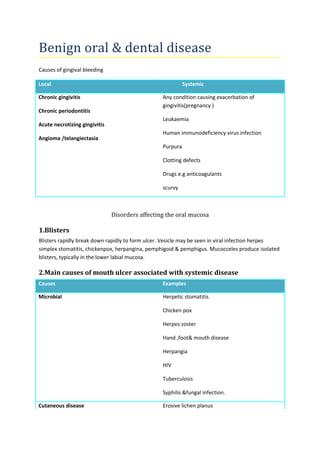
Local Systemic
Chronic gingivitis
Chronic periodontitis
Acute necrotizing gingivitis
Angioma /telangiectasia
Any condition causing exacerbation of
gingivitis(pregnancy )
Leukaemia
Human immunodeficiency virus infection
Purpura
Clotting defects
Drugs e.g anticoagulants
scurvy
Disorders affecting the oral mucosa
1.Blisters
Blisters rapidly break down rapidly to form ulcer. Vesicle may be seen in viral infection herpes
simplex stomatitis, chickenpox, herpangina, pemphigoid & pemphigus. Mucocceles produce isolated
blisters, typically in the lower labial mucosa.
2.Main causes of mouth ulcer associated with systemic disease
Causes Examples
Microbial Herpetic stomatitis
Chicken pox
Herpes zoster
Hand ,foot& mouth disease
Herpangia
HIV
Tuberculosis
Syphilis &fungal infection.
Cutaneous disease Erosive lichen planus
2. Pemphigus
Pemphigoid
Erythema multiforme
Dermatitis herpetiformis
Blood disorder Anaemia
Leukaemia
Neutropenia
Other white cells dyscrasias
Gastrointestinal disease Celiac disease
Crohn’s disease
Ulcerative colitis
Rheumatic disease Lupus erythematosus
Behcet’s syndrome
Sweet’s syndrome
Reitre’s disease
Iatrogenic Drugs ,cytotoxic other agent
Radiotherapy
Disorder of uncertain pathology Angina bullosa haemorrhagica
Hypereosinophilic syndrome
Eosinophilic ulcer
Necrotizing sialometaplasia.
3.Pigmentation
Mucosal pigmentation is most usually seen in darked-skinned races. Others common causes includes
melanotic macules, Addison’s disease, Kaposi’s sarcoma, melanoma.
3. 4. Redness
Redness may be inflammatory or erythroplasia,atropy, petechiae, telangiectasia, haemangiomas or
neoplasm.
a)Inflammatory : candiasis, Herpes simplex stomatitis,lichen planus or mucus membrane
pemphigoid, irradiation ,or chemotherapy.
b) Erythroplasia: represent carcinoma-in-situ or worse.
c)Atrophic: common in lichen planus, lingual depapillation in deficiencies of iron, folate or
vitamin B12 may produce a red tongue, termed glossitis, geographical tongue may also produce red
patches.
d)Oral peteechiae caused by trauma or suction, infectious mononucleosis, rubella, HIV
infection or leukaemia.
5) Telangiectasia: hereditary haemorrhagic telangiectasia, primary biliary cirrhosis or
systemic sclerosis or following radiotherapy,haemangioma.
6) carcinoma: Kaposi’s sarcoma, Wegener’s granulomatasis.
5. Swellings
Trauma , infection, pyogenic granuloma, Chron’s disease,sarcoidosis, angioedema, wegener’s
granulomatosis, amyloidosis.
6) White patches
White patches due to debris (burn, candidiasis, hairy leukaemia,) lichen planus, carcinoma, drugs,
smoking , lupus erythemaosus.
Inherited mucosal disorders
1.Blisters
Epidermolysis bullosa is characterized by blisters which develop where there is trauma & rupture to
produce ulcers .
2.Pigmented lesions
Melanotic maculle is an acquired , solitary, small, flat, brown to brown-black asympmatic
benign lesion, unchanging in character. Melanotic macules are best excised to exclude melanoma.
Naevi approximately half are of the intradermal(intramucosal) types, all pigmented oral
cavity lesions should be excised for histopathology.
Peutz-jegher’s syndrome is a autosomal dominant trait characterized by intestinal polyposis
mucocutananeous melanotic pigmentation.
4. 3.Red lesions
Hereditary haemorrhagic telangiectasia : Oler –rendu-weber sundrome is characterized by
multiple telangiectasia on the lips , perioral skin, oral & nasal mucosa as well as gastrointestinal
tract.
Haemangioma should be excluded with Koposi’s sarcoma.
4.Swelling:
Hereditary angio-oedema(C1 esterase inhibitor deficiency): mimic allergic angio-oedema,
but minor trauma triggers more pronounced odema of the lips, mouth, face &neck region the
extremities & gastrointestinal tract. Odema persist for hours & evens days.
Lymphangioma
Lingual thyroid: although 10% of cadaveric tongue contain thyroid tissue, clinical
presentation is much less common. Occasional a lingual thyroid may produce dysphagia, cough,
pain, & rarely air obstruction. MRI & Radioscanning are important to ensure the presence of normal
thyroid tissue.
5.white & yellow lesions
Fordyce spots: are common. They are sebaceous glands which appear as an yellowish small
grains seen seen beneath buccal or labial mucosa usually inside commissures & sometimes
retromolar regions & upper lip. Fordyce spots are totally benign, often misdiagnosed them as thrush
or lichen planus. No treatment is required.
6. Ulcer
Epidermolysis bullosa is the most common inherited cause of ulceration.
Acquired mucosal disorders
1.Blisters
Blisters rapidly break down rapidly to form ulcer. Vesicle may be seen in viral infection herpes
simplex stomatitis, chickenpox, herpangina, pemphigoid & pemphigus. Mucocceles produce isolated
blisters, typically in the lower labial mucosa.
Recurrent herpes labialis is treated with 5% acyclovir. But pencyclovir more effective.
2. Pigmented lesions
The tongue may be coated with off-white debris in febrile & other illness poor oral hygiene, fasting.
The tongue is often discoloured due to superficial staining from foods, drinks or habits such as
tobacco or betel nuts use. Various medicaments such s chlorhexidine or iron can also cause a black
or brown superficial staining of the tongue.
Occasionally , a brown , a hairy tongue may be caused by drugs that induce xerostomia, lansoprazole
or antibiotic therapy.
5. Treatment consists of patient avoid habit or drugs that stain the tongue,
Brush the tongue with a tooth brush
Use sodium bicarbonate mouth washes,
Chew gum or suck a peach stone
Tropical tretinoin may be effective.
Generalised hyperpigmentation is usually racial in origin, occasionally addison’s disease.
Localized hyperpigmentation : due to naevi, melanoma & Kaposi’s sarcoma.
3. Red lesions
Benign migratory glossits(geographical tongue): it is characterized by map like atrophic red
areas with surrounding borders of increased thickness of filiform papillae. The pattern change from
day to day even within a few hours.
It is necessary to exclude anaemia& DM.
Candidiasis
Dental –related stomatitis: this is the most common form of mild, chronic oral candidiasis. It occur
only beneath a denture. Dentures worn through out the night or dry mouth favour development of
this infection with candida. There is accumulation of microbial plaque on & in the fitting surface of
the denture & underlying mucosa.
Plaque must be removed regularly with disinfectant.
Denture should be left out at night & stored in a antiseptic solution.(chlorhexidine or
hypochloride)
The mucosal infection is eradicated by brushing the palate & using antifungals usually topical
for four weeks.
Acute candidiasis : acute oral candidiasis may complicate long-term corticosteroid or antibiotic
therapy, producing wide spread erythama, soreness& sometimes with thrush.
HIV-associated candidiasis: the clinical presentation is of erythaematous areas generally on the
dorsum of the tongue, palate or buccal mucosa. Lesion on the dorsum of the tongue present as
depapillated areas. Lesion in the palate may have a fingureprint configuration. Antifungal therapy is
indicated.
Median rhomboid glossitis (Central papillary atrophy of the tongue)
This is an uncommon red, depapillated, rhomboidal area in the centre line of the dorsum of the
tongue, anterior to the sulcus terminalis, thought to be associated with candidiasis. Smoking/
dentures/ HIV/ DM predispose. Cessation of smoking & antifungals.
6. Angular cheilitis(angular stomatitis)
Angular cheilitis is a fairly common acute or chronic inflammation of the skin & continuous labial
mucosa at the angles of the mouth, bilaterally. Most cases are in adult & due to mechanical &/or
infective cases but, in children nutritional or immune defects are more prominent causes.
Mechanical factors may contribute in edentulous patients who wear a denture.
Nutritional deficiency- folate, riboflavin iron & general protein malnutrition.
Immunodeficiency –DM, HIV
Infections- candida/ staphylococcus are isolated most cases.
Clinical presentation : angular cheilitis presents as a roughly triangular area of erythaema & oedema
bilaterally , angle of mouth.linear furrow or fissure radiating from the angle of the mouth are seen,
especially in denture wearer.
Treatment ;
Denture induced stomatitis should be treated with an antifugal.miconazole may be a preferred for
candidiasis. It has bacteriostatic action.
New denture which restore facial conture may help.
Any staphylococcus infection should be treatment with fusidic acid cream or ointment.
Burning mouth syndrome (Glossodynia)
This most commonly affects middle-aged & elderly women & is often a medically unexplained
symptom(MUS) but may be neuropathy. Burning mouth syndrome(BMS) may be seen in
DM,deficiency state, with drugs ACE inhibitor, protease inhibitor, cytotoxic drugs,clonazepam).
Amonosymptomatic hpochondriasis or an underlying anxiety about cancer ot other disease. With
excessive tongue activity.
Although the tongue is the most frequent involved, the patient may also occasionally complain of
burning lips, gum or palate. The burning is usually bilateral & relieved by eating & drinking.
Treatment ;
Organic disease excluded, reassurance& psychological treatment (antidepressants).
Causes of burning mouth syndrome;
Local Systemic
Candidiasis
Others infections
Geographical tongue
Psychological
Cancerophobia
Depression
7. Lichen planus
Oral submucous fibrosis
Dentures
Hypochondriasis
Deficiencies states(percinious anaemia, folate
deficiency, iron deficiency)
DM
Drugs (captopril)
Atypical facial pain
The international society defines atypical pain as facial pain not fulfilling other criteria: therefore it is
diagnosis reached by the cxclusion of organic disease.
Atypical facial pain is a constant chronic orofacial discomfort or pain often a dull boring or burning
type & ill-defined location in which there is :
A total lack of objective signs
A negative result from all investigations
No clear explanation as to cause;
Poor response to treatment.
It falls into medically unexplained syndrome most of which have a psychological basis. Atypical facial
pain are often middle aged or elderly & more than 70% of patients are women. The four main group
are those:
1. Extreme stress
2. Personality triat such as hypochindriais
3. Neurosis often depression
4. Psychoses.
Clinical features include a constant discomfort or pain often a deep, dull boring type & mainly upper
jaw. The location of pain unrelated with anatomical distribution of TG nerve pain is poorly localised&
sometimes crosses the midline.
Treatment : the pain reduction achieved by with antidepressants. Cognitive behavioural therapy.
8. Trigeminal neuralgia(tic doloureux)
Trigenial neuralgia causes episodes of unilateral intense, stabbing, electric shock-like pain in the jaw
or the face. TN onset is mainly in the 50-70year age group. The cause is unclear but one hypothesis is
that there may be compression around the trigeminal root in the posterior cranial fossa possibly due
to a cerebral vessels atherosclerosis & therefore less flexible with age & then pressing on the roots
of the TG nerve causing neuronal discharge.
Pain also has the following features;
-distribution along one or more divisions of the TGnerve
-sudden intense,sharp superficial ,stabbing or burning pain
- precipating from trigger areas or by activities such as eating, talking,washing face or
cleaning teeth
-between paroxysms, the patient is entirely asymptomatic.
-no neurological deficit.
-attack are stereotyped in the individual patient
-exclusion of other causes of facial pain by history, examination & investigations
Severe facial pain suggestive of TG pain but facial sensory or motor impairment can result from CVA,
Multple sclerosis, infections or space-occupaying lesion must be cxcluded.
Treatment : carmazepine is the main anticonvulsant used,baclofen, gabapentin, valporate,
clonazepine, lamotrigine or antidepressants.
Acupuncture
Self-hypnosis or meditation.
Neurosurgical procedures.