
Empfohlen
Weitere ähnliche Inhalte
Was ist angesagt?
Was ist angesagt? (20)
Andere mochten auch
Andere mochten auch (13)
Ähnlich wie Respiratory distress in newborn
Ähnlich wie Respiratory distress in newborn (20)
Mehr von All India Institute of Medical Sciences, Bhopal
Mehr von All India Institute of Medical Sciences, Bhopal (20)
Kürzlich hochgeladen
Kürzlich hochgeladen (20)
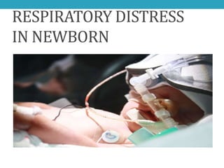
Respiratory distress in newborn
- 2. • A 32 weeks preterm baby born to a GDM mother by LSCS, developed distress, grunting, cyanosis soon after birth. His SPO2 was 90%, hyperoxia test was positive. Downe’s score was 7, silverman-anderson score 8. Urgent CXR was done which showed :
- 3. RDS 3
- 4. Now what next ??? • Baby shifted to NICU and put on warmer. RBS was normal, other blood samples taken. IV fluids were started, ABG was done which showed acidosis. • Baby was put on nasal CPAP, meanwhile survanta was arranged. • Baby then put on venti, surfactant given at 4 hours of life. • Now clinically better.
- 5. SIGNS AND SYMPTOMS • Tachypnea (RR > 60/min) • Nasal flaring • Retraction • Grunting • +/- Cyanosis • +/- Desaturation • Decreased air entry
- 7. RD IN NB - Causes • Pulmonary - Parenchymal - Extraparenchymal • Non Pulmonary - Heart - Metabolic - Brain - Blood - Abdominal
- 8. RD in Newborn– Causes Pulmonary Parenchymal Extraparenchymal * RDS (HMD) * Upper airway obstruction * TTN (RDS II) (Cloanal atresia, stenosis) * Aspiration (Blood * Pneumothorax meconium) * Pleural effusion * PPHN * Cong. Diaph. hernia * Pneumonia * Diaphragmatic paralysis * Pulm. hemorrhage * Pulm. hypoplasia
- 9. RD in Newborn– Causes Extrapulmonary Heart Metabolic Brain Blood Abdominal - CCF - Met. Acidosis - Haemorrhage - Hypovolemia - NEC - PDA - Hypoglycemia - Edema - Hyperviscosity - Pneumo - CCHD - Hypothermia - Drugs - Acute blood peritonium - Vascular - Sepsis - Pain loss - Large mass
- 10. Surgical causes of respiratory distress • Tracheo-esophageal fistula • Diaphragmatic hernia • Lobar emphysema • Pierre -Robin syndrome • Choanal atresia
- 11. Suspect surgical cause • Obvious malformation • Scaphoid abdomen • Frothing • History of aspiration
- 12. A progressively increasing O2 requirement to maintain saturation is also a sensitive indicator of the severity and progress of distress >95% Term baby, pulmonary hypertension (PPHN) 88-94% 28-34 weeks preterm 85-92% Below 28 weeks gestational age Guidelines for monitoring oxygen saturation levels by pulse oximetry MJAFI, Vol. 63, No. 3, 2007 GeneralConsiderations
- 13. Hyperoxia test test Method result diagnosis Hyperoxia 100 % fio2 5-10 min Pao2 increases to > 100 torr Pao2 increases by < 20 torr Parenchymal lung disease PPHN / CCHD Hyperoxia- hypervetilation MV 100 % fio2 & VR 100-150 / min Pao2 increases to > 100 torr w HV Pao2 increases at critical Pco2 No increase in Pao2 with HV Parenchymal lung disease PPHN CCHD
- 14. Downe’s score
- 15. Silverman-Andersen Retraction Scoring Interpretation Score 0-3 = Mild respiratory distress Score 4-6 = Moderate respiratory distress Score > 6 = Impending respiratory failure Score 10 = Severe Respiratory distress GeneralConsiderations
- 16. Investigation Complete Blood Count with a Peripheral blood smear Sepsis screen including C-reactive protein and μ ESR Arterial blood gas (ABG) analysis Blood glucose, Serum calcium Cultures: Blood , Surface swab (where indicated), maternal vaginal swab Chest radiograph with an oro-gastric tube in situ GeneralConsiderations
- 18. RR (bpm Aspiration cong. Pneumonia, sev. HMD CDH cardiac malformation Approx. 6 Hours of age Normal 60 Course of Neonatal Tachypnoea : Etiologic possibilities Source : Baurn DJ, Birth Risks, Nastle Nutrition Workshop, 1993 TTNB HMD
- 19. EvaluationofRD inNB–ClinicalHistory Antenatal History Most likely association * Prematurity, IDMs * HMD * PROM, maternal fever, * Pneumonia Unclean vaginal exams, UTI, diarrhoea * Asphyxia/MSAF * Aspiration * Caesarean delivery * TTN * Polyhydramnios * Pulm. Hypoplasia * Oligohydramnios * TE fistula, CDH * H/o receiving steroids * RDS less * Traumatic/breech delivery * ICH / Phrenic nerve paralysis
- 20. HMD/RDS
- 21. Respiratory Distress Syndrome (RDS) • Also known as Hyaline Membrane Disease (HMD) • Commonest cause of preterm neonatal mortality • RDS occurs primarily in premature infants; its incidence is inversely related to gestational age and birth weight Nelson Textbook of Pediatrics, 18th Ed. Gestational age Percentages Less than 28 wks 60-80% 32-36 wks 15-30% 37-39 wk 5% Term Rare Resp.Dis.Syn.
- 22. ETIOLOGYAND PATHOPHYSIOLOGY. • Surfactant deficiency is the 1O cause of RDS. • Low levels of surfactant cause high surface tension • High surface tension makes it hard to expand the alveoli. • Tendency of affected lungs to become atelectatic at end-expiration when alveolar pressures are too low to maintain alveoli in expansion • Leads to failure to attain an adequate lung inflation and therefore reduced gaseous exchange
- 23. • With advancing gestational age, increasing amounts of phospholipids are synthesized and stored in type II alveolar cells . • Wk 20: start of surfactant production and storage. Does not reach lung surface until later • Wk 28-32: maximal production of surfactant and appears in amniotic fluid • Wk 34-35; mature levels of surfactant in lungs • Quality : The amounts produced or released may be insufficient to meet postnatal demands because of immaturity. • Surfactant inactivating states eg maternal DM may lead to surfactant of lower quality/ immature
- 24. • Rare genetic disorders may cause fatal respiratory distress syndrome eg. • Abnormalities in surfactant protein B and C genes • gene responsible for transporting surfactant across membranes (ABC transporter 3 [ABCA3]) are associated with severe and often lethal familial respiratory disease
- 25. Clinical manifestation • Tachypnea • Nasal flaring • Intercostal, sternal recession • Grunting; closure of glottis during expiration • Cyanosis 25
- 26. Risk factors • Prematurity • Maternal diabetes • Multiple births • Elective cesarean section without labor • Perinatal asphyxia • Cold stress • Genetic disorders
- 27. Decreased risk •Chronic intrauterine stress •Prolonged rupture of membranes •Antenatal steroid prophylaxis
- 28. Structure of lung surfactant major constituents of surfactant are dipalmitoyl phosphatidylcholine (lecithin), phosphatidylglycerol, apoproteins (surfactant proteins SP-A, -B, -C, -D), cholesterol
- 29. Resp.Dis.Syn.
- 30. Resp.Dis.Syn.
- 31. Normal Expiration With Surfactant Surfactant Function Abnormal Respiration Without Surfactant
- 33. Respiratory distress - Management •Monitoring •Supportive - IV fluid - Maintain vital signs - Oxygen therapy - Respiratory support •Specific
- 34. Initial Care • Maintain warmth- cold stress will mimic other causes of distress • Monitor blood glucose levels- assure they are normal • Provide enough oxygen to keep the baby pink
- 35. Is a Clinical diagnosis: respiratory distress occurring soon after birth. Pay attention to risk factors! Pulse Oximetry: aim for SPO2 >85%. ROUTINE! Full blood count and Cultures to check for sepsis: rem culture only positive 40-50% of the time!! gastic aspirates/ buffy smears for GBS Chest radiograph: air bronchogram, reticular/ ground-glass appearance after 6-12 hrs to full opacity later on. Blood gases: hypoxia, hypercapnia, acidosis. Signs of RESP FAILURE determine mgmt eg CPAP vs ventilation etc Electrolytes, glucose, renal and liver function Echocardiogram: diagnosing PDA, determine the direction and degree of shunting, making the diagnosis of pulmonary hypertension and excluding structural cyanotic heart disease
- 36. cxr 36 Shows air bronchograms and reticulonodular shadowing throughout the lung fields (often termed ‘ground glass’ appearance)
- 37. 37
- 39. • Respiratory management • Surfactant replacement therapy • Ventilatory Assistance Oxygen therapy •CPAP ( Nasal, ET, Face-mask ) •Positive pressure ventilation •High-frequency ventilation •ECMO •Liquid ventilation
- 40. BubbleCPAP
- 41. • Avery and Mead in 1959 were the first to demonstrate that surfactant is deficient in the lungs of infants dying of HMD • Fujiwara in 1980 reported the 1st successful clinical trial of tracheal applications of surfactant in infants with RDS • Commercial preparations of surfactant were subsequently approved by the FDA in the USA in 1989 • Pulmonary Surfactants are phospholipids synthesized in the type II cells lining the alveoli
- 42. Prophylactic therapy Extremely preterm <28 wks <1000 gm Not routine in India Rescue therapy Any neonate diagnosed to have RDS Surfactant therapy - Issues Dose 100mg/kg phospholipid Intra tracheal
- 43. Types of Surfactant Natural Surfactants: contain appoproteins SP-B & SP-C • Curosurf (extract of pig lung mince) • Survanta (extract of cow lung mince) • Infasurf (extract of calf lung) Synthetic Surfactants:do not contain proteins • Exocerf • ALEC • Lucinactant (Surfaxin)
- 44. SURFACTANT Surfactant therapy reduces mortality rates most effectively in infants <30 weeks and those of birth weight <1250 gm 44
- 45. Insure technique • Intubation- • surfactant- • extubation to CPAP
- 46. Mode of administration of Surfactant • Dosing may be divided into 2 alliquots and adminitered via a 5-Fr catheter passed in the ET
- 47. Management Prevention: • Tocolytics to inhibit premature labor. • Antenatal corticosteroid therapy: ► They induce surfactant production and accelerate fetal lung maturation. ► Are indicated in pregnant women 24-34 weeks' gestation at high risk of preterm delivery within the next 7 days. ► Optimal benefit begins 24 hrs after initiation of therapy and lasts seven days.
- 48. Diagnosis of lung maturity • Amniotic Fluid Lecithin/Sphingomyelin Ratio • ≥2 suggests lung maturity. • ≤1.5 associated with HMD • Phosphatidyl Glycerol estimation • More specific than L/S ratio • Absence is invariably associated with HMD • Gastric Aspirate Shake Test • Unreliable if gastric aspirate is contaminated with blood or meconium • Serial tests can be done to assess maturity of lungs during course of disease Resp.Dis.Syn.
- 49. Shake test • Take a test tube • Mix 0.5 ml gastric aspirate + 0.5 ml absolute alcohol • Shake for 15 seconds • Allow to stand 15 minutes for interpretation of result
- 50. • NATURAL COURSE • Usually illness peaks in 3 days, then gradual improvement • Improvement is often heralded by spontaneous diuresis and the ability to oxygenate the infant at lower inspired oxygen levels or lower ventilator pressures • Death may occur esp from day2-3
- 51. MORTALITY • Death is rare on the 1st day, • usually occurs between days 2 and 7 • causes are: • alveolar air leaks (interstitial emphysema, pneumothorax), • pulmonary hemorrhage • Intracranial hemorrhage • Late mortality from bronchopulmonary dysplasia
- 52. COMLICATIONS acute Apnea Air leak infection ICH PDA & foramen ovale End-organ hypoxic injury chronic ROP PPH & BPD Neurodevep. impairment
- 53. MAS
- 54. Meconium Aspiration Syndrome Definition MAS is defined as respiratory distress in an infant born through meconium- stained amniotic fluid (MSAF) whose symptoms cannot be otherwise explained J Perinatol. 2008 Dec Mec.Asp.Synd
- 55. Epidemiology •Most common cause of respiratory distress in term and post-term infants. •MSAF observed in 5-25% of all births out of which 10% develop MAS. •One third require ventilator support •10% develop air leaks •5-10% of them have a fatal outcome Mec.Asp.Synd
- 56. What is meconium? • The term was coined by Aristotle from the Greek word “meconium arion” meaning “opium like” • Consists of gastrointestinal, hepatic and pancreatic secretions, cellular debris, swallowed amniotic fluid, lanugo, vernix caseosa and blood • Appear in the fetal intestines by the 10th week of life gradually increasing in amount to reach 200gms at birth Mec.Asp.Synd
- 57. Cause of MSAF • The passage of meconium from the fetus into amnion is prevented by lack of peristalsis(low motilin level) , tonic contraction of the anal sphincter, terminal cap of viscous meconium. • Vagal Stimulation due to in utero hypoxia, acidosis, cord or head compression cause increased peristalsis and a relaxed anal sphincter. • Fetal maturation (post term) causes high motilin level increased peristalsis Mec.Asp.Synd
- 58. Risk factors for MAS • Post term pregnancy • Primigravida • Maternal Anemia • Chorioamnionitis • Prolonged Labour • Fetal Distress • IUGR • Maternal Age >30yrs • Maternal DM • Maternal heavy cigarette smoking • Pre-eclampsia / eclampsia • Oligohydramnios • Antepartum Hemorrhage Mec.Asp.Synd
- 59. Pathophysiology 1. Mechanical obstruction of airways 2. Chemical Pneumonitis 3. Surfactant Inactivation Mec.Asp.Synd
- 60. Mechanical obstruction of airways • With onset of respiration – meconium migrates from central to peripheral airways. • Thick particulate and viscous meconium lead to complete or partial airway obstruction. • Complete obstruction Atelectasis Ventilation-Perfusion (V-Q) mismatch • Partial obstruction Ball-valve – air trapping Obstructive Emphysema Risk of pneumothorax (15 – 33%) Mec.Asp.Synd
- 61. Chemical pneumonitis • Meconium in the airways initiates an inflammatory reaction • Meconium inhibits oxidative burst and phagocytosis by neutrophil increased risk of infection • Meconium induces production of inflammatory cytokines Injury of parenchyma and vascular leakage injury similar to ARDS Mec.Asp.Synd
- 62. Surfactant inactivation • Bilirubin, fatty acid, triglycerides, cholesterol and proteins present in meconium alter phospholipid structure of surfactant reduced surfactant function • Bile has cytotoxic effect on Type II Pneumocytes Reduced surfactant production. Mec.Asp.Synd
- 63. Mec.Asp.Synd
- 64. Clinical Features History • Maternal risk factors present • Term or Post term Infants • Meconium Stained Amniotic fluid (Thick pea soup/Thin) • IUGR. • Many babies are depressed at birth.(in utero aspiration) Mec.Asp.Synd
- 65. Physical examination • Evidence of postmaturity: peeling skin, long fingernails, and decreased vernix. • The vernix, umbilical cord, and nails may be meconium-stained, depending upon how long the infant has been exposed in utero. • In general, nails will become stained after 6 hours and vernix after 12 to 14 hours of exposure . • umbilical cord staining (thick-15min, thin-1hour) Mec.Asp.Synd
- 66. Meconium stained umbilical cord Mec.Asp.Synd
- 68. • Features of respiratory distress within first few hours of birth • The chest typically appears barrel-shaped, with an increased anterior-posterior diameter caused by over inflation. • Auscultation reveals rales and rhonchi -immediately after birth. • Some patients are asymptomatic at birth and develop worsening signs of respiratory distress as the meconium moves from the large airways into the lower tracheobronchial tree. • In case of massive meconium aspiration, meconium pigments may be absorbed from lungs excreted in urine. Urine may appear dark brown in colour. Mec.Asp.Synd
- 69. Diagnosis MAS must be considered in any infant born through MSAF who develops symptoms of Respiratory Distress with typical chest x ray findings Mec.Asp.Synd
- 70. Diagnosis • A chest radiographs is characterized by hyperinflation of the lung field and coarse nodular opacities due to areas of atelectasis and consolidation. • There are coarse irregular patchy infiltrates • A pneumothorax and pneumomediastinum may be present . Mec.Asp.Synd
- 71. Mec.Asp.Synd
- 72. Mec.Asp.Synd
- 73. Mec.Asp.Synd
- 74. Mec.Asp.Synd
- 75. Diagnosis • Arterial blood gas measurements typically show hypoxemia and hypercarbia. • Infants with pulmonary hypertension and right-to-left shunting may have a gradient in oxygenation between preductal and postductal samples. • Echocardiogram for evaluation of Persistent Pulmonary Hypertension. Mec.Asp.Synd
- 76. Management • Prenatal management: Key management lies in prevention during prenatal period. • Identification of high risk pregnancies and close monitoring. Pregnancy that continue past due date, induction as early as 41 weeks may help prevent meconium aspiration. • If there is sign of fetal distress corrective measure should be undertaken or infant should be delivered in timely manner. • Amnioinfusion has no role. Mec.Asp.Synd
- 77. Delivery Room Management Baby delivered through MSAF Intrapartum Suctioning Assess the baby after 10-15 sec Vigorous • HR > 100/min • Spontaneous Respiration • Crying • Reasonable tone No intervention Non Vigorous • Intubate • Tracheal Suction Mec.Asp.Synd
- 78. • When the infant is not vigorous: 1. Place under radiant warmer but delay stimulation. 2. Clear airways as quickly as possible. 3. Intubation and then suction directly to the ET tube. repeat until either ‘‘little meconium is recovered, or until the baby’s heart rate indicates that resuscitation must proceed without delay’’. 4. May also require saline lavage to remove thick particles. 5. After all meconium is sucked out, ventilate the baby with bag and mask. Mec.Asp.Synd
- 79. Postnatal Management • Shift to NICU setup with respiratory support facilities available • Gastric wash with normal saline to reduce gastritis and aspiration of meconium stained products. • Close monitoring for Respiratory distress. • Most infants who develop symptoms will do so in the first 12 hours of life. Mec.Asp.Synd
- 80. Management • Consider CPAP, if FiO2 requirements >0.4; however CPAP mayaggravate air trapping and must be used cautiously. • Mechanical ventilation: in severe cases (paCO2 >60 mmHg orpersistent hypoxemia (paO2 <50 mmHg). • Correct systemic hypotension (hypovolemia, myocardial dysfunction). • Manage PPHN, if present • Manage seizures or renal problems, if present. • Surfactant therapy in infants whose clinical status continue todeteriorate.
- 81. TTNB
- 82. Transient Tachypnea of Newborn • Also called Wet Lung Syndrome or Type II Respiratory Distress Syndrome • The most common cause of neonatal respiratory distress constituting more than 40 percent of cases • 11 per 1,000 live births. • Represents a milder form of HMD or due to failure of drainage of alveolar fluids resulting in pulmonary edema decreased compliance and increased airway resistance Respiratory Distress Tran.Tachy.Newborn
- 83. Risk Factors • Term or Near Term Babies • Male Child • Cesarean Section • Delayed cord clamping or cord milking • Macrosomia • Maternal Sedation • Large amount of IV Fluids to mother during labor • Maternal Asthma • Maternal Diabetes Tran.Tachy.Newborn
- 84. Clinical Features • Tachypnea immediately after birth or within 6hrs after delivery with mild to moderate respiratory distress. • These manifestations usually persist for 12-24hrs, but can last up to 72hrs • Auscultation usually reveals good air entry with or without crackles • Usually maintain good color and are alert. Tran.Tachy.Newborn
- 85. • X-Ray Chest • Hyperinflation of the lungs • Linear streaking at hila due to dilated lymphatics • Interlobar Fluid • Mild Cardiomegaly • Chest x-ray usually shows evidence of clearing by 12-18 hrs with complete resolution by 48-72 hrs Tran.Tachy.Newborn
- 87. Fluid in the inter lobar fissure Tran.Tachy.Newborn
- 88. chestX-ray:Transient Tachypneaof Newborn Fluid in the fissure
- 89. TTN is a clinical diagnosis of exclusion Tran.Tachy.Newborn
- 90. General Management • Provide an adequate nutrion. Infants with sustained RR >60 breaths/min should not be fed orally & should be maintained on gavage feedings for RR 60- 80 breaths/min, and NPO with IV fluids or TPN for more severe tachypnea
- 91. Results from slow absorption of lung fluid Term born by LSCS/IDM /maternal asthma Mild respiratory distress Peaks at about 36 hours of life Resolve spontaneously SUMMARY : Transient Tachypnea of the Newborn
- 92. OTHERS
- 93. Air Leak Syndromes Risk Factors: • MV,MAS, surfactant therapy without decreasing pressure support in ventilated infants • vigorous resuscitation, • prematurity • pneumonia
- 94. Chest x-ray: Right-sided pneumothorax
- 96. Others Pneumomediastinum • It can occur with aggressive ETT insertion, Ryle's feeding tube insertion, lung disease, MV, or chest surgery (e.g., TEF). Pneumopericardium Pneumoperitoneum Subcutaneous emphysema Systemic air embolism
- 97. Chest x-ray with Pneumomediastinum
- 98. Chest x-ray with pneumopericardium
- 100. Management • Conservative therapy: close observation of the degree of respiratory distress as well as oxygen saturation, without any other intervention aiming at spontaneous resolution and absorption of air. • Needle aspiration should be done for suspected cases of pneumothorax with deteriorating general condition until intercostal tube is inserted. • Decompression of air leak according to the type (intercostal tube insertion in case of pneumothorax).
- 101. Ground glass opacity
- 102. X-ray - RDS
- 103. X-ray - MAS
- 104. MAS 104
- 105. X-ray - Pneumothorax
- 106. Pneumothorax 106
- 107. • Right lateral decubitus view of pneumothorax 107