2. History
2
In the 19 th century and early years of the 20 th
century ,there was broad support for the concept that
impulse in nerves acted directly on muscle, resulting in
muscular contraction-”electrical theory”
In 1936 , the Nobel prize in physiology or medicine was
award to sir Henry hallet dale and Otto Loewi “for their
discoveries relating to chemical transmission of nerve
impulses”
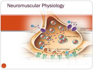
4. Introduction.
4
The nerve synthesizes acetylcholine and
stores it in small, uniformly sized
packages called vesicles.
Stimulation of the nerve causes these vesicles
to migrate to the surface of the nerve,
rupture, and discharge acetylcholine into
the cleft separating the nerve from
muscle.
AChRs in the end plate of the muscle
respond by opening their channels for
influx of sodium ions into the muscle to
depolarize the muscle.
5. 5
The endplate potential created is
continued along the muscle
membrane by the opening of
sodium channels -contraction.
The acetylcholine immediately
detaches from the receptor and is
destroyed- acetylcholinesterase.
Drugs, notably depolarizing
muscle relaxants or nicotine and
carbachol (a synthetic analog of
acetylcholine not destroyed by
acetylcholinesterase), can also act
on these receptors to mimic the
effect of acetylcholine and cause
depolarization of the end plate.
These drugs are therefore called
agonists.
6. 6
NDMRs also act on the receptors, but they prevent acetylcholine
from binding to the receptor and thus prevent depolarization by
agonists. Because these NDMRs prevent the action of agonists
(e.g., acetylcholine, carbachol, succinylcholine), they belong to the
class of compounds known as antagonists at the muscle AChRs.
Other compounds, frequently called reversal drugs or antagonists of
neuromuscular paralysis (e.g., neostigmine, prostigmine), inhibit
acetylcholinesterase and therefore impair the hydrolysis of
acetylcholine. The increased accumulation of acetylcholine can
effectively compete with NDMRs and thereby displace the latter
from the receptor (i.e., law of mass action) and antagonize the
effects of NDMR
7. The Motor Neuron
7
Motor neurons are the nerves that control skeletal muscle activity.
Axons are 10-20µm in diameter and surrounded by a myelin sheath produced
by Schwann cell.
The myelin sheath is interrupted by nodes of Ranvier between which the
action potential jumps causing rapid conduction of the nerve impulse.
8. 8
Each motor neuron connects to several
skeletal muscle fibers to form a motor unit.
As the motor neuron enters a muscle, the
axon divides into telodendria, the ends of
which, the terminal buttons, synapse with
the motor endplate.
The nerve is separated from the surface of
the muscle by a gap of approximately 20
nm, called the junctional cleft or synaptic cleft
The muscle surface is heavily corrugated,
with deep invaginations of the junctional
cleft—the primary and secondary cleft.
The shoulders of the folds are densely populated with AChRs,
approximately 5 million of them in each junction.
9. Because all the muscle cells in a unit are excited by a single
neuron, stimulation of the nerve electrically or by an action
potential originating from the ventral horn or by any agonist,
including depolarizing relaxants (e.g., succinylcholine), causes
all muscle cells in the motor unit to contract synchronously.
Synchronous contraction of the cells in a motor unit is called
fasciculation and is often vigorous enough to be observed through
the skin.
Although most adult human muscles have only one
neuromuscular junction per cell, an important exception is
some of the cells in extra ocular muscles.
9
10. The extraocular muscles are tonic muscles, and, unlike other
mammalian striated muscles, they are multiply innervated with
several neuromuscular junctions strung along the surface of
each muscle fiber.
The ocular muscles slowly contract and relax rather than
quickly as do other striated muscles; they can maintain a
steady contraction, or contracture, the strength of which is
proportional to the stimulus received.
Succinylcholine causes a long-lasting contracture response
that pulls the eye against the orbit and could contribute to an
increase in intraocular fluid pressure.
Succinylcholine-induced contractions of the extraocular
muscles can last as long as 1 to 2 minutes.
Thus succinylcholine probably should not be given to
patients with open eye injuries10
11. Quantal Theory
11
• Once synthesized the molecules of acetylcholine are stored in vesicles within the terminal button,
each vesicle containing approximately 10,000 molecules of acetylcholine.
.
• The release of acetylcholine into the synaptic cleft may be spontaneous or in response to a nerve
impulse
• Spontaneous release of single vesicles of
acetylcholine occurs randomly and results in
miniature endplate potentials of 0.5-1mV.
• These vesicles are loaded with acetylcholine via a magnesium dependent active transport
system in exchange for a hydrogen ion.
12. 12
• Acetylcholine is synthesized from
choline and acetyl-coenzyme A
(acetyl-coA) in the terminal axoplasm
of motor neurons and is catalyzed by
the enzyme cholinacetlytransferase.
• Acetyl-coA is synthesized from
pyruvate in the mitochondria in the
axon terminals.
• Approximately 50% of the choline is extracted from extracellular fluid by a sodium
dependant active transport system, the other 50% is from acetylcholine breakdown at
the neuromuscular junction
• The majority of the choline originates from the diet with hepatic synthesis only
accounting for a small proportion.
• Choline acetyltransferase is produced on the ribosome in the cell body of the motor
neuron.
13. Nerve action potential
13
During a nerve action potential, sodium from outside flows
across the membrane, and the resulting depolarizing voltage
opens the calcium channels, which allows entry of calcium
ions into the nerve and causes acetylcholine to be released.
14. A nerve action potential is the normal activator that releases
the transmitter acetylcholine.
If calcium is not present, then depolarization of the nerve,
even by electrical stimulation, will not produce the release of
transmitter.
An effect of increasing calcium in the nerve ending is also
clinically observed as the so-called posttetanic potentiation,
which occurs after a nerve of a patient paralyzed with an
NDMR is stimulated at high, tetanic frequencies.
Calcium enters the nerve with every stimulus, but it
accumulates during the tetanic period because it cannot be
excreted as quickly as the nerve is stimulated.14
15. Because the nerve ending contains more than the normal
amount of calcium for some time after the tetanus, a
stimulus applied to the nerve during this time causes the
release of more than the normal amount of acetylcholine.
The abnormally large amount of acetylcholine antagonizes
the relaxant and causes the characteristic increase in the size
of the twitch.
The Eaton-Lambert myasthenic syndrome, decreased
function of the calcium channel causes decreased release of
transmitter, which results in inadequate depolarization and
muscle weakness.
15
16. Synaptic vesicles and recycling
Release occurs when calcium ions enter the nerve through
the P channels lined up on the sides of the active zones by
soluble N-ethylmaleimide–sensitive attachment protein receptor
(SNARE) proteins
The SNARE proteins are involved in fusion, docking, and
release of acetylcholine at the active zone.
16
17. Calcium needs to move only a very short distance to
encounter a vesicle and activate the proteins in the vesicle
wall involved in a process known as docking
Calcium may penetrate more deeply than normal into the
nerve or may enter through L channels to activate calcium-
dependent enzymes that break the synapsin links holding the
vesicles to the cytoskeleton, thereby allowing the vesicles to
be moved to the release sites.
Repeated stimulation requires the nerve ending to replenish
its store of vesicles filled with transmitter, a process known
as mobilization.
17
18. Process Of Exocytosis
18 During an action potential and calcium influx, neurotransmitter is released. The
whole process is called exocytosis
20. Acetylcholinesterase
Acetylcholinesterase enzyme is
secreted by the muscle cell but
remains attached to it by thin
collagen threads linking it to
the basement membrane
Acetylcholinesterase is found
in the junctional gap and the
clefts of the postsynaptic folds
and breaks down acetylcholine
within 1 msec of being
released.
20
21. Acetyl cholinesterase
Acetylcholine is removed rapidly from the junctional gap
or synaptic cleft. This is achieved by hydrolysis of
acetylcholine to choline and acetate in a reaction
catalysed by the enzyme acetylcholinesterase (AChE).
Hydrolysis occurs with transfer of the acetyl group to
the serine group resulting in an acetylated molecule of
the enzyme and free choline.
The speed at which this occurs can be gauged by the fact
that approximately 10,000 molecules of acetylcholine
can be hydrolysed per second by a single site.
21
22. The congenital absence of the secreted enzyme leads to
impaired maintenance of the motor neuronal system and
organization of nerve terminal branches.
Congenital abnormalities in cholinesterase function have
been described and result in neuromuscular disorders whose
symptoms and signs usually resemble those of myasthenia
gravis or myasthenic syndromes.
Other acquired diseases involving cholinesterases are related
to chronic inhibition of acetylcholinesterase by
organophosphate pesticides or nerve gas (e.g., sarin)
22
23. Acetylcholine Receptors
AChRs are synthesized in muscle cells and are anchored to the end-
plate membrane by a special 43-kd protein known as rapsyn.
Each nicotinic receptor is a protein comprised of five polypeptide
subunits that form a ring structure around a central, funnel-shaped
pore (the ion channel). The mature adult receptor has two identical α
(alpha) subunits, one β (beta), one δ (delta) and one ε (epsilon)
subunit. In the fetus a γ (gamma) subunit replaces the ε.
23
24. Average 50 million acetylcholine receptors on a normal
endplate, situated on the crests of the junctional folds.
Acetylcholine molecules bind to specific sites on the α
subunits and when both are occupied a conformational
change occurs.
24
25. The channel allows movement of all cations – sodium ,This causes
depolarisation, the cell becomes less negative compared with the
extracellular surroundings. When a threshold of –50mV is achieved
(from a resting potential of –80mV), voltage- gated sodium channels
open, thereby increasing the rate of depolarisation and resulting in an
end plate potential (EPP) Of 50-100mV.
This in turn triggers the muscle action potential that results in
muscle contraction
25
26. Synthesis And Stabilization of AchRs
Muscle tissue is formed from the mesoderm and initially appears as
myoblasts.
Myoblasts fuse to produce myotubes, which therefore have multiple
nuclei. As the myotubes mature, the sarcomere, which is the
contractile element of the muscle consisting of actin and myosin
develops.
Agrin is a protein from the nerve that stimulates postsynaptic
differentiation by activating muscle-specific tyrosine kinase (MuSK), a
tyrosine kinase expressed selectively in muscle.
Agrin, together with neuregulins and other growth factors, induce the
clustering of other critical muscle-derived proteins, including MuSK,
rapsyn proteins, all of which are necessary for maturation and
stabilization of AChRs at the junction26
27. Electrophysiology
Of Neurotransmission
When an agonist occupies both -subunit sites, the proteinα
molecule undergoes a conformational change with a twisting
movement along the central axis of the receptor that results
in the opening of the central channel through which ions can
flow along a concentration gradient.
When the central channel is open, sodium and calcium flow
from the outside of the cell to the inside and potassium flows
from the inside to the outside.
27
28. Action of antagonists such as curare (filled square).
Acetylcholine is in competition with tubocurarine for the
receptor’s recognition site but may also react with
acetylcholinesterase. Tubocurarine is a prototypical
nondepolarizing muscle relaxant. Inhibiting the
acetylcholinesterase enzyme increases the lifetime of
acetylcholine and the probability that it will react with a
receptor. When one of the two binding (recognition) sites is
occupied by curare, the receptor will not open, even if the
other binding site is occupied by acetylcholine.
28
29. classic actions of nondepolarizing
muscle relaxants
Muscle relaxants (e.g., pancuronium, vecuronium )have
similar actions as that of tubocurarine.
Normally, acetylcholinesterase destroys acetylcholine and
removes it from competition for a receptor; therefore
tubocurarine has a better chance of inhibiting transmission.
If, however, an inhibitor of acetylcholinesterase such as
neostigmine is added, then the cholinesterase cannot destroy
acetylcholine.
29
30. Classic Actions Of Depolarizing
Muscle Relaxants
Depolarizing relaxants (e.g., succinylcholine, decamethonium)
initially simulate the effect of acetylcholine and can therefore be
considered agonists.
Structurally, succinylcholine is very similar to the natural ligand
acetylcholine.
Succinylcholine or decamethonium can bind to the receptor,
open the channel, pass current, and depolarize the end plate.
In contrast, the depolarizing relaxants characteristically have a
biphasic action on muscle—an initial contraction, followed by
relaxation lasting from minutes to hours.
30
31. Desensitization Block
The AChR, as a result of its flexibility and the fluidity of the
lipid around it, is capable of existing in a number of
conformational states
Some receptors that bind to agonists, however, do not
undergo the conformational change to open the channel.
Receptors in these states are called desensitized (i.e., they
are not sensitive to the channel-opening actions of agonists).
The receptor macromolecule, 1000 times larger by weight
than most drugs or gases, provides many places at which the
smaller molecules may act.
31
32. Many other drugs used by anesthetists also promote the shift
of receptors from a normal state to a desensitized state.
Desensitization may also be a part of the phenomenon known
as phase II block which is caused by a prolonged administration
of depolarizing relaxants
Drugs That Can Cause or Promote Desensitization of Nicotinic
Cholinergic Receptors
Volatile anesthetics
Halothane
Sevoflurane
Isoflurane
Barbiturates
Thiopental
Pentobarbital
32
34. Channel Block
Local anesthetics and calcium entry blockers prevent the
flow of sodium or calcium through their respective channels,
thus explaining the term channel-blocking drugs.
In a closed-channel block, certain drugs can occupy the
mouth of the channel and prevent ions from passing through
the channel to depolarize the end plate.
In an open-channel block, a drug molecule enters a channel
that has been opened by reaction with acetylcholine but does
not necessarily penetrate all the way through.
34
35. Increasing the concentration of acetylcholine may cause the
channels to open more often and, consequently, become
more susceptible to blockade by use-dependent compounds.
Evidence suggests that neostigmine and related
cholinesterase inhibitors can act as channel-blocking drugs.
Channel block may account for the antibiotic-, cocaine,
quinidine-, piperocaine-, tricyclic antidepressant–,
naltrexone-, naloxone-induced alterations in neuromuscular
function.
35
36. Phase II Block
A phase II block is a complex
phenomenon associated with a typical
fade in muscle during continuous
exposure to depolarizing drugs.
However, fade in muscle during
repetitive nerve stimulation can also be
attributable to postjunctional AChR
block.
The repeated opening of channels
allows a continuous efflux of potassium
and influx of sodium, and the resulting
abnormal electrolyte balance distorts
the function of the junctional
membrane.
As long as the depolarizing drug is
present, the receptor channels remain
open and ion flux through them
remains frequent.36