SlideShare ist ein Scribd-Unternehmen logo
1 von 39
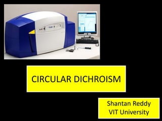
CIRCULAR DICHROISM

               Shantan Reddy
                VIT University
What is it?

• Circular dichroism is the difference in the
  absorption of left-handed circularly polarised
  light (L-CPL) and right-handed circularly
  polarised light (R-CPL).
• Occurs when a molecule contains one or more
  chiral chromophores.

  Circular dichroism = ΔA(λ) = A(λ)LCPL - A(λ)RCPL
           (where λ is the wavelength)
Basics of Polarization


   Linearly
polarised light     Circularly
                  polarised light
Linearly Polarised Light


   In a linearly polarised light oscillations are
  confined to a single plane. All polarised light
states can be described as a sum of two linearly
 polarised states at right angles to each other –
    vertically and horizontally polarised light.
Vertically polarised
        light

                       Horizontally polarised
                               light
Superposition of the plane polarised
              waves
Linearly polarised light

Horizontally and vertically polarised light waves
 of equal amplitude that are in phase with each
 other will produce a resultant light wave which
is linearly polarised at 45˚ and the properties of
the resulting electromagnetic wave depends on
    the intensities and phase difference of the
                    component.


                   1. Linearly Polarised light
Circularly polarised light
• When one of the polarised states is out of phase
  with the other by a quarter-wave, the resultant
  will be a helix and is known as circularly polarised
  light (CPL).
• The optical element that converts between
  linearly polarised light and circularly polarised
  light is termed a quarter-wave plate. A quarter-
  will convert linearly polarised light into circularly
  polarised light by slowing one of the linear
  components of the beam with respect to the
  other so that they are one quarter-wave out of
  phase. This will produce a beam of either left- or
  right-CPL.
2. Left Circularly Polarised light


3. Right Circularly Polarised light
Superposition of circularly polarised
              waves


The superposition of a left circularly polarized
 wave and a right circularly polarized wave of
equal amplitudes and wavelengths is a plane
               polarised wave.
Circular Dichroism

• Some materials possess a special
  property: they absorb left circularly polarized
  light to a different extent than right circularly
  polarized light. This phenomenon is
  called circular dichroism.
• Assume that a plane-polarized light wave
  (blue) traverses a medium that does not
  absorb the left circularly polarized component
  (red) of the wave at all but highly absorbs the
  right circularly polarized component (green).
The intensity of the green component decreases in
comparison to the red one.
The superposition of the two components yields a
resulting field vector that rotates along an ellipsoid
path and is called an elliptically polarized light.
• The direction of rotation of the elliptically
  polarized light is determined by the circular
  component that remains stronger after
  traversing the material.
• Real materials usually absorb both
  components, to a different extent.
• How elliptical the plane-polarized wave
  becomes after traversing the medium is
  determined by the difference between the
  absorptions of the two circularly polarized
  components.
Circular Birefringence

• There are materials having another special
  property: their refraction index is different for left
  and right circularly polarized light. This
  phenomenon is called circular birefringence.
• Assume that a plane-polarized light wave (blue)
  traverses a medium that does not slow down the
  left circularly polarized component (red) of the
  wave at all but slows down the right circularly
  polarized component (green) somewhat. For the
  latter component, the refraction index of the
  material is n=1.05.
This slowdown and the decreased wavelength
is hard to see in the figure because the
refraction index (1.05) is close to 1.0.
• Before the medium, the field vectors of the
  components coincide when they point vertically
  up or down. But after the light exits the
  medium, the superposition of the two circularly
  polarized components is a plane-polarized wave
  with a plane of polarization that is rotated by 36°
  with respect to the original polarization plane.
• Real materials usually have refraction indices
  greater than 1.0 (but not equal) for both circular
  components.
• The angle by which the polarization plane of the
  light exiting the medium rotates with respect to
  the original polarization plane is determined by
  the difference between the refraction indices for
  the two circularly polarized components.
Circular Dichroism & Circular
               Birefringence


The red component traverses the medium
unchanged, but the medium has absorption and a
refraction index with respect to the green component.
• The exiting light is no longer plane-polarized, it is
  not the plane of polarization that gets rotated but
  the big axis of the ellipse of polarization of the
  elliptically polarized light.
• With the appropriate instrument, the ellipticity
  and the angle of rotation of the polarization
  plane of light can be measured. From those
  data, the difference between absorptions and
  refraction indices with respect to left and right
  circularly polarized lights can be calculated.
• Circular dichroism and circular birefringence are
  caused by the asymmetry of the molecular
  structure of matter. The optical activity of
  solutions of biological macromolecules provides
  information about the structural properties of the
  macromolecules.
Physics of CD spectroscopy
• In an optically active sample with a different
  absorbance ‘A’, for the two components the
  amplitude of the stronger absorbed
  component will be smaller than the less
  absorbed component.
• A projection of the resulting amplitude yields
  an ellipse.
• Rotation of the polarization plane by a small
  angle ‘a’ occurs when the phases for the two
  circular components becomes different which
  requires a difference in the refractive index n
  by the effect called circular birefringence.
• The change of optical rotation with
  wavelength is called optical rotatory
  dispersion.
• CD and Optical rotation exist together and
  they are related by Kronig-Krames
  transformation.
• The difference between left and right handed
  absorbance is very small ( in the range of
  0.0001)
• CD is a function of wavelength making it a
  characteristic of molecules.
Data Analysis
Instrumentation


Prism
Nitrogen Purging?

• Removes oxygen from the lamp
  housing, monochromator and the sample
  chamber.
• The presence of oxygen is detrimental for 2
  reasons –
  i) when deep UV light strike oxygen, ozone is
  produced, which degrades optics.
  ii) oxygen absorbs deep UV light which reduces
  the amount available for measurement.
Sample Preparation and
            Measurements
• Additives, Buffers and Stabilizing compounds -
  any compound that absorbs in the region of
  interest (250 -190 nm) should be avoided.
• Protein solution – It should contain only those
  chemicals required to maintain protein
  stability at the lowest conc possible. Any
  additional protein or peptide will contribute to
  the CD signal.
• Data collection – Initial experiments are
  needed to establish the parameters for the
  actual experiment.
Typical Initial Conditions

•   Protein Concentration – 0.5mg/ml
•   Cell path length – 0.5 mm
•   Stabilizers (metal ions, etc.) – minimum
•   Buffer concentration – 5mM or as low as
    possible while maintaining protein stability.
Sample concentration effects

• Optimum absorbance to use is 0.89nm
• For a 1mm path length cell, this absorbance is
  achieved with a protein concentration of 0.1-
  0.3 mg/ml.
CD of proteins and polypeptides

For proteins we will be mainly dealing with the
 absorption in the UV region of the spectrum
originated from such chromophores as peptide
 bond, amino acid side chains (aromatic side
  chains of Phe, Tyr, and Trp have absorption
    bands in the vicinity of 250-320 nm; the
  disulfide group is an inherently asymmetric
   chromophore that can lead to a broad CD
absorption around 250 nm), and any prosthetic
                     groups.
CD of Proteins – Far UV region

         n -> π* centered around 220 nm
         π -> π* centered around 190 nm
         n -> π* involves non-bonding
         electrons of O of the carbonyl
         π -> π* involves the π-electrons
         of the carbonyl
         The intensity and energy of
         these transitions depends on φ
         and ψ (i.e., secondary structure)
Far UV-CD of random coil:
positive at 212 nm (π->π*)
negative at 195 nm (n->π*)
Far UV-CD of β-sheet:
negative at 218 nm (π->π*)
positive at 196 nm (n->π*)
Far UV-CD of α-helix:
exiton coupling of the π->π*
transitions leads to positive (π-
>π*)perpendicular at 192 nm
and negative (π->π*)parallel at
209 nm negative at 222 nm is red
shifted (n->π*)
Far UV CD spectra and Secondary
      Structure of Proteins
              After baseline subtraction we
              are ready to analyze the data.
              Each of the three basic
              secondary structures of a
              polypeptide chain
              (helix, sheet, coil) show a
              characteristic CD spectrum. A
              protein consisting of these
              elements should therefore
              display a spectrum that can
              be deconvoluted into the
              individual contributions.
CD of Proteins – Near UV region
PHENYLALANINES         TYROSINES              TRYPTOPHANS            S-S BONDS


Small extinction       Lower symmetry and     Has the most intense   It has a very broad
coefficient due to     hence intense          absorption band.       band.
high symmetry.         absorption bands.
Absorption maxima      Absorption maxima      Absorption maxima is Absorption maxima
at 254, 256, 262 and   at 276 nm; hydrogen    282 nm               ranges from 250 – 300
267 nm.                bonding to the –OH                          nm
                       group leads to a red
                       shift of up to 4nm
CD in biochemistry

   Used in the understanding of the higher order
    structures of chiral macromolecules such as
proteins and DNA. The reason for this is that the CD
spectrum of a protein or DNA molecule is not a sum
   of the CD spectra of the individual residues or
       bases, but is greatly influenced by the
 3-dimension structure of the macromolecule itself.
   Each structure has a specific circular dichroism
signature, and this can be used to identify structural
 elements and to follow changes in the structure of
              chiral macromolecules.
The most widely studied circular dichroism
signatures are the various secondary structural
elements of proteins such as the α-helix and the β
sheet. This is understood to the point that CD
spectra in the far-UV (below 260nm) can be used to
predict the percentages of each secondary
structural element in the structure of a protein.
There are many algorithms designed for fitting
the circular dichroism spectra of proteins to
provide estimates of secondary structure. The
protein secondary structure CD analysis
software distributed with the Chirascan is CDNN.
Other Applications
• CD is a particularly powerful tool to follow
  dynamic changes in protein structure which
  may result due to the effect of changing
  temperature, pH, ligands, or denaturants etc.
• Circular dichroism can be used to follow the
  kinetics of refolding of the secondary
  structure of a protein using changes in
  denaturant concentration.
• It can also be used to follow the unfolding of
  proteins by thermal denaturation.
• A powerful application of circular dichroism is to
  compare two macromolecules, or the same
  molecule under different conditions, and
  determine if they have a similar structure. This
  can be used simply to ascertain if -
  i) a newly purified protein is correctly folded,
  ii) determine if a mutant protein has folded
  correctly in comparison to the wild-type, or
  iii) for the analysis of biopharmaceutical products
  to confirm that they are still in a correctly folded
  active conformation.
Circular dichroism

Weitere ähnliche Inhalte

Was ist angesagt?

Electron spin resonance(ESR) spectroscopy
Electron spin resonance(ESR) spectroscopyElectron spin resonance(ESR) spectroscopy
Electron spin resonance(ESR) spectroscopyPreeti Choudhary
 
Optical rotatory dispersion & circular dichorism
Optical rotatory dispersion &                    circular dichorismOptical rotatory dispersion &                    circular dichorism
Optical rotatory dispersion & circular dichorismsudha rajput
 
CD & ORD Spectrometer
CD & ORD SpectrometerCD & ORD Spectrometer
CD & ORD Spectrometernaren
 
Circular Dichroism Spectroscopy
Circular Dichroism SpectroscopyCircular Dichroism Spectroscopy
Circular Dichroism SpectroscopyAditya Sharma
 
(Gel Filtration Chromatography)GFC
(Gel Filtration Chromatography)GFC(Gel Filtration Chromatography)GFC
(Gel Filtration Chromatography)GFCAthira athira
 
2 d gel electrophoresis
2 d gel electrophoresis2 d gel electrophoresis
2 d gel electrophoresisAashish Patel
 
MALDI-TOF Mass Spectrometry
MALDI-TOF Mass SpectrometryMALDI-TOF Mass Spectrometry
MALDI-TOF Mass SpectrometryNawaz Shah
 
Differential centrifugation
Differential centrifugationDifferential centrifugation
Differential centrifugationSuganyaPaulraj
 
X-ray Crystallography & Its Applications in Proteomics
X-ray Crystallography & Its Applications in Proteomics X-ray Crystallography & Its Applications in Proteomics
X-ray Crystallography & Its Applications in Proteomics Akash Arora
 
Solid phase peptide synthesis
Solid phase peptide synthesisSolid phase peptide synthesis
Solid phase peptide synthesisABHISHEK SIRSIKAR
 
Analytical centrifugation
Analytical centrifugationAnalytical centrifugation
Analytical centrifugationVarshini3
 
ISOELECTRIC FOCUSING PPT - SLIDE SHARE
ISOELECTRIC FOCUSING PPT - SLIDE SHAREISOELECTRIC FOCUSING PPT - SLIDE SHARE
ISOELECTRIC FOCUSING PPT - SLIDE SHAREAditi Chaturvedi
 
Atomic force microscopy
Atomic force microscopyAtomic force microscopy
Atomic force microscopySonu Bishnoi
 
Isoelectric focussing
Isoelectric focussingIsoelectric focussing
Isoelectric focussingarushe143
 
OPTICAL ROTATORY DISPERSION & CIRCULAR DICHORISM(ORD CD)
OPTICAL ROTATORY DISPERSION & CIRCULAR DICHORISM(ORD CD)OPTICAL ROTATORY DISPERSION & CIRCULAR DICHORISM(ORD CD)
OPTICAL ROTATORY DISPERSION & CIRCULAR DICHORISM(ORD CD)Suvarta Maru
 

Was ist angesagt? (20)

Electron spin resonance(ESR) spectroscopy
Electron spin resonance(ESR) spectroscopyElectron spin resonance(ESR) spectroscopy
Electron spin resonance(ESR) spectroscopy
 
Optical rotatory dispersion & circular dichorism
Optical rotatory dispersion &                    circular dichorismOptical rotatory dispersion &                    circular dichorism
Optical rotatory dispersion & circular dichorism
 
CD & ORD Spectrometer
CD & ORD SpectrometerCD & ORD Spectrometer
CD & ORD Spectrometer
 
Circular Dichroism Spectroscopy
Circular Dichroism SpectroscopyCircular Dichroism Spectroscopy
Circular Dichroism Spectroscopy
 
(Gel Filtration Chromatography)GFC
(Gel Filtration Chromatography)GFC(Gel Filtration Chromatography)GFC
(Gel Filtration Chromatography)GFC
 
2 d gel electrophoresis
2 d gel electrophoresis2 d gel electrophoresis
2 d gel electrophoresis
 
MALDI-TOF Mass Spectrometry
MALDI-TOF Mass SpectrometryMALDI-TOF Mass Spectrometry
MALDI-TOF Mass Spectrometry
 
Radiolabeling technique
Radiolabeling techniqueRadiolabeling technique
Radiolabeling technique
 
Ramachandran plot
Ramachandran plotRamachandran plot
Ramachandran plot
 
Differential centrifugation
Differential centrifugationDifferential centrifugation
Differential centrifugation
 
X-ray Crystallography & Its Applications in Proteomics
X-ray Crystallography & Its Applications in Proteomics X-ray Crystallography & Its Applications in Proteomics
X-ray Crystallography & Its Applications in Proteomics
 
ESR
ESRESR
ESR
 
Solid phase peptide synthesis
Solid phase peptide synthesisSolid phase peptide synthesis
Solid phase peptide synthesis
 
Analytical centrifugation
Analytical centrifugationAnalytical centrifugation
Analytical centrifugation
 
ISOELECTRIC FOCUSING PPT - SLIDE SHARE
ISOELECTRIC FOCUSING PPT - SLIDE SHAREISOELECTRIC FOCUSING PPT - SLIDE SHARE
ISOELECTRIC FOCUSING PPT - SLIDE SHARE
 
Atomic force microscopy
Atomic force microscopyAtomic force microscopy
Atomic force microscopy
 
Isoelectric focussing
Isoelectric focussingIsoelectric focussing
Isoelectric focussing
 
Ord
OrdOrd
Ord
 
Confocal microscopy
Confocal microscopyConfocal microscopy
Confocal microscopy
 
OPTICAL ROTATORY DISPERSION & CIRCULAR DICHORISM(ORD CD)
OPTICAL ROTATORY DISPERSION & CIRCULAR DICHORISM(ORD CD)OPTICAL ROTATORY DISPERSION & CIRCULAR DICHORISM(ORD CD)
OPTICAL ROTATORY DISPERSION & CIRCULAR DICHORISM(ORD CD)
 

Andere mochten auch

MALDI-TOF: Pricinple and Its Application in Biochemistry and Biotechnology
MALDI-TOF: Pricinple  and Its Application in Biochemistry and BiotechnologyMALDI-TOF: Pricinple  and Its Application in Biochemistry and Biotechnology
MALDI-TOF: Pricinple and Its Application in Biochemistry and BiotechnologyDevakumar Jain
 
Optical rotatory dispersion
Optical rotatory dispersionOptical rotatory dispersion
Optical rotatory dispersionSujit Patel
 
Maldi tof ppt adnya
Maldi tof ppt adnyaMaldi tof ppt adnya
Maldi tof ppt adnyaAdnya Desai
 
Ultra violet (UV) spectroscopy, introduction,principle instrumentation,differ...
Ultra violet (UV) spectroscopy, introduction,principle instrumentation,differ...Ultra violet (UV) spectroscopy, introduction,principle instrumentation,differ...
Ultra violet (UV) spectroscopy, introduction,principle instrumentation,differ...mariomS7
 

Andere mochten auch (8)

MALDI-TOF: Pricinple and Its Application in Biochemistry and Biotechnology
MALDI-TOF: Pricinple  and Its Application in Biochemistry and BiotechnologyMALDI-TOF: Pricinple  and Its Application in Biochemistry and Biotechnology
MALDI-TOF: Pricinple and Its Application in Biochemistry and Biotechnology
 
Maldi tof
Maldi tofMaldi tof
Maldi tof
 
Optical rotatory dispersion
Optical rotatory dispersionOptical rotatory dispersion
Optical rotatory dispersion
 
Maldi tof ppt adnya
Maldi tof ppt adnyaMaldi tof ppt adnya
Maldi tof ppt adnya
 
MALDI - TOF
MALDI - TOFMALDI - TOF
MALDI - TOF
 
Flame photometry
Flame photometryFlame photometry
Flame photometry
 
Ultra violet (UV) spectroscopy, introduction,principle instrumentation,differ...
Ultra violet (UV) spectroscopy, introduction,principle instrumentation,differ...Ultra violet (UV) spectroscopy, introduction,principle instrumentation,differ...
Ultra violet (UV) spectroscopy, introduction,principle instrumentation,differ...
 
UV visible spectroscopy
UV visible spectroscopyUV visible spectroscopy
UV visible spectroscopy
 

Ähnlich wie Circular dichroism

Circular dichroism in Biochemistryppt.ppt
Circular dichroism in Biochemistryppt.pptCircular dichroism in Biochemistryppt.ppt
Circular dichroism in Biochemistryppt.pptManjuSingh118444
 
Optical Rotatory Dispersion
Optical Rotatory DispersionOptical Rotatory Dispersion
Optical Rotatory DispersionMahendra G S
 
Circular dichroism & Ord
Circular dichroism & Ord Circular dichroism & Ord
Circular dichroism & Ord Lovnish Thakur
 
Optical rotatory dispersion and circular dichroism presentation
Optical rotatory dispersion and circular dichroism presentationOptical rotatory dispersion and circular dichroism presentation
Optical rotatory dispersion and circular dichroism presentationChaitrali Jadhav
 
Opticalrotatory dispersion
Opticalrotatory dispersionOpticalrotatory dispersion
Opticalrotatory dispersionJagadeesh Babu
 
Electronic circular dichroism(ECD).pdf
Electronic circular dichroism(ECD).pdfElectronic circular dichroism(ECD).pdf
Electronic circular dichroism(ECD).pdfCreative Proteomics
 
Polarization of Light
Polarization of LightPolarization of Light
Polarization of LightHaris Hassan
 
JASCO-CD-Webinar-Part-1-FINAL.pptx
JASCO-CD-Webinar-Part-1-FINAL.pptxJASCO-CD-Webinar-Part-1-FINAL.pptx
JASCO-CD-Webinar-Part-1-FINAL.pptxNIBGE-College
 
Optical rotatory dispersion and circular dichroism
Optical rotatory dispersion and circular dichroismOptical rotatory dispersion and circular dichroism
Optical rotatory dispersion and circular dichroismarimilli haritha
 
Chapter 16 - Infrared Spectroscopy. Overview of infrared spectroscopy
Chapter 16 - Infrared Spectroscopy. Overview of infrared spectroscopyChapter 16 - Infrared Spectroscopy. Overview of infrared spectroscopy
Chapter 16 - Infrared Spectroscopy. Overview of infrared spectroscopysanjayduttainbox
 

Ähnlich wie Circular dichroism (20)

Circular dichroism in Biochemistryppt.ppt
Circular dichroism in Biochemistryppt.pptCircular dichroism in Biochemistryppt.ppt
Circular dichroism in Biochemistryppt.ppt
 
Determination of secondary structure of proteins
Determination of secondary structure of proteinsDetermination of secondary structure of proteins
Determination of secondary structure of proteins
 
Optical Rotatory Dispersion
Optical Rotatory DispersionOptical Rotatory Dispersion
Optical Rotatory Dispersion
 
ORD CD AMU notes.pdf
ORD CD  AMU notes.pdfORD CD  AMU notes.pdf
ORD CD AMU notes.pdf
 
Circular dichroism & Ord
Circular dichroism & Ord Circular dichroism & Ord
Circular dichroism & Ord
 
5. cd and ord jntu pharmacy
5. cd and ord jntu pharmacy5. cd and ord jntu pharmacy
5. cd and ord jntu pharmacy
 
Polarimetry
PolarimetryPolarimetry
Polarimetry
 
Optical rotatory dispersion and circular dichroism presentation
Optical rotatory dispersion and circular dichroism presentationOptical rotatory dispersion and circular dichroism presentation
Optical rotatory dispersion and circular dichroism presentation
 
Opticalrotatory dispersion
Opticalrotatory dispersionOpticalrotatory dispersion
Opticalrotatory dispersion
 
Electronic circular dichroism(ECD).pdf
Electronic circular dichroism(ECD).pdfElectronic circular dichroism(ECD).pdf
Electronic circular dichroism(ECD).pdf
 
polarimetry
polarimetrypolarimetry
polarimetry
 
Prabhakar singh ii sem-paper v-optical methods for determination of molecula...
Prabhakar singh  ii sem-paper v-optical methods for determination of molecula...Prabhakar singh  ii sem-paper v-optical methods for determination of molecula...
Prabhakar singh ii sem-paper v-optical methods for determination of molecula...
 
Polarization of Light
Polarization of LightPolarization of Light
Polarization of Light
 
JASCO-CD-Webinar-Part-1-FINAL.pptx
JASCO-CD-Webinar-Part-1-FINAL.pptxJASCO-CD-Webinar-Part-1-FINAL.pptx
JASCO-CD-Webinar-Part-1-FINAL.pptx
 
2.ir jntu pharmacy
2.ir jntu pharmacy2.ir jntu pharmacy
2.ir jntu pharmacy
 
Ord and cd ppt
Ord and cd pptOrd and cd ppt
Ord and cd ppt
 
Optical rotatory dispersion and circular dichroism
Optical rotatory dispersion and circular dichroismOptical rotatory dispersion and circular dichroism
Optical rotatory dispersion and circular dichroism
 
Chapter 16 - Infrared Spectroscopy. Overview of infrared spectroscopy
Chapter 16 - Infrared Spectroscopy. Overview of infrared spectroscopyChapter 16 - Infrared Spectroscopy. Overview of infrared spectroscopy
Chapter 16 - Infrared Spectroscopy. Overview of infrared spectroscopy
 
Ord and cd
Ord and cdOrd and cd
Ord and cd
 
spect and pet
spect and petspect and pet
spect and pet
 

Circular dichroism

  • 1. CIRCULAR DICHROISM Shantan Reddy VIT University
  • 2. What is it? • Circular dichroism is the difference in the absorption of left-handed circularly polarised light (L-CPL) and right-handed circularly polarised light (R-CPL). • Occurs when a molecule contains one or more chiral chromophores. Circular dichroism = ΔA(λ) = A(λ)LCPL - A(λ)RCPL (where λ is the wavelength)
  • 3. Basics of Polarization Linearly polarised light Circularly polarised light
  • 4. Linearly Polarised Light In a linearly polarised light oscillations are confined to a single plane. All polarised light states can be described as a sum of two linearly polarised states at right angles to each other – vertically and horizontally polarised light.
  • 5. Vertically polarised light Horizontally polarised light
  • 6. Superposition of the plane polarised waves
  • 7. Linearly polarised light Horizontally and vertically polarised light waves of equal amplitude that are in phase with each other will produce a resultant light wave which is linearly polarised at 45˚ and the properties of the resulting electromagnetic wave depends on the intensities and phase difference of the component. 1. Linearly Polarised light
  • 8. Circularly polarised light • When one of the polarised states is out of phase with the other by a quarter-wave, the resultant will be a helix and is known as circularly polarised light (CPL). • The optical element that converts between linearly polarised light and circularly polarised light is termed a quarter-wave plate. A quarter- will convert linearly polarised light into circularly polarised light by slowing one of the linear components of the beam with respect to the other so that they are one quarter-wave out of phase. This will produce a beam of either left- or right-CPL.
  • 9. 2. Left Circularly Polarised light 3. Right Circularly Polarised light
  • 10. Superposition of circularly polarised waves The superposition of a left circularly polarized wave and a right circularly polarized wave of equal amplitudes and wavelengths is a plane polarised wave.
  • 11. Circular Dichroism • Some materials possess a special property: they absorb left circularly polarized light to a different extent than right circularly polarized light. This phenomenon is called circular dichroism. • Assume that a plane-polarized light wave (blue) traverses a medium that does not absorb the left circularly polarized component (red) of the wave at all but highly absorbs the right circularly polarized component (green).
  • 12. The intensity of the green component decreases in comparison to the red one. The superposition of the two components yields a resulting field vector that rotates along an ellipsoid path and is called an elliptically polarized light.
  • 13. • The direction of rotation of the elliptically polarized light is determined by the circular component that remains stronger after traversing the material. • Real materials usually absorb both components, to a different extent. • How elliptical the plane-polarized wave becomes after traversing the medium is determined by the difference between the absorptions of the two circularly polarized components.
  • 14. Circular Birefringence • There are materials having another special property: their refraction index is different for left and right circularly polarized light. This phenomenon is called circular birefringence. • Assume that a plane-polarized light wave (blue) traverses a medium that does not slow down the left circularly polarized component (red) of the wave at all but slows down the right circularly polarized component (green) somewhat. For the latter component, the refraction index of the material is n=1.05.
  • 15. This slowdown and the decreased wavelength is hard to see in the figure because the refraction index (1.05) is close to 1.0.
  • 16. • Before the medium, the field vectors of the components coincide when they point vertically up or down. But after the light exits the medium, the superposition of the two circularly polarized components is a plane-polarized wave with a plane of polarization that is rotated by 36° with respect to the original polarization plane. • Real materials usually have refraction indices greater than 1.0 (but not equal) for both circular components. • The angle by which the polarization plane of the light exiting the medium rotates with respect to the original polarization plane is determined by the difference between the refraction indices for the two circularly polarized components.
  • 17. Circular Dichroism & Circular Birefringence The red component traverses the medium unchanged, but the medium has absorption and a refraction index with respect to the green component.
  • 18. • The exiting light is no longer plane-polarized, it is not the plane of polarization that gets rotated but the big axis of the ellipse of polarization of the elliptically polarized light. • With the appropriate instrument, the ellipticity and the angle of rotation of the polarization plane of light can be measured. From those data, the difference between absorptions and refraction indices with respect to left and right circularly polarized lights can be calculated. • Circular dichroism and circular birefringence are caused by the asymmetry of the molecular structure of matter. The optical activity of solutions of biological macromolecules provides information about the structural properties of the macromolecules.
  • 19. Physics of CD spectroscopy
  • 20. • In an optically active sample with a different absorbance ‘A’, for the two components the amplitude of the stronger absorbed component will be smaller than the less absorbed component. • A projection of the resulting amplitude yields an ellipse. • Rotation of the polarization plane by a small angle ‘a’ occurs when the phases for the two circular components becomes different which requires a difference in the refractive index n by the effect called circular birefringence.
  • 21. • The change of optical rotation with wavelength is called optical rotatory dispersion. • CD and Optical rotation exist together and they are related by Kronig-Krames transformation. • The difference between left and right handed absorbance is very small ( in the range of 0.0001) • CD is a function of wavelength making it a characteristic of molecules.
  • 23.
  • 25. Nitrogen Purging? • Removes oxygen from the lamp housing, monochromator and the sample chamber. • The presence of oxygen is detrimental for 2 reasons – i) when deep UV light strike oxygen, ozone is produced, which degrades optics. ii) oxygen absorbs deep UV light which reduces the amount available for measurement.
  • 26. Sample Preparation and Measurements • Additives, Buffers and Stabilizing compounds - any compound that absorbs in the region of interest (250 -190 nm) should be avoided. • Protein solution – It should contain only those chemicals required to maintain protein stability at the lowest conc possible. Any additional protein or peptide will contribute to the CD signal. • Data collection – Initial experiments are needed to establish the parameters for the actual experiment.
  • 27. Typical Initial Conditions • Protein Concentration – 0.5mg/ml • Cell path length – 0.5 mm • Stabilizers (metal ions, etc.) – minimum • Buffer concentration – 5mM or as low as possible while maintaining protein stability.
  • 28. Sample concentration effects • Optimum absorbance to use is 0.89nm • For a 1mm path length cell, this absorbance is achieved with a protein concentration of 0.1- 0.3 mg/ml.
  • 29. CD of proteins and polypeptides For proteins we will be mainly dealing with the absorption in the UV region of the spectrum originated from such chromophores as peptide bond, amino acid side chains (aromatic side chains of Phe, Tyr, and Trp have absorption bands in the vicinity of 250-320 nm; the disulfide group is an inherently asymmetric chromophore that can lead to a broad CD absorption around 250 nm), and any prosthetic groups.
  • 30. CD of Proteins – Far UV region n -> π* centered around 220 nm π -> π* centered around 190 nm n -> π* involves non-bonding electrons of O of the carbonyl π -> π* involves the π-electrons of the carbonyl The intensity and energy of these transitions depends on φ and ψ (i.e., secondary structure)
  • 31. Far UV-CD of random coil: positive at 212 nm (π->π*) negative at 195 nm (n->π*) Far UV-CD of β-sheet: negative at 218 nm (π->π*) positive at 196 nm (n->π*) Far UV-CD of α-helix: exiton coupling of the π->π* transitions leads to positive (π- >π*)perpendicular at 192 nm and negative (π->π*)parallel at 209 nm negative at 222 nm is red shifted (n->π*)
  • 32. Far UV CD spectra and Secondary Structure of Proteins After baseline subtraction we are ready to analyze the data. Each of the three basic secondary structures of a polypeptide chain (helix, sheet, coil) show a characteristic CD spectrum. A protein consisting of these elements should therefore display a spectrum that can be deconvoluted into the individual contributions.
  • 33. CD of Proteins – Near UV region PHENYLALANINES TYROSINES TRYPTOPHANS S-S BONDS Small extinction Lower symmetry and Has the most intense It has a very broad coefficient due to hence intense absorption band. band. high symmetry. absorption bands. Absorption maxima Absorption maxima Absorption maxima is Absorption maxima at 254, 256, 262 and at 276 nm; hydrogen 282 nm ranges from 250 – 300 267 nm. bonding to the –OH nm group leads to a red shift of up to 4nm
  • 34. CD in biochemistry Used in the understanding of the higher order structures of chiral macromolecules such as proteins and DNA. The reason for this is that the CD spectrum of a protein or DNA molecule is not a sum of the CD spectra of the individual residues or bases, but is greatly influenced by the 3-dimension structure of the macromolecule itself. Each structure has a specific circular dichroism signature, and this can be used to identify structural elements and to follow changes in the structure of chiral macromolecules.
  • 35. The most widely studied circular dichroism signatures are the various secondary structural elements of proteins such as the α-helix and the β sheet. This is understood to the point that CD spectra in the far-UV (below 260nm) can be used to predict the percentages of each secondary structural element in the structure of a protein.
  • 36. There are many algorithms designed for fitting the circular dichroism spectra of proteins to provide estimates of secondary structure. The protein secondary structure CD analysis software distributed with the Chirascan is CDNN.
  • 37. Other Applications • CD is a particularly powerful tool to follow dynamic changes in protein structure which may result due to the effect of changing temperature, pH, ligands, or denaturants etc. • Circular dichroism can be used to follow the kinetics of refolding of the secondary structure of a protein using changes in denaturant concentration. • It can also be used to follow the unfolding of proteins by thermal denaturation.
  • 38. • A powerful application of circular dichroism is to compare two macromolecules, or the same molecule under different conditions, and determine if they have a similar structure. This can be used simply to ascertain if - i) a newly purified protein is correctly folded, ii) determine if a mutant protein has folded correctly in comparison to the wild-type, or iii) for the analysis of biopharmaceutical products to confirm that they are still in a correctly folded active conformation.