Right Ventricular Cardiomyopathy in the Rat - Is There an Association with Gavage?
•Als PPTX, PDF herunterladen•
0 gefällt mir•525 views

Right Ventricular Cardiomyopathy in the Rat - Is There an Association with Gavage?
Empfohlen

Empfohlen
Weitere ähnliche Inhalte
Was ist angesagt?
Was ist angesagt? (19)
Andere mochten auch
Andere mochten auch (20)
Ähnlich wie Right Ventricular Cardiomyopathy in the Rat - Is There an Association with Gavage?
Ähnlich wie Right Ventricular Cardiomyopathy in the Rat - Is There an Association with Gavage? (20)
Mehr von EPL, Inc.
Mehr von EPL, Inc. (14)
Kürzlich hochgeladen
Kürzlich hochgeladen (20)
Right Ventricular Cardiomyopathy in the Rat - Is There an Association with Gavage?
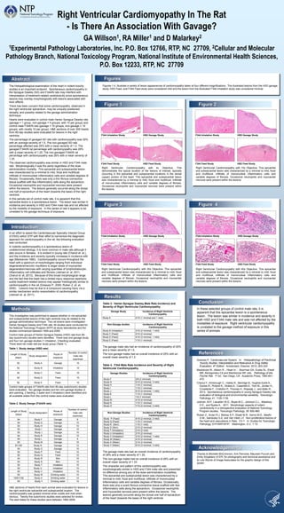
- 1. Greaves P. Cardiovascular System. In: Histopathology of Preclinical Toxicity Studies. Interpretation and Relevance in Drug Safety Evaluation, 4th Edition. Amsterdam, Elsevier; 2012; 263-324 Mackenzie W., Allison R ., Heart In: : Boorman GA, Eustis SL, Elwell MR, Montgomery CA and MacKenzie WF, eds. Pathology of the Fischer Rat. 1st ed. San Diego, CA: Academic Press; 1990:461- 472. Chanut F., Kimbrough C., Hailey R., Berridge B., Hughes-Earle A., Davies R., Roland K., Stokes A., Casartelli A., York M., Jordan H., Crivellente F., Cristofori P., Thomas H., Klapwijk J., and Adler R., 2013. Spontaneous cardiomyopathy in young Sprague-Dawley rats: evaluation of biological and environmental variability. Toxicologic Pathology, 41: 1126-1136. Jokinen, M.P., Lieuallen W.G., Boyle M.C., Johnson C.L., Malarkey D.E., and Nyska A., 2011. Morphologic aspects of rodent cardiotoxicity in a retrospective evaluation of National Toxicology Program studies. Toxicologic Pathology, 39: 850-860. Ruben Z., Arceo R.J, Bishop S.P., Elwell M.R., Kerns W.D., Mesfin G.M., Sandusky G.E. and Van Vleet J.F. Non-proliferative lesions of the heart and vasculature in Rats. CV-1. In: Guides for Toxicologic Pathology, STP/ARP/AFIP. Washington, D.C. 1-10. Right Ventricular Cardiomyopathy In The Rat - Is There An Association With Gavage? GA Willson1, RA Miller1 and D Malarkey2 1Experimental Pathology Laboratories, Inc. P.O. Box 12766, RTP, NC 27709, 2Cellular and Molecular Pathology Branch, National Toxicology Program, National Institute of Environmental Health Sciences, P.O. Box 12233, RTP, NC 27709 Figure 3 The histopathological examination of the heart in rodent toxicity studies is an important endpoint. Spontaneous cardiomyopathy in the Sprague Dawley (SD) and F344/N rats may interfere with interpretation of treatment-related cardiotoxicity since spontaneous lesions may overlap morphologically with lesions associated with toxic effects. There has been concern that some cardiomyopathy, observed in the right ventricular epicardium, may be uniquely positioned, dorsally, and possibly related to the gavage administration technique. Hearts were evaluated in control male Harlan Sprague Dawley rats (gavage = 1 group, non-gavage = 4 groups; with 10 per group) and control male F344/N rats (gavage = 10 groups, non-gavage = 10 groups; with mostly 10 per group). H&E sections of over 200 hearts from 90-day studies were evaluated for lesions in the right ventricle. The percentage of gavaged SD rats with cardiomyopathy was 20% with an average severity of 1.5. The non-gavaged SD rats percentage affected was 25% with a mean severity of 1.2. The gavaged F344/N rat percentage with cardiomyopathy was 28% with a mean severity of 1.25. The non-gavaged F344/N rat percentage with cardiomyopathy was 26% with a mean severity of 1.33. The observed cardiomyopathy was similar in HSD and F344 male rats. Morphologically it was the same regardless of the dose administration modality. This epicardial and subepicardial lesion was characterized by a minimal to mild, focal and multifocal, infiltrate of mononuclear inflammatory cells and variable degrees of fibrosis. Occasionally, there was only a thin fibrous connective tissue scaffold with few inflammatory cells along the epicardium. Occasional neutrophils and myocardial necrosis were present within the lesions. The lesions generally occurred along the dorsal one half of epicardium of the heart (towards the base) of the right ventricle. In this sample set of control male rats, it is apparent that this epicardial lesion is a spontaneous lesion. The lesion was similar in incidence and severity in HSD and F344 male rats and not affected by the modality of exposure. In this series of rats it appears to be unrelated to the gavage technique of exposure. Table 3. Harlan Sprague Dawley Male Rats Incidence and Severity of Right Ventricular Cardiomyopathy The gavage male rats had an incidence of cardiomyopathy of 20% and a mean severity of 1.5. The non-gavage males had an overall incidence of 25% with an overall mean severity of 1.2. Table 4. F344 Male Rats Incidence and Severity of Right Ventricular Cardiomyopathy The gavage male rats had an overall incidence of cardiomyopathy of 28% and a mean severity of 1.25. The non-gavage males had an overall incidence of 26% with an overall mean severity of 1.33. The character and pattern of this cardiomyopathy was morphologically similar in HSD and F344 male rats and presented no difference among any of the dose administration modalities. This epicardial and subepicardial lesion was characterized by a minimal to mild, focal and multifocal, infiltrate of mononuclear inflammatory cells and variable degrees of fibrosis. Occasionally, there was only a scant fibrous connective tissue scaffold with few inflammatory cells along the epicardium. Occasional neutrophils and myocardial necrosis were present within the lesions. The lesions generally occurred along the dorsal one half of epicardium of the heart (towards the base) of the right ventricle. Figure 2 Results In an effort to assist the CardioVascular Specialty Interest Group (CVSIG) within STP with their effort to harmonize the diagnostic approach for cardiomyopathy in the rat, the following evaluation was conducted. In rodents cardiomyopathy is a spontaneous lesion of undetermined etiology. It is more common in male rats although it also occurs in females. It is evident in young rats (Chanut et. al.), and the incidence and severity typically increases in incidence with age (Mackenzie 1990). Cardiomyopathy occurs throughout the heart with a spectrum of morphologies ranging from individual degenerative/necrotic myofibers to larger areas of myocardial degeneration/necrosis with varying quantities of lymphohistiocytic inflammatory cell infiltrates and fibrosis (Jokinen et. al, 2011; Chanut et. al, 2013). Because of this broad morphological range and the fact that the heart has a limited injury response repertoire, subtle treatment-related lesions may appear histologically similar to cardiomyopathy in the rat (Greaves P., 2000; Ruben Z. et. al, 2000). Lesions may be due to a compound causing injury via a similar mechanism and/or exacerbation of cardiomyopathy (Jokinen et. al, 2011). Introduction Figure 1 Figure 4 References This investigation was performed to assess whether or not epicardial and subepicardial lesions of the right ventricle may be related to the gavage administration technique. Two rat strains were investigated - Harlan Sprague Dawley and F344 rats. All studies were conducted for the National Toxicology Program (NTP) at study laboratories and the studies commenced between 1994 and 2009. Control male groups of Harlan Sprague Dawley (HSD) rats from 90- day (subchronic) studies were identified. There was one gavage study and four non-gavage studies (1 inhalation, 3 feeding) evaluated. There were ten male rats per study group (Table 1). Table 1. Study design (HSD rats) Control male groups of F344/N rats from 90-day (subchronic) studies were evaluated. Ten gavage studies and ten non-gavage studies (2 skin painting, 2 feeding, 3 water and 3 inhalation) were identified and all available slides from the control males were evaluated. Table 2. Study Design (F344/N rats) H&E sections of hearts from each animal were evaluated for lesions in the right ventricular epicardial and subepicardial location. The cardiomyopathy was graded minimal when subtle and mild when obvious. Twenty-five subchronic studies were selected for review. The start dates for these studies were between 1994-2009. Methods Abstract Right Ventricular Cardiomyopathy with 4x Objective. This demonstrates the typical location of the lesions of interest, typically occurring in the epicardial and subepicardial locations in the dorsal (upper) portion of the heart. This epicardial and subepicardial lesion was characterized by a minimal to mild, focal and multifocal, infiltrate of mononuclear inflammatory cells and variable degrees of fibrosis. Occasional neutrophils and myocardial necrosis were present within the lesions. Right Ventricular Cardiomyopathy with 10x Objective. This epicardial and subepicardial lesion was characterized by a minimal to mild, focal and multifocal, infiltrate of mononuclear inflammatory cells and variable degrees of fibrosis. Occasional neutrophils and myocardial necrosis were present within the lesions. Right Ventricular Cardiomyopathy with 20x Objective. This epicardial and subepicardial lesion was characterized by a minimal to mild, focal and multifocal, infiltrate of mononuclear inflammatory cells and variable degrees of fibrosis. Occasional neutrophils and myocardial necrosis were present within the lesions. Right Ventricular Cardiomyopathy with 40x Objective. This epicardial and subepicardial lesion was characterized by a minimal to mild, focal and multifocal, infiltrate of mononuclear inflammatory cells and variable degrees of fibrosis. Occasional neutrophils and myocardial necrosis were present within the lesions. In these selected groups of control male rats, it is apparent that this epicardial lesion is a spontaneous lesion. The lesion was similar in incidence and severity in both HSD and F344 male rats and was not affected by the modalities of exposure. Right ventricular cardiomyopathy is unrelated to the gavage method of exposure in this series of animals. Conclusion Length of Study (days) Study designation Route of exposure Number of control male rats examined 92 Study A Gavage 10 92 Study B Inhalation 10 92 Study C Feed 10 92 Study D Feed 10 92 Study E Feed 10 Length of Study (days) Study designation Route of exposure Number of control male rats examined 94 Study F Gavage 9 94 Study G Gavage 10 93 Study H Gavage 10 93 Study I Gavage 10 93 Study J Gavage 10 94 Study K Gavage 10 93 Study L Gavage 9 93 Study M Gavage 10 91/150 Study N Gavage 12 93 Study O Gavage 10 92 Study P Feed 10 92 Study Q Feed 10 93 Study R Skin 10 93 Study S Skin 10 93 Study T Inhalation 10 93 Study U Inhalation 10 93 Study V Inhalation 10 93 Study W Drinking water 10 15/91/150 Study X Drinking water 14 93 Study Y Drinking water 10 Gavage Study Incidence of Right Ventricle Cardiomyopathy Study A 2/10 (1 minimal and 1 mild) Non-Gavage studies Incidence of Right Ventricle Cardiomyopathy Study B (Inhalation) 3/10 (2 minimal, 1 mild) Study C (Feed) 2/10 (2 minimal) Study D (Feed) 4/10 (3 minimal, 1 mild) Study E (Feed) 1/10 (1 minimal) Gavage Studies Incidence of Right Ventricle Cardiomyopathy Study F 2/9 (2 minimal) Study G 5/10 (2 minimal, 3 mild) Study H 1/10 (1 minimal) Study I 1/10 (1 minimal) Study J 5/10 (4 minimal, 1 mild) Study K 1/10 (1 minimal) Study L 1/9 (1 minimal) Study M 4/10 (4 minimal) Study N 1/12 (1 minimal) Study O 7/10 (4 minimal, 3 mild) Non-Gavage Studies Incidence of Right Ventricle Cardiomyopathy Study P (Feed) 4/10 (2 minimal, 2 mild) Study Q (Feed) 1/10 (1 minimal) Study R (Skin) 1/10 (1 mild) Study S (Skin) 2/10 (2 minimal) Study T (Inhalation) 2/10 (2 mild) Study U (Inhalation) 2/10 (2 minimal) Study V (Inhalation) 3/10 (2 minimal, 1 mild) Study W (Water) 4/10 (4 minimal) Study X (Water) 2/14 (2 minimal) Study Y (Water) 6/10 (3 minimal, 3 mild) Figures 1-4. Illustrate a variety of lesion appearances of cardiomyopathy taken at four different magnifications. The illustrated lesions from the HSD gavage study, HSD Feed, and F344 Feed study were considered mild and the lesion from the illustrated F344 inhalation study was considered minimal. Figures F344 Inhalation Study HSD Gavage Study F344 Feed Study HSD Feed Study F344 Inhalation Study HSD Gavage Study F344 Feed Study HSD Feed Study F344 Inhalation Study HSD Gavage Study F344 Feed Study HSD Feed Study F344 Inhalation Study HSD Gavage Study F344 Feed Study HSD Feed Study Thanks to Michelle McCrimmon, Kim Pernicka, Maureen Puccini and Emily Singletary of EPL for photography and technical assistance and to Lois Wyrick of Image Associates for the graphic design of this poster. Acknowledgements