Comparative anatomy of integumentary systems in vertebrates
•Download as PPTX, PDF•
8 likes•12,082 views

The document discusses the integumentary system across different chordate groups. It describes the key features of skin in protochordates, cyclostomes, fish, amphibians, reptiles, birds, and mammals. The integumentary system generally consists of an outer epidermis layer and inner dermis layer, with variations in features like keratinization, glands, scales/feathers/hair, and pigment cells across groups. The skin serves protective, sensory, and excretory functions for chordates.

Recommended
More Related Content
What's hot
What's hot (20)
Similar to Comparative anatomy of integumentary systems in vertebrates
Similar to Comparative anatomy of integumentary systems in vertebrates (20)
More from Govt.college,Nagda, ujjain.M.P
More from Govt.college,Nagda, ujjain.M.P (20)
Recently uploaded
Recently uploaded (20)
Comparative anatomy of integumentary systems in vertebrates
- 1. Dr. P.B.Reddy M.Sc,M.Phil,Ph.D, FIMRF,FICER,FSLSc,FISZS,FISQEM PG DEPARTMENT OF ZOOLOGY GOVERTNAMENT PG COLLEGE, RATLAM.M.P reddysirr@gmail.com Comparative anatomy integumentary system
- 4. The integumentary system comprises the skin and its appendages. Skin + derivatives= Integument. It aims to protect the body from various kinds of damage, such as loss of water or damages from outside. The integumentary system in chordates includes hair, scales, feathers, hooves, and nails. It may serve to water proof, and protect the deeper tissues. Excrete wastes, and regulate body temperature. It is the attachment site for sensory receptors to detect pain, sensation, pressure, and temperature. Integumentary system
- 5. Functions (Jack of all trades and Master of none) Maintains the body's equilibrium. The acts as the body's first line of defense. Regulates the body temperature change, and homeostasis. Protect the body's internal living tissues and organs. Protect against invasion by infectious organisms. Protect the body from dehydration. Help excrete waste materials through perspiration. Act as a receptor for touch, pressure, pain, heat, and cold (Tactile receptors) Protect the body against sunburns by secreting melanin. Generate vitamin D through exposure to ultraviolet light. Store water, fat, glucose, vitamin D. Maintenance of the body shape and outline. Formation of new cells from stratum germinativum to repair minor injuries Protect from UV rays. It distinguishes, separates, and protects the organism from its surroundings. Some invertebrates and frogs respire (cutaneous) using the outer layer
- 6. Skin The skin is the largest organ of the body. In humans, it accounts for about 12 to 15 percent of total body weight and covers 1.5-2m2 of surface area. The is composed of two major layers of tissue: the epidermis and dermis. (The hypodermis or subcutaneous layer is not part of the skin) The epidermis is the outermost layer, providing the initial barrier to the external environment. It is separated from the dermis by the basement membrane. The epidermis contains melanocytes and gives color to the skin. The deepest layer of epidermis also contains nerve endings. Beneath this, the dermis comprises two sections, the papillary and reticular layers, and contains connective tissues, vessels, glands, follicles, hair roots, sensory nerve endings, and muscular tissue. The deepest layer, the hypodermis, is primarily made up of adipose tissue. Substantial collagen bundles anchor the dermis to the hypodermis in a way that permits most areas of the skin to move freely over the deeper tissue layers.
- 7. The epidermis, derived from somatic ectoderm, The epidermis is the top layer of skin and made up of epithelial cells. It does not contain blood vessels. Its main functions are protection, absorption of nutrients, and homeostasis. It provides a barrier to infection from environmental pathogens and regulates the amount of water. In structure, it consists of a keratinized stratified squamous epithelium; four types of cells: keratinocytes, melanocytes, Merkel cells, and Langerhans cells. Keratin is a waterproofing protein. Millions of dead keratinocytes rub off daily (ecdysis or moulting). The layers of the epidermis include the stratum basale (the deepest portion of the epidermis), stratum spinosum, stratum granulosum, stratum lucidum, and stratum corneum (the most superficial portion of the epidermis). Fishes and amphibians have a mucus layer for bacterial and mechanical Epidermis The epidermis of fish and of most amphibians consists entirely of live cells, with only minimal quantities of keratin in the cells of the superficial layer. It is generally permeable, and in the case of many amphibians, may actually be a major respiratory organ.
- 8. Dermis The dermis is the middle layer of skin, composed of dense irregular connective tissue and areolar connective tissue . Collagen with elastin are arranged in a diffusely bundled and woven pattern. The dermis has two layers. One is the papillary layer which is the superficial layer and consists of the areolar connective tissue. The other is the reticular layer which is the deep layer of the dermis and consists of the dense irregular connective tissue. These layers serve to give elasticity to the integument, allowing stretching and conferring flexibility, while also resisting distortions, wrinkling, and sagging. The dermal layer provides a site for the endings of blood vessels and nerves. Many chromatophores are also stored in this layer, as are the bases of integumental structures such as hair, feathers, and glands.
- 9. Epidermal Derivatives of the Integument A) Keratin Structures New epidermal cells are formed continuously in the lower layers of the epidermis. In terrestrial vertebrates, new epidermal cells push more superficial ones to the stratum corneum, the outer-most epithelial layer. In the process of self-destruction, these exterior epidermal cells accumulate protein products called keratin. Keratinized or cornified skin serves to prevent water escape and to protect against friction and direct mechanical stimulation (e.g. calluses in humans). The production of all of the following structures involves keratinization: Epidermal Scales: a continuous layer of repetitious thickenings of the stratum corneum; These scales may be shed entirely or in small flakes. Examine preserved specimen of snake and dried specimens of bird legs and feet. Claws and Talons: curved, laterally compressed keratinized projections from the tips of digits. See dried specimen of cat claws and bird talons. The possible functions of these structures Hooves: enlarged keratinized plates found on the ends of ungulate digits. Examine the hooves of pig and horse. Nails: keratinized epithelial cells produced at the name base by pushing the existing nail forward. Protect from mechanical injury and stabilize skin for better grasping. Found only in primates. Horns: a tough, cornified layer of the integument covers horns. Their core, however, is bone, of dermal origin. Horns are found in bovines (cattle, antelope, sheep, goats, bison, wildebeest). They are retained year-round and grow throughout the animal’s lifetime. Baleen: found in some whales, is a series of keratinized plates that arise from oral epithelium. These sheets hang from the palate along its length. use would the sieve-like action of these plates be. Beaks: epidermal structures, jaws are covered by keratinized sheaths in birds. Feathers: are believed to have evolved from reptilian scales. Columns of epidermal cells project into the skin initially to form an invagination called the feather follicle. Hair: just as in feathers, there is an initial in growth of epidermal cells to form the hair follicle, followed by an outward growth of keratinized cells to form the hair shaft. Both feather and hair possess dermal papillae, shafts, an inner pulp and columns of specialized keratinized cells. Hair is characteristic of mammals.
- 10. B) Glands (unicellular and multicellular) Epidermal glands are formed from the Malpighian layer of the epidermis. They arise from the epidermis and often penetrate the dermis. These are lined by cuboidal or columnar cells. Specialized to secrete specific products (oil, sweat, milk, etc.). These cells are derived by an infolding of the epidermis. In many cases they retain a connection to the stratum corneum whereby their secretions can be released at the skin surface. Hair follicles, sebaceous glands, sweat glands, apocrine glands, and mammary glands are considered epidermal glands or epidermal appendages, because they develop as down growths or diverticula of the epidermis into the dermis.
- 11. COMPARISION The fundamental structure of skin in all the vertebrates is the same but there are certain variations in different classes. 1. Protochordata: In Branchiostoma the skin is simple without keratin. The epidermis is single layered made of tall or columnar cells. These are ciliated in Balanoglossus. Epidermis has numerous unicellular gland cells which secrete a thin cuticle in Branchiostoma. Dermis (corium) is gelatinous in Amphioxus.
- 12. 2. Cyclostomata: Epidermis is multilayered (stratified) but has no keratin. It has three types of unicellular gland cells- mucous glands secrete mucous, club cells probably scab-forming cells, and granular cells are of unknown function. Below the epidermis is the cutis formed of collagen and elastic fibers. Star-shaped pigment cells are also present in the cutis. 3. Pisces: The epidermis has several layers of simple and thin cells, but there is no dead stratum corneum. The outermost cells are nucleated and living. The stratum Malpighi fills the outer layers of cells which have some keratin. Unicellular goblet or mucous gland cells are found in the epidermis, as in all aquatic animals. The mucus makes the skin slimy reducing friction between body surface and water, protects the skin from bacteria and fungi, and assists in the control of osmosis. Multicellular epidermal glands like poison glands and light producing organs (photophores) may also be found. The epidermis rests on a delicate basement membrane.
- 13. The mucus makes the skin slimy reducing friction between body surface and water, protects the skin from bacteria and fungi, and assists in the control of osmosis. Multicellular epidermal glands like poison glands and light producing organs (photophores) may also be found. The epidermis rests on a delicate basement membrane. The dermis contains connective tissue, smooth muscles, blood vessels, nerves, lymph vessels, and collagen fibers. The connective tissue fibers are generally not arranged at right angles, but run parallel to the surface. Scales are embedded in the dermis and projected above the epidermal surface. These are of five types. The elasmobranches have placoid scales, Chondrostei and Holostei have ganoid scales, while most Teleostei have cycloid and ctenoid scales lodged in pouches of the dermis. Extinct Crossopterygii had cosmoid scales. Many bony fishes show more brilliant colours than any other group of animals. The colours of fishes are due to chromatophores and iridocytes. (a) Chromatophores in the dermis are derived from neural crest cells. They contain pigments which not only produce colours but also cause variations of colours. Chromatophores containing brown or black pigment are known as melanophores and those containing red, yellow, or orange pigment are collectively called lipophores. (b) Iridocytes or guanophores are reflecting cells. They have no pigment but contain crystals of guanin. They lie in the dermis and cause iridescence. Iridocytes reflect light from guanin crystals to produce white or silvery colours if the iridocytes are below the scales, if the iridocytes are above the scales they cause iridescent hues. By combinations of chromatophores and iridocytes various colours are produced, e.g., blue is produced by reflection from iridocytes, the blue combines with yellow pigment to produce green.
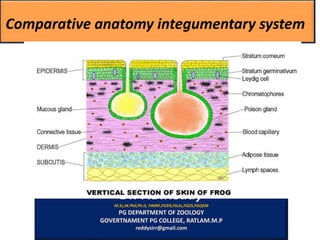
- 14. 4. Amphibia: The epidermis is multilayered. The stratum corneum made of flattened, highly keratinized cells. Such a dead layer appears first in amphibians. The stratum corneum is an adaptation to terrestrial life. It not only protects the body but prevents any excessive loss of moisture. In ecdysis, the stratum corneum is cast off in fragments or as a whole in some. The dermis is relatively thin, it is made of two layers, an upper loose stratum spongiosum and a lower dense and compact stratum compactum. Connective tissue fibers run both vertically and horizontally. There are two kinds of glands, they are multicellular mucous glands and poison glands in the dermis, but they are derivatives of the epidermis. The mucous glands produce mucus which not only forms a slimy protective covering but also helps in respiration. The poison glands found in toads as parotid glands produce a mild but unpleasant poison which is protective, keep the enemies away. In the upper part of the dermis are chromatophores which have black melanophores and yellow lipophores, these produce the colour of the skin.
- 15. The ability of the skin for changing colour to blend with the environment is well developed. Skin of labyrinthodontia, the stem Amphibia had a armour of dermal scales. Bony dermal scales are found embedded in the skin of some Gymnophiona (Apoda) and a few tropical toads. These scales are absent in modern Amphibia. The skin is sensitive to light in amphibians, especially in cave-dwelling forms. It is an important organ of respiration, and also enables the frog to respire under water for long periods, during hibernation or aestivation it is the only organ of respiration. The skin is loose being attached to muscles only at certain places by connective tissue septa which mark the boundaries of subcutaneous lymph spaces.
- 16. Reptilia: The integument is thick and dry. It prevents any loss of water. It has almost no glands, this is an adaptation to prevent evaporation of water. The epidermis has a well-developed stratum corneum and well adapted to a terrestrial life. The epidermis produces horny scales. Scales are shed periodically in fragments or cast in a single slough, as in snakes and some lizards. The scales often form spines or crests. Below the epidermal scales are dermal bony plates. These are retained for life and are not shed off.
- 17. The dermis is thick and has an upper loose connective tissue layer and a lower layer tella subjunctiva and separating the two is a horizontal layer of fibrous connective tissue. The upper layer has an abundance of chromatophores in snakes and lizards like fishes and amphibians. Many lizards and snakes have elaborate colour patterns, for concealment or as warning colours. In Calotes, the chromatophores have no nerve fibers, they are controlled by hormones of the posterior lobe of the pituitary. In Chameleon, chromatophores are controlled by the autonomic nervous system. Reptiles essentially lack skin glands. Many lizards have glands near the cloaca. Musk glands in the throat and cloacal opening of crocodilians function during courtship. Generation glands found recently are associated with periodic shedding of the skin.
- 18. 6. Aves: The integument is thin, loose, dry and devoid of glands except a uropygial gland at the base of the tail whose secreted oil is used for preening the feathers, especially in aquatic birds. The stratified epidermis is delicate, except on shanks and feet where it is thick and forms epidermal scales. The claws, spurs and horny sheaths of beaks are also the modifications of stratum corneum of epidermis. Claws and beaks are variously modified in birds according to habitat. The rest of the body has a protective covering of epidermal feathers which are evolved from epidermal scales. Feathers protect and insulate the body, i.e, keep the body warm.
- 19. The dermis is thin and has interlacing connective tissue fibres, abundant muscle fibres for moving feathers, blood vessels and nerves. The dermis forms an upper vascular and spongy layer and a lower compact layer. The dermis also contains fat cells. The skin has no chromatophores. Pigment found in melanocytes migrates into feathers and scales. Colour patterns of birds are vivid; They are for concealment, recognition, and sexual stimulation. The colours are mainly produced by reflection and refraction of light from surface of feathers.
- 20. 7. Mammalia: The skin (Fig. 41.19) is elastic and waterproof and is much thicker than in other vertebrates, especially the dermis is very thick and tough and is used for making leather. The epidermis is thickest in mammals and is differentiated into five layers- stratum corneum, stratum lucidum, stratum granulosum, stratum spinosum and stratum germinativum or Malpighian layer.
- 21. The outer layer of stratum corneum containing keratin, its cells lose their nuclei, but the cells are not dead as believed before. They secrete several hormones, one of which represses the mitotic activities of the Malpighian layer. In places of friction, such as soles and palms, the stratum corneum is very thick. Stratum corneum is variously modified in various mammals to form epidermal scales, bristles, hairs, claws, nails, hoofs and horns etc., Below the stratum corneum is a refractive stratum lucidum in certain regions only. The stratum lucidum is now known as a barrier layer. It prevents passage of substances into or out of the body. Stratum lucidum contains a chemical known as eleidin. Keratohyalin and eleidin are intermediate products in the formation of keratin. Below this is a stratum granulosum which is having darkly-staining granules of keratohyalin. Below the stratum granulosum is a stratum spinosum whose cells are held together by spiny intercellular bridges, each bridge has two arms in close contact, one arm arising from each cell. Lastly there is a stratum germinativum or Malpighian layer which rests on a thin basement membrane. The Malpighian layer forms new cells continuously which move towards the surface and become flat and keratinised till the stratum corneum has flat, cornified cells made only of keratin. This layer is sloughed off continuously and replaced by new cells. There are no mucous glands in the epidermis of mammals. The keratin from the epidermis at ends of digits forms claws, nails
- 22. The dermis is best developed in mammals. The upper part of the dermis in contact with the epidermis is the papillary layer which is made of elastic and collagen fibers with capillaries in between and lower part of the dermis is a reticular layer having elastic and collagen fibers. In both layers there are blood vessels, nerves, smooth muscles, certain glands, tactile corpuscles, and connective tissue fibers extending in all directions. Below the dermis the subcutaneous tissue has a layer of fat cells forming adipose tissue which helps to maintain body heat. no pigment-bearing chromatophores in mammals. In man some branching dendritic cells or melanoblasts lie between the epidermis and dermis, they contain pigment. The epidermis forms hairs, sudorific glands, sebaceous glands and mammary glands.
- 23. Sebaceous glands are out pushings of the wall of hair follicle and produce an oily substance which keeps the hair supple and prevents its wetting in water. It also lubricates the skin. In the dermis are present coiled sudorific or sweat glands, which occur all over except lips and glans penis. Mammary glands are modified sebaceous glands, but in monotremes they are modified sudorific glands. They are functional only in females for producing milk for the young. Mucous glands are not found in mammals.