SlideShare a Scribd company logo
1 of 11
Download to read offline
Section VII – Nuclear Radiology


                                               Figure 1A


                                                Figure 1B


269. A 21-year-old man presented one and one half years after chemotherapy and radiation therapy for nodular
     scerosing Hodgkin’s Disease involving the mediastinum. He underwent a CT scan of the chest (Figure 1A).
     On the same day as the follow-up CT examination, the patient also underwent positron emission tomography
     (PET) with F-18 fluorodeoxyglucose (FDG) (Figure 1B). Which one of the following is the MOST likely
     diagnosis?
      A. Recurrent Hodgkin’s disease
      B.   Post-therapy fibrosis
      C. Transformation to high-grade non-Hodgkin’s lymphoma
      D. Sarcoidosis
      E.   Rebound thymic hyperplasia

                                   Diagnostic In-Training Exam 2003                                        1
Section VII – Nuclear Radiology
Question #269
Findings: The CT scan of the chest at the time of presentation (Figure 1A) demonstrates a small soft tissue mass
in the anterior mediastinum in the midline, anterior to the great vessels. The repeat CT obtained 15 months later
(Figure 1B) appears essentially unchanged, with a persistent soft tissue mass in the anterior mediastinum. The FDG
PET scan performed at the time of the follow-up CT (Figure 1C) is normal, without evidence of lymphadenopathy
in the neck, chest, abdomen, or pelvis. In particular, there is no increased FDG uptake in the region of the anterior
mediastinal mass noted on the CT studies.

Rationales:
A) Incorrect. In the case of Hodgkin’s disease resistant to chemotherapy, persistent and/or more extensive
   lymphadenopathy within the chest would be anticipated, as well as possible other new sites of involvement.
   None of these findings are evident in the current case, in which the PET scan is normal.
B) Correct. A residual soft tissue mass on CT secondary to post-treatment fibrosis is the most likely explanation
   for the findings in this case. FDG PET imaging is more specific than CT for the detection of recurrence in
   lymphoma. The absence of increased FDG uptake in the patient’s persistent anterior mediastinal mass is strong
   evidence against the presence of residual or recurrent active disease
C) Incorrect. De-differentiation into anaplastic lymphoma is not common in Hodgkin’s disease in young adults,
   and represents a far less likely explanation for the findings in this case than post-treatment fibrosis and scarring.
D) Incorrect. The findings in this case are not suggestive of active sarcoidosis. Like other forms of granulomatous
   disease, sarcoidosis is a potential cause for a false positive FDG PET scan, not a false negative study. Therefore,
   if the mediastinal mass on the CT studies was due to sarcoidosis, then a focal area of increased FDG uptake in
   this region would be expected. Furthermore, the distribution of adenopathy on the CT studies is not typical
   for sarcoidosis, which usually involves the paratracheal and hilar regions, and may affect the pulmonary
   parenchyma.
E) Incorrect. Thymic hypertrophy can occur following treatment for lymphoma of the chest, particularly in young
   adult patients. The location of the CT findings is reasonable for thymic hypertrophy, although the configuration
   of the soft tissue mass is not classic in appearance. Furthermore, in such cases there is commonly mild to
   moderate increased FDG uptake within the thymus gland, a finding not present in this case. Thus, while not
   excluded, thymic hypertrophy is a less likely etiology for the findings in this case than post-treatment fibrosis
   and scarring.

Citations:
Jerusalem G, et. al.: Blood 1999;94:429-433.


2                                       American College of Radiology
Section VII – Nuclear Radiology


                                                    Figure 2


270. A 44-year-old woman underwent I-131 whole body imaging, 7 days following administration of a therapeutic
     dose of I-131 sodium iodide for a well-differentiated thyroid carcinoma (Figure 2). Based upon the appear-
     ance of the scan, which one of the following is CORRECT?
      A. The tumor most likely is a follicular carcinoma
      B.   The patient has diffuse hepatic metastases
      C. The therapeutic I-131 dose should have been 30 mCi.
      D. The patient is a candidate for adjunct chemotherapy
      E.   A follow-up whole body I-131 scan should be obtained in 3 years


                                   Diagnostic In-Training Exam 2003                                          3
Section VII – Nuclear Radiology
Question #270
Findings: Post-therapy whole body I-131 images demonstrate multiple foci of functioning thyroid tissue in the
thyroid bed, as well as an additional focus located more superiorly in the neck, to the left of midline. Additional
foci of uptake consistent with skeletal metastases are noted in the region of the right shoulder, both proximal
femora (right > left) and in the mid-thoracic and lower lumbar spine. Small foci of abnormal tracer uptake are
noted in the soft tissue superolateral to the right hip and in the lower abdomen, to the right of midline posteriorly.
These latter lesions could also represent additional skeletal metastases.

Rationales:
A) Correct. The patient has evidence of widely metastatic well-differentiated thyroid carcinoma, involving the
   thyroid bed, cervical lymph nodes, and skeleton. Statistically, the most likely histologic type to produce these
   findings is follicular carcinoma. Papillary and mixed papillary-follicular carcinoma is more common, but less
   commonly metastasizes to the skeleton. Anaplastic and medullary carcinoma are less well differentiated, and
   are unlikely to accumulate I-131.
B) Incorrect. The scan demonstrates mild diffuse hepatic uptake. This finding is typically seen on post-therapy
   imaging, especially in patients with significant amounts of residual functioning thyroid tissue present. It is
   most often secondary to hepatic uptake of iodinated thyroid hormone, a normal metabolic process. Hepatic
   metastases are uncommon in well-differentiated thyroid carcinoma, and when present, are more likely to
   demonstrate an irregular or focal distribution, which is not evident in this case.
C) Incorrect. The patient has definite evidence of skeletal metastases, indicative of advanced Stage IV disease.
   This study is not consistent with Stage I disease.
D) Incorrect. Most authorities agree that patients with well-differentiated thyroid carcinoma and evidence of
   residual or recurrent disease should be treated with I-131 sodium iodide as long as there is evidence of I-131
   uptake by the lesions, which indicates the potential for cure or at least significant reduction in the extent of
   disease. This recommendation is particularly true in patients with widespread disease, as in the current case.
   I-131 is not only efficacious in such patients, but is also associated with significantly lower morbidity than
   external beam radiotherapy. I-131 therapy can also be efficacious in patients with well-differentiated thyroid
   carcinoma and negative diagnostic I-131 whole body scans, but elevated serum thyroglobulin levels. In such
   patients, I-131 uptake by metastases may be evident only on post-therapy images. External beam radiotherapy
   is appropriately reserved for patients whose lesions do not actively take up I-131, who have undifferentiated
   histologic types of thyroid cancer or who demonstrate evidence of de-differentiation of the tumor.
E) Incorrect. In general, patients are maintained on thyroid hormone replacement, followed up with repeat
   diagnostic whole body I-131 scans annually and monitored by serum thyroglobulin levels between scans.
   Treating no more frequently than annually is usually sufficient for disease control, and reduces the likelihood
   of hematologic or pulmonary toxicity. In cases in which there is clinical evidence of rapidly progressive disease,
   the interval between scans can be reduced to 6 months. In a patient with widespread metastases, such as the
   present case, a three-year follow-up interval is far too long, and would be inappropriate, potentially permitting
   significant disease progression.

Citations:
Mettler FA, Jr. and Guiberteau MJ: Essentials of Nuclear Medicine Imaging, 4th ed., W. B. Saunders Co.,
  Philadelphia, 1998, pp. 121-125.


4                                       American College of Radiology
Section VII – Nuclear Radiology


                                                  Figure 3A


                                                  Figure 3B

271. A 20-year-old woman had massive intracranial bleeding during surgery for resection of a craniopharyngioma,
     and is now comatose. Radionuclide flow studies and delayed brain imaging using Tc-99m glucoheptonate
     were performed following surgery (Figures 3A and 3B) and repeated the following day (Figures 3C and 3D).
     Based upon these images, which one of the following is CORRECT?
      A. Both studies demonstrate findings consistent with brain death
      B.   Neither study demonstrates findings consistent with brain death
      C. Radionuclide findings of brain death are present only on the second study
      D. Both studies are indeterminate for the presence of brain death
      E.   Follow-up magnetic resonance imaging is necessary to confirm the presence of brain death

                                  Diagnostic In-Training Exam 2003                                            5
Section VII – Nuclear Radiology


                  Figure 3C


                  Figure 3D


6          American College of Radiology
Section VII – Nuclear Radiology
Question #271
Findings: The first study (Figures 3A and 3B) demonstrates severely reduced intracerebral perfusion, with no
visualization of the intracerebral vessels and only questionable visualization of the sagittal sinus on the flow study.
However, the delayed static images demonstrate definite visualization of the sagittal and both transverse sinuses.
The repeat study performed the following day (Figures 3C and 3D) demonstrates interval deterioration in
cerebral perfusion. The flow study again demonstrates no definite evidence of intracerebral perfusion, now without
visualization of the intracerebral vessels or the dural sinuses. A “hot nose” sign is also present, another indirect
indication of severely reduced perfusion of the internal carotid circulation as compared to the external carotid
circulation. A focal area of increased uptake is noted in the posterior scalp region on the static images, most likely
secondary to inflammation and dependent edema in this region. There has been interval disappearance of sagittal
and transverse sinus visualization, with only questionable faint activity noted in the region of the posterior aspect
of the sagittal sinus.

Rationales:
A) Incorrect. While cerebral perfusion is significantly impaired on both studies, the visualization of the dural
   sinuses on the first study is not consistent with the radionuclide diagnosis of brain death.
B) Incorrect. As noted above, the first study is not diagnostic of brain death. However, the second study
   demonstrates no evidence of intracerebral vessel visualization nor definite visualization of the dural sinuses,
   and demonstrates the corroborative “hot nose” sign. Thus, the 2nd study is consistent with brain death.
C) Correct. As discussed in A and B above, the second study is consistent with brain death, representing an
   interval change compared to the study performed on the previous day.
D) Incorrect. The second study is consistent with brain death. The findings of the radionuclide brain scan are
   correlated with other clinical data (physical findings, EEG, etc.) in order to make a final determination of
   clinical brain death.
E) Incorrect. Confirmation of brain death is done by correlating the brain scan findings with other clinical data,
   primarily physical findings, and the EEG. Magnetic resonance imaging is not a sensitive indicator of brain
   death.

Citations:
Sandler MP, Coleman RE, Wackers FJTh, Patton JA, Gottschalk A and Hoffer PB (eds.): Diagnostic Nuclear
  Medicine, 3rd Ed., Williams & Wilkins, Baltimore, 1996, pp. 1091-1097.


                                    Diagnostic In-Training Exam 2003                                                  7
Section VII – Nuclear Radiology


                                                   Figure 4A


                                                   Figure 4B


272. A 60-year-old woman presented with hypercalcemia and a history of a renal calculus. Tc-99m sestamibi
     images of the neck and mediastinum were obtained at 30 minutes and 3 hours post-injection, including
     (clockwise from upper left) anterior neck, anterior neck with “cold” sternal notch marker, anterior neck and
     chest, pinhole neck, left anterior oblique neck and chest and right anterior oblique neck and chest (Figures 4A
     and 4B). Which one of the following is the LEAST likely etiology for the findings in this case?
      A. Thyroid adenoma
      B.   Parathyroid hyperplasia
      C. Parathyroid adenoma
      D. Papillary carcinoma of the thyroid
      E.   Colloid goiter


8                                      American College of Radiology
Section VII – Nuclear Radiology
Question #272
Findings: Tc-99m sestamibi images of the neck and mediastinum demonstrate normal uptake by the thyroid and
salivary glands on the initial 30-minute post-injection images (Figure 4A). Normal left ventricular myocardial
activity and hepatic activity are also evident. There is a superimposed focal area of increased tracer uptake noted
overlying the left lower pole of the thyroid. The 3 hour delayed images (Figure 4B) demonstrate substantial washout
of activity from the thyroid, with a persistent focus of increased uptake overlying the left lower pole region.

Rationales:
A) Incorrect. A focal thyroid adenoma is a common cause of a false positive radionuclide parathyroid scan. In this
   clinical setting, a parathyroid adenoma is more likely, but a thyroid adenoma cannot be excluded based on the
   imaging findings in this case alone.
B) Incorrect. Parathyroid hyperplasia typically produces multiple foci of less striking increased tracer uptake in the
   neck, in the expected locations of the parathyroid glands, with the intensity of uptake usually less than is present
   in this case. Nonetheless, it is definitely in the differential diagnosis for focally increased sestamibi uptake in the
   neck, especially in a patient with clinical evidence of hyperparathyroidism.
C) Incorrect. The findings in this case are classic for a focal parathyroid adenoma, which is in fact the most likely
   diagnosis in this case. A solitary parathyroid adenoma is found to be the cause of primary hyperparathyroidism
   in approximately 85% of cases.
D) Incorrect. Papillary carcinoma of the thyroid is another cause for false positive Tc-99m sestamibi parathyroid
   scintigraphy studies. The clinical setting of the case again is more suggestive of parathyroid adenoma, but a
   thyroid malignancy cannot entirely be excluded.
E) Correct. Of all the choices indicated, colloid goiter is the least likely cause for the findings in this case.
   A colloid goiter is typically a multifocal process, associated with thyroid enlargement and inhomogeneous
   uptake on early images. In addition, colloid goiter is unlikely to be associated with focal area(s) of significantly
   increased sestamibi uptake, as is present in this case.

Citations:
Sandler MP, Coleman RE, Wackers FJTh, Patton JA, Gottschalk A and Hoffer PB (eds.): Diagnostic Nuclear
  Medicine, 3rd Ed., Williams & Wilkins, Baltimore, 1996, pp. 998-1011.


                                    Diagnostic In-Training Exam 2003                                                   9
Section VII – Nuclear Radiology


                                                  Figure 5


273. A 55-year-old woman presented with abdominal pain and weight loss. You are presented with 48-hour
     anterior and posterior whole body In-111 pentetreotide images (Figure 5). Which one of the following is the
     MOST likely diagnosis?
      A. Pheochromocytoma
      B.   Carcinoid tumor of the small bowel
      C. Non-Hodgkin’s lymphoma
      D. Islet cell carcinoma
      E.   Adenocarcinoma of the colon


10                                    American College of Radiology
Section VII – Nuclear Radiology
Question #273
Findings: Forty-eight hour whole body In-111 pentetreotide images demonstrate multiple focal areas of increased
tracer uptake within both lobes of the liver, consistent with multiple focal neuroendocrine tumor hepatic metastases.
There is also a large focal area of increased uptake in the epigastric region, just to the right of midline, in the
expected location of the body of the pancreas. No other focal lesions are demonstrated in the abdomen or in the
chest.

Rationales:
A) Incorrect. Pheochromocytoma is a neoplasm that may demonstrate increased uptake of octreotide analogs,
   and the focal lesion in the right upper quadrant could conceivably represent a right adrenal lesion. However,
   widespread hepatic metastases are not as common as in carcinoid, islet cell carcinoma, or small cell lung
   carcinoma, and pheochromocytoma is therefore a less likely etiology for the findings demonstrated.
B) Incorrect. Carcinoid is a highly octreotide-avid neoplasm in general, and often produces multiple hepatic
   metastases as well. The primary lesion is most often located within the small bowel or lung, neither of which
   areas demonstrate focal lesions in this case. Therefore, while not unlikely, this diagnosis is less likely than an
   islet cell pancreatic tumor.
C) Incorrect. Again, small cell lung carcinomas may demonstrate neuroendocrine features, and may have significant
   somatostatin receptor activity. However, the pattern of involvement in this case within the liver and epigastric
   region, without a focal pulmonary or mediastinal lesion, make small cell lung carcinoma less likely than islet cell
   tumor or carcinoid.
D) Correct. Islet cell carcinoma is the most likely diagnosis in this case. The focal lesion in the epigastric region
   is secondary to uptake within the primary neoplasm in the body of the pancreas. Multiple hepatic metastases
   containing somatostatin receptor activity are also common in this entity. There is no evidence of focal lesions
   within the lungs or mediastinum to suggest small cell carcinoma of the lung and no focal extrahepatic abdominal
   lesions in the region of the small bowel to suggest a primary lesion in that location.
E) Incorrect. Medullary carcinoma of the thyroid is often associated with somatostatin receptor activity, but less so
   than carcinoid or islet cell pancreatic tumors. There is also no focal increased tracer uptake in the neck to suggest
   a primary thyroid lesion, and widespread hepatic metastases are less common than in carcinoid or islet cell
   tumor.

Citations:
Mettler FA, Jr. and Guiberteau MJ: Essentials of Nuclear Medicine Imaging, 4th ed., W. B. Saunders Co.,
  Philadelphia, 1998, pp. 379-382.


                                    Diagnostic In-Training Exam 2003                                                11

More Related Content

What's hot

23205004
2320500423205004
23205004radgirl
 
23204980
2320498023204980
23204980radgirl
 
23204955
2320495523204955
23204955radgirl
 
23204943
2320494323204943
23204943radgirl
 
23204992
2320499223204992
23204992radgirl
 
23204916
2320491623204916
23204916radgirl
 
Radiology most important signs sample questions based on neet pg , usmle, pla...
Radiology most important signs sample questions based on neet pg , usmle, pla...Radiology most important signs sample questions based on neet pg , usmle, pla...
Radiology most important signs sample questions based on neet pg , usmle, pla...Medico Apps
 
Previous year question on bone cyst based on neet pg, usmle, plab and fmge or...
Previous year question on bone cyst based on neet pg, usmle, plab and fmge or...Previous year question on bone cyst based on neet pg, usmle, plab and fmge or...
Previous year question on bone cyst based on neet pg, usmle, plab and fmge or...Medico Apps
 
23204946
2320494623204946
23204946radgirl
 
Paediatric abdominal masses
Paediatric abdominal massesPaediatric abdominal masses
Paediatric abdominal massesairwave12
 
Cystic fibrosis sample questions based on neet pg , usmle, plab and fmge patt...
Cystic fibrosis sample questions based on neet pg , usmle, plab and fmge patt...Cystic fibrosis sample questions based on neet pg , usmle, plab and fmge patt...
Cystic fibrosis sample questions based on neet pg , usmle, plab and fmge patt...Medico Apps
 
23204907
2320490723204907
23204907radgirl
 
Conjunctival MALT Lymphoma
Conjunctival MALT LymphomaConjunctival MALT Lymphoma
Conjunctival MALT LymphomaRyan Alfonso
 
23204922
2320492223204922
23204922radgirl
 
NERRS CHEST answers 2012
NERRS CHEST answers 2012NERRS CHEST answers 2012
NERRS CHEST answers 2012NERRS
 
Ewings sarcoma_UTSAV
Ewings sarcoma_UTSAVEwings sarcoma_UTSAV
Ewings sarcoma_UTSAVUtsav Agrawal
 
Externalbeam rt in ews3.12.20 - frida yseminar-finallll
Externalbeam rt in ews3.12.20    - frida yseminar-finallllExternalbeam rt in ews3.12.20    - frida yseminar-finallll
Externalbeam rt in ews3.12.20 - frida yseminar-finallllPRARABDH95
 
Neuroblastoma imaging
Neuroblastoma imagingNeuroblastoma imaging
Neuroblastoma imagingPrasunDas31
 

What's hot (20)

23205004
2320500423205004
23205004
 
23204980
2320498023204980
23204980
 
23204955
2320495523204955
23204955
 
23204943
2320494323204943
23204943
 
23204992
2320499223204992
23204992
 
23204916
2320491623204916
23204916
 
Radiology most important signs sample questions based on neet pg , usmle, pla...
Radiology most important signs sample questions based on neet pg , usmle, pla...Radiology most important signs sample questions based on neet pg , usmle, pla...
Radiology most important signs sample questions based on neet pg , usmle, pla...
 
Previous year question on bone cyst based on neet pg, usmle, plab and fmge or...
Previous year question on bone cyst based on neet pg, usmle, plab and fmge or...Previous year question on bone cyst based on neet pg, usmle, plab and fmge or...
Previous year question on bone cyst based on neet pg, usmle, plab and fmge or...
 
23204946
2320494623204946
23204946
 
Paediatric abdominal masses
Paediatric abdominal massesPaediatric abdominal masses
Paediatric abdominal masses
 
Cystic fibrosis sample questions based on neet pg , usmle, plab and fmge patt...
Cystic fibrosis sample questions based on neet pg , usmle, plab and fmge patt...Cystic fibrosis sample questions based on neet pg , usmle, plab and fmge patt...
Cystic fibrosis sample questions based on neet pg , usmle, plab and fmge patt...
 
23204907
2320490723204907
23204907
 
Ewing Sarcoma
Ewing SarcomaEwing Sarcoma
Ewing Sarcoma
 
Conjunctival MALT Lymphoma
Conjunctival MALT LymphomaConjunctival MALT Lymphoma
Conjunctival MALT Lymphoma
 
23204922
2320492223204922
23204922
 
NERRS CHEST answers 2012
NERRS CHEST answers 2012NERRS CHEST answers 2012
NERRS CHEST answers 2012
 
Ewings sarcoma_UTSAV
Ewings sarcoma_UTSAVEwings sarcoma_UTSAV
Ewings sarcoma_UTSAV
 
Ewings sarcoma
Ewings sarcomaEwings sarcoma
Ewings sarcoma
 
Externalbeam rt in ews3.12.20 - frida yseminar-finallll
Externalbeam rt in ews3.12.20    - frida yseminar-finallllExternalbeam rt in ews3.12.20    - frida yseminar-finallll
Externalbeam rt in ews3.12.20 - frida yseminar-finallll
 
Neuroblastoma imaging
Neuroblastoma imagingNeuroblastoma imaging
Neuroblastoma imaging
 

Viewers also liked

6 Radionuclide and Hybrid Imaging Dr. Muhammad Bin Zulfiqar Grainger and Allison
6 Radionuclide and Hybrid Imaging Dr. Muhammad Bin Zulfiqar Grainger and Allison6 Radionuclide and Hybrid Imaging Dr. Muhammad Bin Zulfiqar Grainger and Allison
6 Radionuclide and Hybrid Imaging Dr. Muhammad Bin Zulfiqar Grainger and AllisonDr. Muhammad Bin Zulfiqar
 
Donatas Bedulskis: „PR 2.0 spindesys ir skurdas“
Donatas Bedulskis: „PR 2.0 spindesys ir skurdas“Donatas Bedulskis: „PR 2.0 spindesys ir skurdas“
Donatas Bedulskis: „PR 2.0 spindesys ir skurdas“bestmarketing
 
記者,你為什麼不反叛?
記者,你為什麼不反叛?記者,你為什麼不反叛?
記者,你為什麼不反叛?bibliobank
 
2008 Directors Reportpdf
2008 Directors Reportpdf2008 Directors Reportpdf
2008 Directors Reportpdfandy biggin
 
23205011
2320501123205011
23205011radgirl
 
Ten steps to plan a presentation
Ten steps to plan a presentationTen steps to plan a presentation
Ten steps to plan a presentationLee Bandy
 
DiffCalculus: September 17, 2012
DiffCalculus: September 17, 2012DiffCalculus: September 17, 2012
DiffCalculus: September 17, 2012Carlos Vázquez
 
Eventum Premo Projects for Auto
Eventum Premo Projects for Auto Eventum Premo Projects for Auto
Eventum Premo Projects for Auto Eventum Premo
 
Trade with China. AsstrA Presentation 2009
Trade with China. AsstrA Presentation 2009Trade with China. AsstrA Presentation 2009
Trade with China. AsstrA Presentation 2009Pavel Red'ko
 
مكتبات الجمعيات الأهلية و المؤسسات الخاصة بمحافظة الإسكندرية
مكتبات الجمعيات الأهلية و المؤسسات الخاصة بمحافظة الإسكندريةمكتبات الجمعيات الأهلية و المؤسسات الخاصة بمحافظة الإسكندرية
مكتبات الجمعيات الأهلية و المؤسسات الخاصة بمحافظة الإسكندريةEssam Obaid
 
Chapter 2 slideshow
Chapter 2 slideshowChapter 2 slideshow
Chapter 2 slideshowmarshalls1
 
Achieving Mastery Through Gamification
Achieving Mastery Through GamificationAchieving Mastery Through Gamification
Achieving Mastery Through GamificationPete Baikins
 

Viewers also liked (20)

6 Radionuclide and Hybrid Imaging Dr. Muhammad Bin Zulfiqar Grainger and Allison
6 Radionuclide and Hybrid Imaging Dr. Muhammad Bin Zulfiqar Grainger and Allison6 Radionuclide and Hybrid Imaging Dr. Muhammad Bin Zulfiqar Grainger and Allison
6 Radionuclide and Hybrid Imaging Dr. Muhammad Bin Zulfiqar Grainger and Allison
 
Donatas Bedulskis: „PR 2.0 spindesys ir skurdas“
Donatas Bedulskis: „PR 2.0 spindesys ir skurdas“Donatas Bedulskis: „PR 2.0 spindesys ir skurdas“
Donatas Bedulskis: „PR 2.0 spindesys ir skurdas“
 
記者,你為什麼不反叛?
記者,你為什麼不反叛?記者,你為什麼不反叛?
記者,你為什麼不反叛?
 
#Curation Restart Education Project . Top 10 #edtech20 tools who will change ...
#Curation Restart Education Project . Top 10 #edtech20 tools who will change ...#Curation Restart Education Project . Top 10 #edtech20 tools who will change ...
#Curation Restart Education Project . Top 10 #edtech20 tools who will change ...
 
2008 Directors Reportpdf
2008 Directors Reportpdf2008 Directors Reportpdf
2008 Directors Reportpdf
 
Pasti
PastiPasti
Pasti
 
Signe
SigneSigne
Signe
 
23205011
2320501123205011
23205011
 
Ten steps to plan a presentation
Ten steps to plan a presentationTen steps to plan a presentation
Ten steps to plan a presentation
 
DiffCalculus: September 17, 2012
DiffCalculus: September 17, 2012DiffCalculus: September 17, 2012
DiffCalculus: September 17, 2012
 
Magic Of Colours
Magic Of ColoursMagic Of Colours
Magic Of Colours
 
Eventum Premo Projects for Auto
Eventum Premo Projects for Auto Eventum Premo Projects for Auto
Eventum Premo Projects for Auto
 
Trade with China. AsstrA Presentation 2009
Trade with China. AsstrA Presentation 2009Trade with China. AsstrA Presentation 2009
Trade with China. AsstrA Presentation 2009
 
C sharp
C sharpC sharp
C sharp
 
مكتبات الجمعيات الأهلية و المؤسسات الخاصة بمحافظة الإسكندرية
مكتبات الجمعيات الأهلية و المؤسسات الخاصة بمحافظة الإسكندريةمكتبات الجمعيات الأهلية و المؤسسات الخاصة بمحافظة الإسكندرية
مكتبات الجمعيات الأهلية و المؤسسات الخاصة بمحافظة الإسكندرية
 
Chapter 2 slideshow
Chapter 2 slideshowChapter 2 slideshow
Chapter 2 slideshow
 
Achieving Mastery Through Gamification
Achieving Mastery Through GamificationAchieving Mastery Through Gamification
Achieving Mastery Through Gamification
 
U R A Brand! 2
U R A Brand! 2U R A Brand! 2
U R A Brand! 2
 
Ageing consumers
Ageing consumersAgeing consumers
Ageing consumers
 
Bottom Up Marketing 到第一線找行銷點子
Bottom Up Marketing 到第一線找行銷點子Bottom Up Marketing 到第一線找行銷點子
Bottom Up Marketing 到第一線找行銷點子
 

Similar to 23204958

Disseminated lung metastases in thyroid cancer
Disseminated lung metastases in thyroid cancerDisseminated lung metastases in thyroid cancer
Disseminated lung metastases in thyroid cancerJacoblix
 
23205016
2320501623205016
23205016radgirl
 
Positron Emission Tomography And Assessment Of Cancer Therapy
Positron Emission Tomography And Assessment Of Cancer TherapyPositron Emission Tomography And Assessment Of Cancer Therapy
Positron Emission Tomography And Assessment Of Cancer Therapyfondas vakalis
 
Intramedullary Diffuse Astrocytoma of The Spinal Cord in a 3-Year-Old Patient...
Intramedullary Diffuse Astrocytoma of The Spinal Cord in a 3-Year-Old Patient...Intramedullary Diffuse Astrocytoma of The Spinal Cord in a 3-Year-Old Patient...
Intramedullary Diffuse Astrocytoma of The Spinal Cord in a 3-Year-Old Patient...CrimsonPublishersTNN
 
Primary Hemangiopericytoma in Parietal Bone: Literature Review and Case Report
Primary Hemangiopericytoma in Parietal Bone: Literature Review and Case ReportPrimary Hemangiopericytoma in Parietal Bone: Literature Review and Case Report
Primary Hemangiopericytoma in Parietal Bone: Literature Review and Case Reportkomalicarol
 
Primary Small Cell Neuroendocrine Carcinoma of the Petrous Apex: A Report of ...
Primary Small Cell Neuroendocrine Carcinoma of the Petrous Apex: A Report of ...Primary Small Cell Neuroendocrine Carcinoma of the Petrous Apex: A Report of ...
Primary Small Cell Neuroendocrine Carcinoma of the Petrous Apex: A Report of ...IJBNT Journal
 
Pediatric Meningiomas- A Case Report and Review of the Literature_ Crimson Pu...
Pediatric Meningiomas- A Case Report and Review of the Literature_ Crimson Pu...Pediatric Meningiomas- A Case Report and Review of the Literature_ Crimson Pu...
Pediatric Meningiomas- A Case Report and Review of the Literature_ Crimson Pu...Crimsonpublisherssmoaj
 
Desmoplastic Ganglioglioma. Epidemiology and Molecular Pathogenesis
Desmoplastic Ganglioglioma. Epidemiology and Molecular PathogenesisDesmoplastic Ganglioglioma. Epidemiology and Molecular Pathogenesis
Desmoplastic Ganglioglioma. Epidemiology and Molecular PathogenesisNeuroAcademy
 
Primary Epithelioid Hemangioendothelioma of the Kidney Pelvis
Primary Epithelioid Hemangioendothelioma of the Kidney PelvisPrimary Epithelioid Hemangioendothelioma of the Kidney Pelvis
Primary Epithelioid Hemangioendothelioma of the Kidney Pelvissemualkaira
 
Primary Epithelioid Hemangioendothelioma of the Kidney Pelvis
Primary Epithelioid Hemangioendothelioma of the Kidney PelvisPrimary Epithelioid Hemangioendothelioma of the Kidney Pelvis
Primary Epithelioid Hemangioendothelioma of the Kidney Pelvissuppubs1pubs1
 
Primary Epithelioid Hemangioendothelioma of the Kidney Pelvis
Primary Epithelioid Hemangioendothelioma of the Kidney PelvisPrimary Epithelioid Hemangioendothelioma of the Kidney Pelvis
Primary Epithelioid Hemangioendothelioma of the Kidney PelvisJohnJulie1
 
Primary Epithelioid Hemangioendothelioma of the Kidney Pelvis
Primary Epithelioid Hemangioendothelioma of the Kidney PelvisPrimary Epithelioid Hemangioendothelioma of the Kidney Pelvis
Primary Epithelioid Hemangioendothelioma of the Kidney Pelvissuppubs1pubs1
 
Sonodynamic and photodynamic therapy in advanced breast carcinoma: a report o...
Sonodynamic and photodynamic therapy in advanced breast carcinoma: a report o...Sonodynamic and photodynamic therapy in advanced breast carcinoma: a report o...
Sonodynamic and photodynamic therapy in advanced breast carcinoma: a report o...Renkang Hospital
 
Coma Due to Wernicke Encephalopathy During Neoadjuvant Chemotherapy for Breas...
Coma Due to Wernicke Encephalopathy During Neoadjuvant Chemotherapy for Breas...Coma Due to Wernicke Encephalopathy During Neoadjuvant Chemotherapy for Breas...
Coma Due to Wernicke Encephalopathy During Neoadjuvant Chemotherapy for Breas...semualkaira
 
Annals of Clinical and Medical Case Reports - Acmcasereport
Annals of Clinical and Medical Case Reports - AcmcasereportAnnals of Clinical and Medical Case Reports - Acmcasereport
Annals of Clinical and Medical Case Reports - Acmcasereportsemualkaira
 
Papillary Glioneuronal Tumor of Third Ventricle Endoscopically Treated: Case ...
Papillary Glioneuronal Tumor of Third Ventricle Endoscopically Treated: Case ...Papillary Glioneuronal Tumor of Third Ventricle Endoscopically Treated: Case ...
Papillary Glioneuronal Tumor of Third Ventricle Endoscopically Treated: Case ...semualkaira
 

Similar to 23204958 (20)

Disseminated lung metastases in thyroid cancer
Disseminated lung metastases in thyroid cancerDisseminated lung metastases in thyroid cancer
Disseminated lung metastases in thyroid cancer
 
23205016
2320501623205016
23205016
 
PET.pptx
PET.pptxPET.pptx
PET.pptx
 
medulloblastoma
medulloblastomamedulloblastoma
medulloblastoma
 
Positron Emission Tomography And Assessment Of Cancer Therapy
Positron Emission Tomography And Assessment Of Cancer TherapyPositron Emission Tomography And Assessment Of Cancer Therapy
Positron Emission Tomography And Assessment Of Cancer Therapy
 
Intramedullary Diffuse Astrocytoma of The Spinal Cord in a 3-Year-Old Patient...
Intramedullary Diffuse Astrocytoma of The Spinal Cord in a 3-Year-Old Patient...Intramedullary Diffuse Astrocytoma of The Spinal Cord in a 3-Year-Old Patient...
Intramedullary Diffuse Astrocytoma of The Spinal Cord in a 3-Year-Old Patient...
 
Austin Journal of Radiology
Austin Journal of Radiology Austin Journal of Radiology
Austin Journal of Radiology
 
Primary Hemangiopericytoma in Parietal Bone: Literature Review and Case Report
Primary Hemangiopericytoma in Parietal Bone: Literature Review and Case ReportPrimary Hemangiopericytoma in Parietal Bone: Literature Review and Case Report
Primary Hemangiopericytoma in Parietal Bone: Literature Review and Case Report
 
Foramen magnum papilloma case report and review of the literature
Foramen magnum papilloma case report and review of the literatureForamen magnum papilloma case report and review of the literature
Foramen magnum papilloma case report and review of the literature
 
Primary Small Cell Neuroendocrine Carcinoma of the Petrous Apex: A Report of ...
Primary Small Cell Neuroendocrine Carcinoma of the Petrous Apex: A Report of ...Primary Small Cell Neuroendocrine Carcinoma of the Petrous Apex: A Report of ...
Primary Small Cell Neuroendocrine Carcinoma of the Petrous Apex: A Report of ...
 
Pediatric Meningiomas- A Case Report and Review of the Literature_ Crimson Pu...
Pediatric Meningiomas- A Case Report and Review of the Literature_ Crimson Pu...Pediatric Meningiomas- A Case Report and Review of the Literature_ Crimson Pu...
Pediatric Meningiomas- A Case Report and Review of the Literature_ Crimson Pu...
 
Desmoplastic Ganglioglioma. Epidemiology and Molecular Pathogenesis
Desmoplastic Ganglioglioma. Epidemiology and Molecular PathogenesisDesmoplastic Ganglioglioma. Epidemiology and Molecular Pathogenesis
Desmoplastic Ganglioglioma. Epidemiology and Molecular Pathogenesis
 
Primary Epithelioid Hemangioendothelioma of the Kidney Pelvis
Primary Epithelioid Hemangioendothelioma of the Kidney PelvisPrimary Epithelioid Hemangioendothelioma of the Kidney Pelvis
Primary Epithelioid Hemangioendothelioma of the Kidney Pelvis
 
Primary Epithelioid Hemangioendothelioma of the Kidney Pelvis
Primary Epithelioid Hemangioendothelioma of the Kidney PelvisPrimary Epithelioid Hemangioendothelioma of the Kidney Pelvis
Primary Epithelioid Hemangioendothelioma of the Kidney Pelvis
 
Primary Epithelioid Hemangioendothelioma of the Kidney Pelvis
Primary Epithelioid Hemangioendothelioma of the Kidney PelvisPrimary Epithelioid Hemangioendothelioma of the Kidney Pelvis
Primary Epithelioid Hemangioendothelioma of the Kidney Pelvis
 
Primary Epithelioid Hemangioendothelioma of the Kidney Pelvis
Primary Epithelioid Hemangioendothelioma of the Kidney PelvisPrimary Epithelioid Hemangioendothelioma of the Kidney Pelvis
Primary Epithelioid Hemangioendothelioma of the Kidney Pelvis
 
Sonodynamic and photodynamic therapy in advanced breast carcinoma: a report o...
Sonodynamic and photodynamic therapy in advanced breast carcinoma: a report o...Sonodynamic and photodynamic therapy in advanced breast carcinoma: a report o...
Sonodynamic and photodynamic therapy in advanced breast carcinoma: a report o...
 
Coma Due to Wernicke Encephalopathy During Neoadjuvant Chemotherapy for Breas...
Coma Due to Wernicke Encephalopathy During Neoadjuvant Chemotherapy for Breas...Coma Due to Wernicke Encephalopathy During Neoadjuvant Chemotherapy for Breas...
Coma Due to Wernicke Encephalopathy During Neoadjuvant Chemotherapy for Breas...
 
Annals of Clinical and Medical Case Reports - Acmcasereport
Annals of Clinical and Medical Case Reports - AcmcasereportAnnals of Clinical and Medical Case Reports - Acmcasereport
Annals of Clinical and Medical Case Reports - Acmcasereport
 
Papillary Glioneuronal Tumor of Third Ventricle Endoscopically Treated: Case ...
Papillary Glioneuronal Tumor of Third Ventricle Endoscopically Treated: Case ...Papillary Glioneuronal Tumor of Third Ventricle Endoscopically Treated: Case ...
Papillary Glioneuronal Tumor of Third Ventricle Endoscopically Treated: Case ...
 

More from radgirl

23205062
2320506223205062
23205062radgirl
 
23205059
2320505923205059
23205059radgirl
 
23205056
2320505623205056
23205056radgirl
 
23205042
2320504223205042
23205042radgirl
 
23205052
2320505223205052
23205052radgirl
 
23205045
2320504523205045
23205045radgirl
 
23205048
2320504823205048
23205048radgirl
 
23205039
2320503923205039
23205039radgirl
 
23205036
2320503623205036
23205036radgirl
 
23205032
2320503223205032
23205032radgirl
 
23205029
2320502923205029
23205029radgirl
 
23205025
2320502523205025
23205025radgirl
 
23204998
2320499823204998
23204998radgirl
 
23205008
2320500823205008
23205008radgirl
 
23205001
2320500123205001
23205001radgirl
 
23204995
2320499523204995
23204995radgirl
 
23204989
2320498923204989
23204989radgirl
 
23204986
2320498623204986
23204986radgirl
 
23204983
2320498323204983
23204983radgirl
 

More from radgirl (19)

23205062
2320506223205062
23205062
 
23205059
2320505923205059
23205059
 
23205056
2320505623205056
23205056
 
23205042
2320504223205042
23205042
 
23205052
2320505223205052
23205052
 
23205045
2320504523205045
23205045
 
23205048
2320504823205048
23205048
 
23205039
2320503923205039
23205039
 
23205036
2320503623205036
23205036
 
23205032
2320503223205032
23205032
 
23205029
2320502923205029
23205029
 
23205025
2320502523205025
23205025
 
23204998
2320499823204998
23204998
 
23205008
2320500823205008
23205008
 
23205001
2320500123205001
23205001
 
23204995
2320499523204995
23204995
 
23204989
2320498923204989
23204989
 
23204986
2320498623204986
23204986
 
23204983
2320498323204983
23204983
 

Recently uploaded

Call Girls Service Jaipur {9521753030} ❤️VVIP RIDDHI Call Girl in Jaipur Raja...
Call Girls Service Jaipur {9521753030} ❤️VVIP RIDDHI Call Girl in Jaipur Raja...Call Girls Service Jaipur {9521753030} ❤️VVIP RIDDHI Call Girl in Jaipur Raja...
Call Girls Service Jaipur {9521753030} ❤️VVIP RIDDHI Call Girl in Jaipur Raja...Sheetaleventcompany
 
All Time Service Available Call Girls Marine Drive 📳 9820252231 For 18+ VIP C...
All Time Service Available Call Girls Marine Drive 📳 9820252231 For 18+ VIP C...All Time Service Available Call Girls Marine Drive 📳 9820252231 For 18+ VIP C...
All Time Service Available Call Girls Marine Drive 📳 9820252231 For 18+ VIP C...Arohi Goyal
 
Russian Call Girls Service Jaipur {8445551418} ❤️PALLAVI VIP Jaipur Call Gir...
Russian Call Girls Service  Jaipur {8445551418} ❤️PALLAVI VIP Jaipur Call Gir...Russian Call Girls Service  Jaipur {8445551418} ❤️PALLAVI VIP Jaipur Call Gir...
Russian Call Girls Service Jaipur {8445551418} ❤️PALLAVI VIP Jaipur Call Gir...parulsinha
 
Call Girls Jaipur Just Call 9521753030 Top Class Call Girl Service Available
Call Girls Jaipur Just Call 9521753030 Top Class Call Girl Service AvailableCall Girls Jaipur Just Call 9521753030 Top Class Call Girl Service Available
Call Girls Jaipur Just Call 9521753030 Top Class Call Girl Service AvailableJanvi Singh
 
8980367676 Call Girls In Ahmedabad Escort Service Available 24×7 In Ahmedabad
8980367676 Call Girls In Ahmedabad Escort Service Available 24×7 In Ahmedabad8980367676 Call Girls In Ahmedabad Escort Service Available 24×7 In Ahmedabad
8980367676 Call Girls In Ahmedabad Escort Service Available 24×7 In AhmedabadGENUINE ESCORT AGENCY
 
(Low Rate RASHMI ) Rate Of Call Girls Jaipur ❣ 8445551418 ❣ Elite Models & Ce...
(Low Rate RASHMI ) Rate Of Call Girls Jaipur ❣ 8445551418 ❣ Elite Models & Ce...(Low Rate RASHMI ) Rate Of Call Girls Jaipur ❣ 8445551418 ❣ Elite Models & Ce...
(Low Rate RASHMI ) Rate Of Call Girls Jaipur ❣ 8445551418 ❣ Elite Models & Ce...parulsinha
 
Call Girls Hyderabad Just Call 8250077686 Top Class Call Girl Service Available
Call Girls Hyderabad Just Call 8250077686 Top Class Call Girl Service AvailableCall Girls Hyderabad Just Call 8250077686 Top Class Call Girl Service Available
Call Girls Hyderabad Just Call 8250077686 Top Class Call Girl Service AvailableDipal Arora
 
Call Girls Madurai Just Call 9630942363 Top Class Call Girl Service Available
Call Girls Madurai Just Call 9630942363 Top Class Call Girl Service AvailableCall Girls Madurai Just Call 9630942363 Top Class Call Girl Service Available
Call Girls Madurai Just Call 9630942363 Top Class Call Girl Service AvailableGENUINE ESCORT AGENCY
 
Coimbatore Call Girls in Coimbatore 7427069034 genuine Escort Service Girl 10...
Coimbatore Call Girls in Coimbatore 7427069034 genuine Escort Service Girl 10...Coimbatore Call Girls in Coimbatore 7427069034 genuine Escort Service Girl 10...
Coimbatore Call Girls in Coimbatore 7427069034 genuine Escort Service Girl 10...chennailover
 
Night 7k to 12k Navi Mumbai Call Girl Photo 👉 BOOK NOW 9833363713 👈 ♀️ night ...
Night 7k to 12k Navi Mumbai Call Girl Photo 👉 BOOK NOW 9833363713 👈 ♀️ night ...Night 7k to 12k Navi Mumbai Call Girl Photo 👉 BOOK NOW 9833363713 👈 ♀️ night ...
Night 7k to 12k Navi Mumbai Call Girl Photo 👉 BOOK NOW 9833363713 👈 ♀️ night ...aartirawatdelhi
 
Trichy Call Girls Book Now 9630942363 Top Class Trichy Escort Service Available
Trichy Call Girls Book Now 9630942363 Top Class Trichy Escort Service AvailableTrichy Call Girls Book Now 9630942363 Top Class Trichy Escort Service Available
Trichy Call Girls Book Now 9630942363 Top Class Trichy Escort Service AvailableGENUINE ESCORT AGENCY
 
Top Rated Hyderabad Call Girls Erragadda ⟟ 9332606886 ⟟ Call Me For Genuine ...
Top Rated  Hyderabad Call Girls Erragadda ⟟ 9332606886 ⟟ Call Me For Genuine ...Top Rated  Hyderabad Call Girls Erragadda ⟟ 9332606886 ⟟ Call Me For Genuine ...
Top Rated Hyderabad Call Girls Erragadda ⟟ 9332606886 ⟟ Call Me For Genuine ...chandars293
 
Call Girls Hosur Just Call 9630942363 Top Class Call Girl Service Available
Call Girls Hosur Just Call 9630942363 Top Class Call Girl Service AvailableCall Girls Hosur Just Call 9630942363 Top Class Call Girl Service Available
Call Girls Hosur Just Call 9630942363 Top Class Call Girl Service AvailableGENUINE ESCORT AGENCY
 
Top Quality Call Girl Service Kalyanpur 6378878445 Available Call Girls Any Time
Top Quality Call Girl Service Kalyanpur 6378878445 Available Call Girls Any TimeTop Quality Call Girl Service Kalyanpur 6378878445 Available Call Girls Any Time
Top Quality Call Girl Service Kalyanpur 6378878445 Available Call Girls Any TimeCall Girls Delhi
 
Low Rate Call Girls Bangalore {7304373326} ❤️VVIP NISHA Call Girls in Bangalo...
Low Rate Call Girls Bangalore {7304373326} ❤️VVIP NISHA Call Girls in Bangalo...Low Rate Call Girls Bangalore {7304373326} ❤️VVIP NISHA Call Girls in Bangalo...
Low Rate Call Girls Bangalore {7304373326} ❤️VVIP NISHA Call Girls in Bangalo...Sheetaleventcompany
 
Call Girls in Delhi Triveni Complex Escort Service(🔝))/WhatsApp 97111⇛47426
Call Girls in Delhi Triveni Complex Escort Service(🔝))/WhatsApp 97111⇛47426Call Girls in Delhi Triveni Complex Escort Service(🔝))/WhatsApp 97111⇛47426
Call Girls in Delhi Triveni Complex Escort Service(🔝))/WhatsApp 97111⇛47426jennyeacort
 
Call Girl In Pune 👉 Just CALL ME: 9352988975 💋 Call Out Call Both With High p...
Call Girl In Pune 👉 Just CALL ME: 9352988975 💋 Call Out Call Both With High p...Call Girl In Pune 👉 Just CALL ME: 9352988975 💋 Call Out Call Both With High p...
Call Girl In Pune 👉 Just CALL ME: 9352988975 💋 Call Out Call Both With High p...chetankumar9855
 
Most Beautiful Call Girl in Bangalore Contact on Whatsapp
Most Beautiful Call Girl in Bangalore Contact on WhatsappMost Beautiful Call Girl in Bangalore Contact on Whatsapp
Most Beautiful Call Girl in Bangalore Contact on WhatsappInaaya Sharma
 
Call Girls Vasai Virar Just Call 9630942363 Top Class Call Girl Service Avail...
Call Girls Vasai Virar Just Call 9630942363 Top Class Call Girl Service Avail...Call Girls Vasai Virar Just Call 9630942363 Top Class Call Girl Service Avail...
Call Girls Vasai Virar Just Call 9630942363 Top Class Call Girl Service Avail...GENUINE ESCORT AGENCY
 

Recently uploaded (20)

Call Girls Service Jaipur {9521753030} ❤️VVIP RIDDHI Call Girl in Jaipur Raja...
Call Girls Service Jaipur {9521753030} ❤️VVIP RIDDHI Call Girl in Jaipur Raja...Call Girls Service Jaipur {9521753030} ❤️VVIP RIDDHI Call Girl in Jaipur Raja...
Call Girls Service Jaipur {9521753030} ❤️VVIP RIDDHI Call Girl in Jaipur Raja...
 
All Time Service Available Call Girls Marine Drive 📳 9820252231 For 18+ VIP C...
All Time Service Available Call Girls Marine Drive 📳 9820252231 For 18+ VIP C...All Time Service Available Call Girls Marine Drive 📳 9820252231 For 18+ VIP C...
All Time Service Available Call Girls Marine Drive 📳 9820252231 For 18+ VIP C...
 
Russian Call Girls Service Jaipur {8445551418} ❤️PALLAVI VIP Jaipur Call Gir...
Russian Call Girls Service  Jaipur {8445551418} ❤️PALLAVI VIP Jaipur Call Gir...Russian Call Girls Service  Jaipur {8445551418} ❤️PALLAVI VIP Jaipur Call Gir...
Russian Call Girls Service Jaipur {8445551418} ❤️PALLAVI VIP Jaipur Call Gir...
 
Call Girls Jaipur Just Call 9521753030 Top Class Call Girl Service Available
Call Girls Jaipur Just Call 9521753030 Top Class Call Girl Service AvailableCall Girls Jaipur Just Call 9521753030 Top Class Call Girl Service Available
Call Girls Jaipur Just Call 9521753030 Top Class Call Girl Service Available
 
8980367676 Call Girls In Ahmedabad Escort Service Available 24×7 In Ahmedabad
8980367676 Call Girls In Ahmedabad Escort Service Available 24×7 In Ahmedabad8980367676 Call Girls In Ahmedabad Escort Service Available 24×7 In Ahmedabad
8980367676 Call Girls In Ahmedabad Escort Service Available 24×7 In Ahmedabad
 
🌹Attapur⬅️ Vip Call Girls Hyderabad 📱9352852248 Book Well Trand Call Girls In...
🌹Attapur⬅️ Vip Call Girls Hyderabad 📱9352852248 Book Well Trand Call Girls In...🌹Attapur⬅️ Vip Call Girls Hyderabad 📱9352852248 Book Well Trand Call Girls In...
🌹Attapur⬅️ Vip Call Girls Hyderabad 📱9352852248 Book Well Trand Call Girls In...
 
(Low Rate RASHMI ) Rate Of Call Girls Jaipur ❣ 8445551418 ❣ Elite Models & Ce...
(Low Rate RASHMI ) Rate Of Call Girls Jaipur ❣ 8445551418 ❣ Elite Models & Ce...(Low Rate RASHMI ) Rate Of Call Girls Jaipur ❣ 8445551418 ❣ Elite Models & Ce...
(Low Rate RASHMI ) Rate Of Call Girls Jaipur ❣ 8445551418 ❣ Elite Models & Ce...
 
Call Girls Hyderabad Just Call 8250077686 Top Class Call Girl Service Available
Call Girls Hyderabad Just Call 8250077686 Top Class Call Girl Service AvailableCall Girls Hyderabad Just Call 8250077686 Top Class Call Girl Service Available
Call Girls Hyderabad Just Call 8250077686 Top Class Call Girl Service Available
 
Call Girls Madurai Just Call 9630942363 Top Class Call Girl Service Available
Call Girls Madurai Just Call 9630942363 Top Class Call Girl Service AvailableCall Girls Madurai Just Call 9630942363 Top Class Call Girl Service Available
Call Girls Madurai Just Call 9630942363 Top Class Call Girl Service Available
 
Coimbatore Call Girls in Coimbatore 7427069034 genuine Escort Service Girl 10...
Coimbatore Call Girls in Coimbatore 7427069034 genuine Escort Service Girl 10...Coimbatore Call Girls in Coimbatore 7427069034 genuine Escort Service Girl 10...
Coimbatore Call Girls in Coimbatore 7427069034 genuine Escort Service Girl 10...
 
Night 7k to 12k Navi Mumbai Call Girl Photo 👉 BOOK NOW 9833363713 👈 ♀️ night ...
Night 7k to 12k Navi Mumbai Call Girl Photo 👉 BOOK NOW 9833363713 👈 ♀️ night ...Night 7k to 12k Navi Mumbai Call Girl Photo 👉 BOOK NOW 9833363713 👈 ♀️ night ...
Night 7k to 12k Navi Mumbai Call Girl Photo 👉 BOOK NOW 9833363713 👈 ♀️ night ...
 
Trichy Call Girls Book Now 9630942363 Top Class Trichy Escort Service Available
Trichy Call Girls Book Now 9630942363 Top Class Trichy Escort Service AvailableTrichy Call Girls Book Now 9630942363 Top Class Trichy Escort Service Available
Trichy Call Girls Book Now 9630942363 Top Class Trichy Escort Service Available
 
Top Rated Hyderabad Call Girls Erragadda ⟟ 9332606886 ⟟ Call Me For Genuine ...
Top Rated  Hyderabad Call Girls Erragadda ⟟ 9332606886 ⟟ Call Me For Genuine ...Top Rated  Hyderabad Call Girls Erragadda ⟟ 9332606886 ⟟ Call Me For Genuine ...
Top Rated Hyderabad Call Girls Erragadda ⟟ 9332606886 ⟟ Call Me For Genuine ...
 
Call Girls Hosur Just Call 9630942363 Top Class Call Girl Service Available
Call Girls Hosur Just Call 9630942363 Top Class Call Girl Service AvailableCall Girls Hosur Just Call 9630942363 Top Class Call Girl Service Available
Call Girls Hosur Just Call 9630942363 Top Class Call Girl Service Available
 
Top Quality Call Girl Service Kalyanpur 6378878445 Available Call Girls Any Time
Top Quality Call Girl Service Kalyanpur 6378878445 Available Call Girls Any TimeTop Quality Call Girl Service Kalyanpur 6378878445 Available Call Girls Any Time
Top Quality Call Girl Service Kalyanpur 6378878445 Available Call Girls Any Time
 
Low Rate Call Girls Bangalore {7304373326} ❤️VVIP NISHA Call Girls in Bangalo...
Low Rate Call Girls Bangalore {7304373326} ❤️VVIP NISHA Call Girls in Bangalo...Low Rate Call Girls Bangalore {7304373326} ❤️VVIP NISHA Call Girls in Bangalo...
Low Rate Call Girls Bangalore {7304373326} ❤️VVIP NISHA Call Girls in Bangalo...
 
Call Girls in Delhi Triveni Complex Escort Service(🔝))/WhatsApp 97111⇛47426
Call Girls in Delhi Triveni Complex Escort Service(🔝))/WhatsApp 97111⇛47426Call Girls in Delhi Triveni Complex Escort Service(🔝))/WhatsApp 97111⇛47426
Call Girls in Delhi Triveni Complex Escort Service(🔝))/WhatsApp 97111⇛47426
 
Call Girl In Pune 👉 Just CALL ME: 9352988975 💋 Call Out Call Both With High p...
Call Girl In Pune 👉 Just CALL ME: 9352988975 💋 Call Out Call Both With High p...Call Girl In Pune 👉 Just CALL ME: 9352988975 💋 Call Out Call Both With High p...
Call Girl In Pune 👉 Just CALL ME: 9352988975 💋 Call Out Call Both With High p...
 
Most Beautiful Call Girl in Bangalore Contact on Whatsapp
Most Beautiful Call Girl in Bangalore Contact on WhatsappMost Beautiful Call Girl in Bangalore Contact on Whatsapp
Most Beautiful Call Girl in Bangalore Contact on Whatsapp
 
Call Girls Vasai Virar Just Call 9630942363 Top Class Call Girl Service Avail...
Call Girls Vasai Virar Just Call 9630942363 Top Class Call Girl Service Avail...Call Girls Vasai Virar Just Call 9630942363 Top Class Call Girl Service Avail...
Call Girls Vasai Virar Just Call 9630942363 Top Class Call Girl Service Avail...
 

23204958

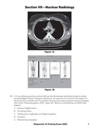
  • 1. Section VII – Nuclear Radiology Figure 1A Figure 1B 269. A 21-year-old man presented one and one half years after chemotherapy and radiation therapy for nodular scerosing Hodgkin’s Disease involving the mediastinum. He underwent a CT scan of the chest (Figure 1A). On the same day as the follow-up CT examination, the patient also underwent positron emission tomography (PET) with F-18 fluorodeoxyglucose (FDG) (Figure 1B). Which one of the following is the MOST likely diagnosis? A. Recurrent Hodgkin’s disease B. Post-therapy fibrosis C. Transformation to high-grade non-Hodgkin’s lymphoma D. Sarcoidosis E. Rebound thymic hyperplasia Diagnostic In-Training Exam 2003 1
  • 2. Section VII – Nuclear Radiology Question #269 Findings: The CT scan of the chest at the time of presentation (Figure 1A) demonstrates a small soft tissue mass in the anterior mediastinum in the midline, anterior to the great vessels. The repeat CT obtained 15 months later (Figure 1B) appears essentially unchanged, with a persistent soft tissue mass in the anterior mediastinum. The FDG PET scan performed at the time of the follow-up CT (Figure 1C) is normal, without evidence of lymphadenopathy in the neck, chest, abdomen, or pelvis. In particular, there is no increased FDG uptake in the region of the anterior mediastinal mass noted on the CT studies. Rationales: A) Incorrect. In the case of Hodgkin’s disease resistant to chemotherapy, persistent and/or more extensive lymphadenopathy within the chest would be anticipated, as well as possible other new sites of involvement. None of these findings are evident in the current case, in which the PET scan is normal. B) Correct. A residual soft tissue mass on CT secondary to post-treatment fibrosis is the most likely explanation for the findings in this case. FDG PET imaging is more specific than CT for the detection of recurrence in lymphoma. The absence of increased FDG uptake in the patient’s persistent anterior mediastinal mass is strong evidence against the presence of residual or recurrent active disease C) Incorrect. De-differentiation into anaplastic lymphoma is not common in Hodgkin’s disease in young adults, and represents a far less likely explanation for the findings in this case than post-treatment fibrosis and scarring. D) Incorrect. The findings in this case are not suggestive of active sarcoidosis. Like other forms of granulomatous disease, sarcoidosis is a potential cause for a false positive FDG PET scan, not a false negative study. Therefore, if the mediastinal mass on the CT studies was due to sarcoidosis, then a focal area of increased FDG uptake in this region would be expected. Furthermore, the distribution of adenopathy on the CT studies is not typical for sarcoidosis, which usually involves the paratracheal and hilar regions, and may affect the pulmonary parenchyma. E) Incorrect. Thymic hypertrophy can occur following treatment for lymphoma of the chest, particularly in young adult patients. The location of the CT findings is reasonable for thymic hypertrophy, although the configuration of the soft tissue mass is not classic in appearance. Furthermore, in such cases there is commonly mild to moderate increased FDG uptake within the thymus gland, a finding not present in this case. Thus, while not excluded, thymic hypertrophy is a less likely etiology for the findings in this case than post-treatment fibrosis and scarring. Citations: Jerusalem G, et. al.: Blood 1999;94:429-433. 2 American College of Radiology
  • 3. Section VII – Nuclear Radiology Figure 2 270. A 44-year-old woman underwent I-131 whole body imaging, 7 days following administration of a therapeutic dose of I-131 sodium iodide for a well-differentiated thyroid carcinoma (Figure 2). Based upon the appear- ance of the scan, which one of the following is CORRECT? A. The tumor most likely is a follicular carcinoma B. The patient has diffuse hepatic metastases C. The therapeutic I-131 dose should have been 30 mCi. D. The patient is a candidate for adjunct chemotherapy E. A follow-up whole body I-131 scan should be obtained in 3 years Diagnostic In-Training Exam 2003 3
  • 4. Section VII – Nuclear Radiology Question #270 Findings: Post-therapy whole body I-131 images demonstrate multiple foci of functioning thyroid tissue in the thyroid bed, as well as an additional focus located more superiorly in the neck, to the left of midline. Additional foci of uptake consistent with skeletal metastases are noted in the region of the right shoulder, both proximal femora (right > left) and in the mid-thoracic and lower lumbar spine. Small foci of abnormal tracer uptake are noted in the soft tissue superolateral to the right hip and in the lower abdomen, to the right of midline posteriorly. These latter lesions could also represent additional skeletal metastases. Rationales: A) Correct. The patient has evidence of widely metastatic well-differentiated thyroid carcinoma, involving the thyroid bed, cervical lymph nodes, and skeleton. Statistically, the most likely histologic type to produce these findings is follicular carcinoma. Papillary and mixed papillary-follicular carcinoma is more common, but less commonly metastasizes to the skeleton. Anaplastic and medullary carcinoma are less well differentiated, and are unlikely to accumulate I-131. B) Incorrect. The scan demonstrates mild diffuse hepatic uptake. This finding is typically seen on post-therapy imaging, especially in patients with significant amounts of residual functioning thyroid tissue present. It is most often secondary to hepatic uptake of iodinated thyroid hormone, a normal metabolic process. Hepatic metastases are uncommon in well-differentiated thyroid carcinoma, and when present, are more likely to demonstrate an irregular or focal distribution, which is not evident in this case. C) Incorrect. The patient has definite evidence of skeletal metastases, indicative of advanced Stage IV disease. This study is not consistent with Stage I disease. D) Incorrect. Most authorities agree that patients with well-differentiated thyroid carcinoma and evidence of residual or recurrent disease should be treated with I-131 sodium iodide as long as there is evidence of I-131 uptake by the lesions, which indicates the potential for cure or at least significant reduction in the extent of disease. This recommendation is particularly true in patients with widespread disease, as in the current case. I-131 is not only efficacious in such patients, but is also associated with significantly lower morbidity than external beam radiotherapy. I-131 therapy can also be efficacious in patients with well-differentiated thyroid carcinoma and negative diagnostic I-131 whole body scans, but elevated serum thyroglobulin levels. In such patients, I-131 uptake by metastases may be evident only on post-therapy images. External beam radiotherapy is appropriately reserved for patients whose lesions do not actively take up I-131, who have undifferentiated histologic types of thyroid cancer or who demonstrate evidence of de-differentiation of the tumor. E) Incorrect. In general, patients are maintained on thyroid hormone replacement, followed up with repeat diagnostic whole body I-131 scans annually and monitored by serum thyroglobulin levels between scans. Treating no more frequently than annually is usually sufficient for disease control, and reduces the likelihood of hematologic or pulmonary toxicity. In cases in which there is clinical evidence of rapidly progressive disease, the interval between scans can be reduced to 6 months. In a patient with widespread metastases, such as the present case, a three-year follow-up interval is far too long, and would be inappropriate, potentially permitting significant disease progression. Citations: Mettler FA, Jr. and Guiberteau MJ: Essentials of Nuclear Medicine Imaging, 4th ed., W. B. Saunders Co., Philadelphia, 1998, pp. 121-125. 4 American College of Radiology
  • 5. Section VII – Nuclear Radiology Figure 3A Figure 3B 271. A 20-year-old woman had massive intracranial bleeding during surgery for resection of a craniopharyngioma, and is now comatose. Radionuclide flow studies and delayed brain imaging using Tc-99m glucoheptonate were performed following surgery (Figures 3A and 3B) and repeated the following day (Figures 3C and 3D). Based upon these images, which one of the following is CORRECT? A. Both studies demonstrate findings consistent with brain death B. Neither study demonstrates findings consistent with brain death C. Radionuclide findings of brain death are present only on the second study D. Both studies are indeterminate for the presence of brain death E. Follow-up magnetic resonance imaging is necessary to confirm the presence of brain death Diagnostic In-Training Exam 2003 5
  • 6. Section VII – Nuclear Radiology Figure 3C Figure 3D 6 American College of Radiology
  • 7. Section VII – Nuclear Radiology Question #271 Findings: The first study (Figures 3A and 3B) demonstrates severely reduced intracerebral perfusion, with no visualization of the intracerebral vessels and only questionable visualization of the sagittal sinus on the flow study. However, the delayed static images demonstrate definite visualization of the sagittal and both transverse sinuses. The repeat study performed the following day (Figures 3C and 3D) demonstrates interval deterioration in cerebral perfusion. The flow study again demonstrates no definite evidence of intracerebral perfusion, now without visualization of the intracerebral vessels or the dural sinuses. A “hot nose” sign is also present, another indirect indication of severely reduced perfusion of the internal carotid circulation as compared to the external carotid circulation. A focal area of increased uptake is noted in the posterior scalp region on the static images, most likely secondary to inflammation and dependent edema in this region. There has been interval disappearance of sagittal and transverse sinus visualization, with only questionable faint activity noted in the region of the posterior aspect of the sagittal sinus. Rationales: A) Incorrect. While cerebral perfusion is significantly impaired on both studies, the visualization of the dural sinuses on the first study is not consistent with the radionuclide diagnosis of brain death. B) Incorrect. As noted above, the first study is not diagnostic of brain death. However, the second study demonstrates no evidence of intracerebral vessel visualization nor definite visualization of the dural sinuses, and demonstrates the corroborative “hot nose” sign. Thus, the 2nd study is consistent with brain death. C) Correct. As discussed in A and B above, the second study is consistent with brain death, representing an interval change compared to the study performed on the previous day. D) Incorrect. The second study is consistent with brain death. The findings of the radionuclide brain scan are correlated with other clinical data (physical findings, EEG, etc.) in order to make a final determination of clinical brain death. E) Incorrect. Confirmation of brain death is done by correlating the brain scan findings with other clinical data, primarily physical findings, and the EEG. Magnetic resonance imaging is not a sensitive indicator of brain death. Citations: Sandler MP, Coleman RE, Wackers FJTh, Patton JA, Gottschalk A and Hoffer PB (eds.): Diagnostic Nuclear Medicine, 3rd Ed., Williams & Wilkins, Baltimore, 1996, pp. 1091-1097. Diagnostic In-Training Exam 2003 7
  • 8. Section VII – Nuclear Radiology Figure 4A Figure 4B 272. A 60-year-old woman presented with hypercalcemia and a history of a renal calculus. Tc-99m sestamibi images of the neck and mediastinum were obtained at 30 minutes and 3 hours post-injection, including (clockwise from upper left) anterior neck, anterior neck with “cold” sternal notch marker, anterior neck and chest, pinhole neck, left anterior oblique neck and chest and right anterior oblique neck and chest (Figures 4A and 4B). Which one of the following is the LEAST likely etiology for the findings in this case? A. Thyroid adenoma B. Parathyroid hyperplasia C. Parathyroid adenoma D. Papillary carcinoma of the thyroid E. Colloid goiter 8 American College of Radiology
  • 9. Section VII – Nuclear Radiology Question #272 Findings: Tc-99m sestamibi images of the neck and mediastinum demonstrate normal uptake by the thyroid and salivary glands on the initial 30-minute post-injection images (Figure 4A). Normal left ventricular myocardial activity and hepatic activity are also evident. There is a superimposed focal area of increased tracer uptake noted overlying the left lower pole of the thyroid. The 3 hour delayed images (Figure 4B) demonstrate substantial washout of activity from the thyroid, with a persistent focus of increased uptake overlying the left lower pole region. Rationales: A) Incorrect. A focal thyroid adenoma is a common cause of a false positive radionuclide parathyroid scan. In this clinical setting, a parathyroid adenoma is more likely, but a thyroid adenoma cannot be excluded based on the imaging findings in this case alone. B) Incorrect. Parathyroid hyperplasia typically produces multiple foci of less striking increased tracer uptake in the neck, in the expected locations of the parathyroid glands, with the intensity of uptake usually less than is present in this case. Nonetheless, it is definitely in the differential diagnosis for focally increased sestamibi uptake in the neck, especially in a patient with clinical evidence of hyperparathyroidism. C) Incorrect. The findings in this case are classic for a focal parathyroid adenoma, which is in fact the most likely diagnosis in this case. A solitary parathyroid adenoma is found to be the cause of primary hyperparathyroidism in approximately 85% of cases. D) Incorrect. Papillary carcinoma of the thyroid is another cause for false positive Tc-99m sestamibi parathyroid scintigraphy studies. The clinical setting of the case again is more suggestive of parathyroid adenoma, but a thyroid malignancy cannot entirely be excluded. E) Correct. Of all the choices indicated, colloid goiter is the least likely cause for the findings in this case. A colloid goiter is typically a multifocal process, associated with thyroid enlargement and inhomogeneous uptake on early images. In addition, colloid goiter is unlikely to be associated with focal area(s) of significantly increased sestamibi uptake, as is present in this case. Citations: Sandler MP, Coleman RE, Wackers FJTh, Patton JA, Gottschalk A and Hoffer PB (eds.): Diagnostic Nuclear Medicine, 3rd Ed., Williams & Wilkins, Baltimore, 1996, pp. 998-1011. Diagnostic In-Training Exam 2003 9
  • 10. Section VII – Nuclear Radiology Figure 5 273. A 55-year-old woman presented with abdominal pain and weight loss. You are presented with 48-hour anterior and posterior whole body In-111 pentetreotide images (Figure 5). Which one of the following is the MOST likely diagnosis? A. Pheochromocytoma B. Carcinoid tumor of the small bowel C. Non-Hodgkin’s lymphoma D. Islet cell carcinoma E. Adenocarcinoma of the colon 10 American College of Radiology
  • 11. Section VII – Nuclear Radiology Question #273 Findings: Forty-eight hour whole body In-111 pentetreotide images demonstrate multiple focal areas of increased tracer uptake within both lobes of the liver, consistent with multiple focal neuroendocrine tumor hepatic metastases. There is also a large focal area of increased uptake in the epigastric region, just to the right of midline, in the expected location of the body of the pancreas. No other focal lesions are demonstrated in the abdomen or in the chest. Rationales: A) Incorrect. Pheochromocytoma is a neoplasm that may demonstrate increased uptake of octreotide analogs, and the focal lesion in the right upper quadrant could conceivably represent a right adrenal lesion. However, widespread hepatic metastases are not as common as in carcinoid, islet cell carcinoma, or small cell lung carcinoma, and pheochromocytoma is therefore a less likely etiology for the findings demonstrated. B) Incorrect. Carcinoid is a highly octreotide-avid neoplasm in general, and often produces multiple hepatic metastases as well. The primary lesion is most often located within the small bowel or lung, neither of which areas demonstrate focal lesions in this case. Therefore, while not unlikely, this diagnosis is less likely than an islet cell pancreatic tumor. C) Incorrect. Again, small cell lung carcinomas may demonstrate neuroendocrine features, and may have significant somatostatin receptor activity. However, the pattern of involvement in this case within the liver and epigastric region, without a focal pulmonary or mediastinal lesion, make small cell lung carcinoma less likely than islet cell tumor or carcinoid. D) Correct. Islet cell carcinoma is the most likely diagnosis in this case. The focal lesion in the epigastric region is secondary to uptake within the primary neoplasm in the body of the pancreas. Multiple hepatic metastases containing somatostatin receptor activity are also common in this entity. There is no evidence of focal lesions within the lungs or mediastinum to suggest small cell carcinoma of the lung and no focal extrahepatic abdominal lesions in the region of the small bowel to suggest a primary lesion in that location. E) Incorrect. Medullary carcinoma of the thyroid is often associated with somatostatin receptor activity, but less so than carcinoid or islet cell pancreatic tumors. There is also no focal increased tracer uptake in the neck to suggest a primary thyroid lesion, and widespread hepatic metastases are less common than in carcinoid or islet cell tumor. Citations: Mettler FA, Jr. and Guiberteau MJ: Essentials of Nuclear Medicine Imaging, 4th ed., W. B. Saunders Co., Philadelphia, 1998, pp. 379-382. Diagnostic In-Training Exam 2003 11