3. Until the early 1990s, the only
method for diagnosing disease in the
intracranial arteries was the invasive
angiography
Angiography has 4% risk of minor
stroke,1% risk of major stroke and
0.1% risk of death.
Introduction
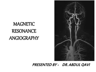
4. MR ANGIOGRAPHY
NON INVASIVE
NO CONTRAST REQUIRED
NON IONISING
INVASIVE
NEPHROTOXIC CONTRAST
IONISING RADIATION
CONVENTIONAL ANGIO
5. Vascular structures of brain can be imaged by 4 means:
1. DSA: gold standard. Invasive and risk of nephrotoxic contrast,
ionising radiation
2. Vascular ultrasound: least invasive, can be done bedside, cost
effective. best choice for imaging vessels close to skin surface.
Drawback: limited anatomic coverage, deep vessels cant be imaged,
operator dependent, requires skill
3. CT angio: main drawbacks are contrast use and radiation exposure,
calcifications are overestimated. It is preferred for aorta and
coronaries
4. MRA : is non invasive, no radiation exposure. Preferred for carotids
and intracranial vessels as MRI brain can also be obtained and is
widely used in neurological disorders
DSA-USG-CTA-MRA
6. 1. To evaluate conditions of the carotid arteries such as:
Stenotic / occlusive disease in symptomatic patients (e.g., TIA or
CVA)
Stenotic / occlusive disease in asymptomatic members
who are candidates for carotid endarterectomy surgery
when a Doppler Scan is abnormal.
Aneurysms.
Cervicocranial arterial dissection in members with suggestive
signs or symptoms (e.g., unilateral headache , oculosympathetic
palsy, amaurosis fugax, and symptoms of focal brain ischemia)
Indications of MRA
7. 2. To rule out intracranial aneurysm (ICA), including aneurysms of the
Circle of Willis, in persons who are thought to be at higher risk (e.g.,
history of ICA in a first-degree relative or presence of polycystic
kidney disease);
3. As a follow-up study for a known arteriovenous malformation (AVM),
and for a known non-ruptured intracranial aneurysm that is greater
than 3 mm in size;
4. To evaluate patients with signs/symptoms highly suggestive of
leaking/ruptured aneurysm or AVM (i.e., sudden explosive headache,
stiff neck, blood in the cerebral spinal fluid);
5. To definitively establish presence of stenoses or other abnormalities
of the vertebrobasilar system in patients with symptoms highly
suggestive of vertebrobasilar syndrome (binocular vision loss,
positional vertigo, dysarthria, dysphagia, diplopia).
Indications of MRA contd..
9. BASIC PRINCIPLES
PRINCIPLES Protons are excited using GRE pulse sequence
TOF MRA- “manipulating magnitude of magnetization”
Vascular contrast in TOF MRA is due to difference in
the magnitude of the inflowing protons in the blood
and the surrounding stationary protons.
No contrast agent injected
Motion artifact
Difficulty with slow flow
10. PC MRA- “manipulating phase of magnetization”
directional flow encoding magnetic field gradient
causing difference in the phase/orientation
In PC MRA bipolar gradient is applied and if the
proton is stationary there is no phase shift. However if
the protons are moving, a phase shift occurs. The
faster the proton moves greater the phase shift.
Phase is proportional to velocity
Allows quantification of blood flow and velocity
More time consuming
11. CE MRA -
Uses parameters typical of 3D TOF MRA but gadolinium
contrast is also given.
Data are acquired after contrast bolus infusion ( Gad.
0.1-.2 mmol/kg).
Unlike, time-of-flight (TOF) or phase contrast (PC)
imaging, the signals of the blood in CEMRA is based on
the intrinsic T1 signal of blood and rather less on flow
effects; therefore, this technique is less flow sensitive
It can be performed within seconds
Nephrogenic systemic fibrosis is rare but serious
complication.
12. • In 2DFT technique, multiple thin sections of body are studied
individually and even slow flow is identified
• In 3DFT technique , a large volume of tissue is studied ,which can be
subsequently partitioned into individual slices, hence high resolution
can be obtained and flow artifacts are minimised, and less likely to be
affected by loops and tortusity of vessels
• MOTSA(multiple overlapping thin slab acquisition): prevents proton
saturation across the slab. This technique have advantage of both 2D
and 3D studies. It is better than 3D TOF MRA in correctly identifying
vascular loops and tortusity, and have lesser chances of
overestimating carotid stenosis.
2D and 3D fourier transform
49. Initial screening test is noninvasive , either DS or MRA.
TOF MRA is less sensitive DS (75%Vs 87%) but more specific (88%Vs
46%).
Concordance in TOF and DS is more sensitive(96%) and specific
(85%) than either test alone.(Johnston, and Goldstein et al 2001)
2D TOF MRA over estimates the degree of stenosis.
3D TOF MRA is less likely to overestimate stenosis.
Combination of 2D and 3D TOF MRA results in greater specificity.
‘FLOW GAP’- segmental dropout , When the stenosis is more than
70%. ( Heiserman JE et al 1996)
3D CEMRA have greater anatomical coverage in terms of surface
morphology, carotid bifurcation, near occlusion
3D CE MRA is more sensitive(94.6% vs 91.2%) and specific ( 88.3%vs
91.9%) than TOF MRA for high grade ICA stenosis.
Carotid atherosclerotic narrowing
54. Catheter angiography has been traditionally used for diagnosis of
carotid dissection.
MRI +fat saturation along with 3D TOF MRA characterise dissecting
hematoma, associated pseudo-aneurysm & length and caliber of
residual patent lumen.
MRI with MRA is currently the investigation of choice for suspected
dissection.
Not nearly as helpful in vertebral artery dissections
Carotid or Vertebral artery dissection
58. 3D TOF is now accepted as a non-invasive screening tool for
familial aneurysmal disease.
Sensitivity is greater for detecting aneurysms > 3mm (94%) than
aneurysms < 3mm (38%). ( White et al 2000)
Overall inferior to DSA and misses aneurysm <3mm.(Adams et al
2000)
3D CE MRA is superior to TOF MRA .It is method of choice for
evaluation of giant cerebral aneurysms.
CE MRA shows promise in the follow-up of treated intracranial
aneurysms.
Aneurysms
65. Secondary role to digital subtraction angiography.
The typical AVM appears on spin-echo MRI as a cluster
of focal round lesions or serpentine areas of signal void
3D CE MRA is superior to 3D TOF MRA and equivalent to
subtraction angiography in 70-90%of cases in depicting
AVM components.
A-V Malformations
68. Acute ischemic stroke
A novel application of MRA is to guide acute stroke
intervention potentially.
MRA can be a predictor of clinical outcome in acute
ischemic pt. undergoing thrombolysis with IV rtPA in
window period. ( Marks et al 2008).
The Boston scale ( BASIS) is a classification tool to
help predict outcomes in acute stroke by using MRA
study. ( Torres-Mozqueda et al 2008)
81. Summary
1. Two different approaches to MRA are
commonly used: Time-of-Flight (TOF-MRA) &
Phase Contrast (PC-MRA)
2. TOF-MRA is easy to implement and is robust but
has difficulty with slow flow
3. 3D TOF can be combined with fast imaging
methods and Gd contrast agents to obtain
improved depiction of vascular structures
82. Summary
4. PC-MRA requires more time to acquire
more images but can result in high
resolution, fewer flow related artifacts,
and quantitative measurement of flow
5. Phase-contrast MRI may provide the most
accurate, noninvasive method for
measuring blood flow in vivo
87. Characteristics CT-angiography MR-angiography
Spatial resolution Good Fair
Size of vessel upto 0.6mm 1.0-1.5mm
Tortuous vessel better visualized -
Time seconds minutes
Risk of radiation exposure yes no
Use of contrast always +/-
Detection of calcification excellent -
CT-angiography vs MR-angiography:
ferromagnetic substance indicated not indicated
Editor's Notes
Intracranial Vascular Diseases are major source of death and
disability
Clinical Policy Bulletin:Aetna Magnetic Resonance Angiography (MRA) and Magnetic Resonance Venography (MRV)
Note: As MRA is considered an alternative to angiography for evaluation of the carotids, a subsequent angiography would only be considered medically necessary if there was a significant discrepancy between the findings of Duplex ultrasonography and MRA that would impact on surgical planning.
Disadv of MRA high cost , cant identify small vs,susceptibility to complex fow, claustrophobia, not good for root of neck and aortic arch
Prevalence of missing or hypo-plastic vesselsof the circle of Willis according to a study byKrabbe-Hartmann et al. (Krabbe-Hartkamp, van derGrond et al. 1998). For example, the posterior and anteriorcirculation are not connected in 10.7% subjects(out of 150 subjects of the study), while 28.7% haveone missing posterior communicating artery. No distinction
is made between left and right side configurations.
Magnetic resonance techniques to measure distribution of cerebral bloodflow
M. Günther1,21mediri GmbH, Heidelberg, Germany; 2Neurologische Klinik, Universitätsklinikum Mannheim, Universität Heidelberg,Germany. Applied Cardiopulmonary Pathophysiology 13: 212-218, 2009
a: a single ACoA. The ICA bifurcates (arrow) into theA1 segment of the ACA, and the MCA. b: two (or
more) ACoAs. c: medial artery of the corpus callosum(MACC, arrow) arises from the ACoA. d: fusion of theanterior cerebral arteries over a short distance. e: anteriorcerebral arteries form a common trunk which splitsdistally into two A2 segments. f: MCA originates fromthe ICA as two separate trunks. g: hypoplasia or absenceof an anterior communication. h: one A1 segment ishypoplastic or absent, the other A1 segment gives rise toboth A2 segments. i: hypoplasia or absence of an ICA,
the contralateral A1 segment gives rise to both A2 andsupplies retrograde flow to the ipsilateral A1, which, inturn, gives rise to the ipsilateral MCA (both anteriorcerebral arteries and both MCA are supplied by a singleICA). j: hypoplasia or absence of an anterior communication.The MCA arises as two separate trunks. Variants a–f are complete; variants g–j are incomplete.
Anatomical variations in the posterior part ofthe circle of Willis.
a: bilateral PCoA present. b: a posterior cerebral arteryoriginates predominantly from the internal carotidartery; this variant is known as a unilateral fetal-typeposterior cerebral artery (FTP, arrows); the PCoA onthe other side is present. c: bilateral FTP, with bothP1 segments patent. d: unilateral PCoA present. e: hypoplasiaor absence of both PCoAs and isolation of the
anterior and posterior parts of the circle at this level.f: unilateral FTP, and hypoplasia or absence of the P1
segment. g: unilateral FTP, and hypoplasia or absenceof the contralateral PCoA. h: unilateral FTP and hypoplasia
or absence of both the P1 and PCoA. i: bilateralFTP with hypoplasia or absence of both P1 segments.j: bilateral FTP with hypoplasia or absence of one P1segment. Variants a–c are complete, whereas variants d–j are incomplete.
Combination of 2D and 3D TOF MRA results in greater specificity.
TOF MRA is less sensitive DS (75%Vs 87%) but more specific (88%Vs 46%).
‘FLOW GAP’- segmental dropout , When the stenosis is more than 70%.
2D TOF study with normal or nearnormal findings effectively excludes the possibility of severe(70-99%) stenosis. The most accurate results are obtainedwhen short TE and small voxel size are used. 2). If there is a flow gap, poorly shown surface
morphology, or findings indeterminate for near occlusionversus occlusion involving the bifurcation and cervical
internal carotid, then a time-resolved 3D CE-MRA is done, which can better demontrate ulcerationHigh resolution 3T MRI can better demonstrate unstable plaque with lipid core.Typically, a 3D TOF study coveringthe vertebral-basilar system from the C2 level to the tipof the basilar artery is done.The 3D CE-MRA techniques can displayboth the origins and distal intracranial portions of thevertebrals in a single acquisition and are particularly usefulin evaluating vertebral arterysegments with partial orcomplete signal loss caused by slow flow and in-planesaturation effects
Carotid stenosis, high-resolution three-dimensional (3D) time-of-flight (TOF) versus two-dimensional (2D) TOF. The 2D TOF magnetic resonance angiography (A) shows complete signal loss, implying tight stenosis in the proximal internal carotid artery. High-resolution 3D TOF (B) demonstrates only moderate narrowing, which matches the catheter angiogram (C).
Not nearly as helpful in vertebral artery dissections
55 Ovid scott Spontaneous carotid artery dissection. A: Spin echo image demonstrating normal flow void in the left carotid artery and a halo of hyperintensity from a mural hematoma (arrow). B: Individual partitions from the three-dimensional Fourier transform time-of-flight magnetic resonance angiography demonstrating the normal hyperintense flow signal in the left internal carotid artery with a less hyperintense crescent-shaped rim, representing mural hematoma (arrow). C: Maximum intensity projection (MIP) image of the internal carotid arteries demonstrating the surrounding slight hyperintense, but less than the flowing blood, around the left internal carotid artery (arrow), representing methemoglobin in the vessel wall that has been incorporated into the image by the MIP algorithm.
Catheter angiogram (A), and two-dimensional Fourier transform (2DFT) time-of-flight (TOF) (B) and three-dimensional Fourier transform (3DFT) TOF (C) magnetic resonance angiographs (MRAs) of the carotid arteries. Notice the pseudoaneurysm in a patient who had a carotid artery dissection 1 month prior (arrow). The lower spatial resolution of the 2D TOF MRA image of the carotid artery pseudoaneurysm does not define the neck of the aneurysm completely (open arrow). The 3D TOF MRA image better demonstrates the association of the aneurysm to the carotid artery.
MRA is useful as non invasive tool f/u of coiled aneurysm
3DTOF MRA show spherical aneurysm at Acom A
3D reconstruction maximum intensity projection[MIP] of magnetic resonance angiography [MCA] of the circleof Willis reveals bilobed aneurysm at the right MCAbifurcation (arrow).
Arteriovenous malformation (AVM). (A) Longrepetition time-echo spin-echo mri show a wedge-shaped cluster of vessels in tlie left temporal lobeseen as linear and round areas of signal void. Note enlargeddraining vein {arrow).
B) Magnetic resonance angiogram formedby a 3dtof circle of Willis shows an AVM (small blackarrows) in the left temporal lobe. Feeding vessels arise from the leftmiddle cerebral artery {arrowheads). A large medially drainingvein is seen (probably a peri mesencephalic vein; large arrow).
23 yr young female presented with post partum rt hemiparesis with altered sensorium, on improvement had aphasia,
T2 and flair show multiple hyperintensities in lt parietooccipital region, corrosponding ,with restrictn on DWI and low ADC value s/o infarctn, corrosponding MRA , lt MCA not visualised s/o thrombosis.
Multiple aneurysms in a 54-year-old woman. (a) Oblique DSA image of the left carotid artery depicts a 6.5-mm-diameter spherical aneurysm (arrowhead) at the trifurcation of the left middle cerebral artery. (b) Coronal DSA image of the right carotid artery demonstrates a bilobate aneurysm (two spheres with 2-mm diameter each [solid arrows]) located close to the trifurcation of the right middle cerebral artery and pointing caudally. A 3-mm-diameter spherical aneurysm (open arrow) is also depicted, on a branch of the right middle cerebral artery close to the insula. (c-e) Aneurysms are labeled as in a and b. (c) Targeted coronal MIP image from coronal 3D contrast-enhanced T1-weighted arterial phase images (5.8/1.6) depicts all three aneurysms clearly. (d) Coronal MIP image from MT TONE images (31/3.4) demonstrates the bilobate lesion, but it was depicted less accurately (as grade 1). (e) Coronal phase-contrast (14/6.7) image does not depict the bilobate lesion.