

Empfohlen
Weitere ähnliche Inhalte
Was ist angesagt?
Was ist angesagt? (20)
Andere mochten auch
Ähnlich wie Dyspnoea - dr.kkl
Ähnlich wie Dyspnoea - dr.kkl (20)
Mehr von Kyaw Swar Aung
Dyspnoea - dr.kkl
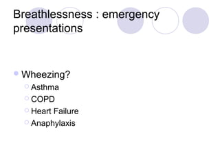
- 1. Breathlessness : emergency presentations Wheezing? Asthma COPD HeartFailure Anaphylaxis
- 2. Stridor? (Upper airway obstruction) Foreignbody or tumour Acute epiglottitis Anaphylaxis Trauma, eg laryngeal fracture Crepitations? Heart failure Pneumonia Bronchiectasis Fibrosis
- 3. Chest clear? Pulmonary embolism Hyperventilation Metabolic acidosis, eg diabetic ketoacidosis (DKA) Anaemia Drugs, eg: salicylates Shock (may cause air hunger) Pneumocystis pneumonia Central causes Others Pneumothorax – pain, increased resonance Pleural effusion – 'stony dullness'
- 4. Priority management of acute breathlessness
- 5. Acute breathlessness Oxygen, ECG monitor, Check BP, Listen over lungs, IV cannula, Nebulized salbutamol if wheeze Sign of tension Decompress with large-bore needle, 2nd pneumothorax intercostal space in mid-clavicular line Major arrhythmia? Treat No Clinical assessment, Chest X-ray, Arterial blood gases, 12 lead ECG Chest X-ray clear Chest X-ray abnormal Consider: - Acute asthma - Exacerbation of COPD - Upper airways obstruction - Pulmonary embolism Specific diagnosis and -Pre-radiological pneumonia treatment - Sepsis syndrome
- 6. Urgent investigations in acute breathlessness Chest X-ray Arterial blood gases and pH if oxygen saturation is <90% or diagnosis is unclear ECG(except in patients under 40 with pneumothorax or acute asthma) Full blood count Creatinine, sodium, potassium and glucose Echocardiogram if: Suspected cardiac tamponade Suspected surgically correctable cause of pulmonary oedema
- 7. Features pointing to a diagnosis in the breathless patient
- 8. Diagnosis Features Acute asthma Wheeze with reduced peak flow rate Previous similar episodes responding to bronchodilator therapy Diurnal and seasonal variation in symptoms Symptoms provoked by allergen exposure or exercise Sleep disturbance by breathlessness and wheeze Pulmonary oedema Cardiac disease Abnormal ECG Bilateral interstitial or alveolar shadowing on chest x-ray
- 9. Pneumonia Fever Productive cough Pleuritic chest pain Focal shadowing on chest X-ray Exacerbation of chronic obstructive pulmonary Increase in sputum volume, tenacity disease or purulence Previous chronic bronchitis: sputum production daily for 3 months of the year, for 2 or more consecutive years Wheeze with reduced peak flow rate
- 10. Pulmonary Pleuritic or non-pleuritic chest embolism pain Haemoptysis Risk factors for venous thromboembolism present (signs of DVT commonly absent) Sudden breathlessness in young Pneumothorax otherwise fit adult Breathlessness following invasive procedure e.g subclavian vein puncture Pleuritic chest pain Visceral pleural line on chest x-ray, with absent lung markings between this line and the chest wall
- 11. Cardiac tamponade Raised JVP Pulsus paradoxus > 20mmHg Enlarged cardiac silhouette on chest X-ray Known carcinoma of bronchus or breast Laryngeal History of smoke inhalation or the ingestion of obstruction corrosives Palatal or tongue oedema Anaphylaxis
- 12. Tracheobronchial Stridor (inspiratory noise) or mnophonic obstruction wheeze (expiratory 'squeak') Known carcinoma of the bronchus History of inhaled foreign body PaCo2>5 kPa in the absence of chronic obstructive pulmonary disease Wheeze unresponsive to bronchodilators
- 13. Large pleural Distinguished from pulmonary consolidation effusion on the chest x-ray by: Shadowing higher laterally than medially Shadowing does not conform to that of a lobe or segment No air bronchogram Trachea and mediastinum pushed to opposite side
- 14. Arterial blood gases and pH in breathlessness with a normal chest X-ray Disorder PaO2 PaCO2 PHa Acute asthma Normal/low Low High May be Normal or Acute exacerbation of COPD Usually low high low Normal/low (without pre-existing Pulmonary embolism cardiopulmonary disease) Low High Pre-radiological pneumonia Low Low High Sepsis syndrome Normal/low Low Low Metabolic acidosis Normal Low Low Hyperventilation without organic disease High/normal Low High