SlideShare ist ein Scribd-Unternehmen logo
1 von 115
HEPATOCELLULAR CARCINOMA (HCC):
MANAGEMENT
Pratap Sagar Tiwari
Part 1: Managing Advanced HCC (past, present & future perspectives)
Part 2: Radiotherapy in HCC (is there any role ?)
Part 3: Locoregional Therapies in HCC (guidelines & beyond)
Part 4: Surgery in HCC (limit and limitations ?)
Part 5: Liver transplantation (HCC Perspective)
PART 3
LOCOREGIONAL THERAPIES IN HCC
(GUIDELINES & BEYOND)
Clipart :http://www.scientificanimations.com/wiki-images/
HCC MX: OVERVIEW
Meets criteria for LT
?
Resectable ?
Locally
Advanced?
Advance HCC ? End stage HCC?
LT Surgery
Locoregional
Therapies
Chemotherapy BSC
LOCOREGIONAL THERAPY MODALITIES
Transarterial Chemoembolisation
(TACE)Per cutaneous Ethanol Injection (PEI)
Ablation therapies Vascular Intervention Therapies
Combination therapies/ Future DirectivesIrreversible Electroporation (IRE)
Microwave Ablation (MWA)
Radiofrequency ablation(RFA)
High-intensity focused ultrasound
(HIFU)
Hepatic artery infusion chemotherapy
(HAIC)
Transarterial Radioembolisation (TARE)
• Laser-induced thermotherapy
• Cryoablation
• Percutaneous acetic acid injection
LOCOREGIONAL THERAPY MODALITIES
Per cutaneous Ethanol Injection (PEI)
Ablation therapies
PERCUTANEOUS ETHANOL ABLATION/INJECTION(PEI)
 PEI for treatment of hepatic tumors was first described by Sugiura et al. [1] in 1983.
 PEI was recommended as the standard percutaneous treatment for early-stage, non-surgical
HCC in the 2001 guidelines of the EASL [2].
 PEI has been reported to induce chemical coagulative necrosis in 70-80% of HCC lesions ≤30
mm in diameter [3-5], 95% of tumours <2 cm[6–8], but 50% when of 3–5 cm diameter [6–8],
with survival of >50% at 5 years [9]. The best candidates are in Child-Pugh A stage with small
HCC [10].
 The ablation is limited to the lesion itself and may be suboptimal for the presence of internal
septa: residual nests of vital neoplasia inside or just close to the pseudocapsule are hence
detected in about one-third of cases during follow-up [11].
References are at the end of the slides.
PEI: THE PROCEDURE
 The procedure is discussed with the pt/patient’s family. Alternative treatments are discussed with
the pt so an informed choice can be made. The pt is instructed to fast for 8 hr before the
procedure, and a coagulation profile is checked. A combination of IV midazolam and fentanyl is
used for sedation and analgesia.
 The lesion or lesions are visualized via USG or CT . A single 20- or 22-gauge needle is inserted
into the deep portion of the lesion, or more than one needle is inserted if the lesion is >3 cm. It
is important to make as few passes as possible into the lesion because of possible reflux of
alcohol back through excessive needle tracks .
Lee MJ, et al. Percutaneous Ethanol Injection for the Treatment of Hepatic Tumors: Indications, Mechanism of Action, Technique, and Efficacy.AJR 1995;164:215-220
PEI: THE PROCEDURE
 Usually, most hepatologists inject approx 8-10 ml of alcohol per treatment session per lesion. However,
larger volumes of alcohol have been injected, depending on the pt’s tolerance.
 First, alcohol is injected in the deep part of the lesion, and then the needle is slowly withdrawn to the
more superficial part while alcohol continues to be injected. In this way, it is hoped that ethanol will be
distributed evenly through the lesion.
 After alcohol injection, the needle(s) are left in place for 2-3 min so reflux of alcohol along the track is
reduced. Reflux of alcohol along the needle track can lead to significant pain from penitoneal
irritation.
 Alternatively, gelatin sponge can be injected as the needle is withdrawn to prevent reflux of alcohol
Lee MJ, et al. Percutaneous Ethanol Injection for the Treatment of Hepatic Tumors: Indications, Mechanism of Action, Technique, and Efficacy.AJR 1995;164:215-220
PEI: THE PROCEDURE/RESPONSE
 The injection is repeated once or twice a week for up to six to eight sessions, depending on the
tumor size.
 The therapeutic effect of PEI can be evaluated by contrast CT scan.
 A CT scan was performed 1–3 days after the procedure to evaluate technique effectiveness (14).
Complete ablation was defined as hypoattenuation of the entire tumour without contrast
enhancement[1]. When the presence of unablated tumour portions was suspected, a few more
procedures were performed.
 Color Doppler USG and MRI are alternative imaging techniques for assessing the response to
PEI.[2,3](done every 4 months in a largest study[4]
1. Ebara M, Kita K, Sugiura N, et al. Therapeutic effect of percutaneous ethanol injection on small hepatocellular carcinoma: evaluation with CT. Radiology 1995;
195:371–377.
2. Lencioni R, Caramella D, Bartolozzi C. Hepatocellular carcinoma: use of color Doppler US to evaluate response to treatment with percutaneous ethanol injection.
Radiology 1995; 194:113–118.
3. Bartolozzi C, Lencioni R, Caramella D, et al. Treatment of hepatocellular carcinoma with percutaneous ethanol injection: evaluation with contrast-enhanced MR
imaging. AJR Am J Roentgenol 1994; 162:827–831.
4. Shiina S, et al. Percutaneous ethanol injection for hepatocellular carcinoma: 20-year outcome and prognostic factors. Liver Int. 2012 Oct; 32(9): 1434–1442.
PEI: INDICATIONS/CONTRAINDICATIONS
 It is generally agreed that pts with HCCs 3 cm or smaller and three or fewer in number are the
best candidates for PEI, although many centers perform PEI for HCCs up to 5 cm.[1-3]
 PEI is CI in the presence of gross ascites, severe thrombocytopenia, or coagulopathy (platelet
count <40,000/mm3, INR >1.75, in a study[5])because of a high risk of bleeding.
 Large infiltrative tumors, thrombosis in the main portal or hepatic vein, and extrahepatic
metastasis are also considered CIs in most centers.
 Pts with tumors on the surface of the liver are not favorable candidates for PEI because the
injected ethanol can leak back into the peritoneal cavity, and there is also a higher risk of tumor
implantation into the peritoneal cavity.[4]
1. Ebara M, Ohto M, Sugiura N, et al. Percutaneous ethanol injection for treatment of small hepatocellular carcinoma. Study of 95 patients. J Gastroenterol Hepatol 1990; 5:616–626.
2. Livraghi T, Bolondi L, Lazzaroni S, et al. Percutaneous ethanol injection in the treatment of hepatocellular carcinoma in cirrhosis: A study on 207 patients. Cancer 1992; 69:925–929.
3. Di Stasi M, Buscarini L, Livraghi T, et al. Percutaneous ethanol injection in the treatment of hepatocellular carcinoma. A multicenter survey of evaluation practices and complication rates. Scand J
Gastroenterol 1997; 32:1168–1173.
4. Shina S, Tagawa K, Unuma T, et al. Percutaneous ethanol injection therapy for hepatocellular carcinoma. A histopathologic study. Cancer 1991; 68:1524–1530.
5. brunello F, et al. Radiofrequency ablation versus ethanol injection for early hepatocellular carcinoma: A randomized controlled trial. Scandinavian Journal of Gastroenterology, 2008; 43: 727735
PEI: COMPLICATIONS
 The MC AES include fever, pain, and a feeling of alcohol intoxication. These are generally
transient and may only require symptomatic therapy.
 Depending on the site of the hepatic lesion, serious complications may occur.
 When a lesion is subdiaphragmatic, the pleura may be transgressed, with possible resulting
complications such as pneumothorax, pleural effusion, or hemothorax.
 Intrapenitoneal hemorrhage has been reported in three of 190 pts by Shiina et al. [1].
 Frequency of tumor seeding should theoretically be low because of the cytotoxic effect of
ethanol on any tumor cells that may find their way into the percutaneous track [2].
1. Shiina S, Niwa Y, Omata M. Percutaneous ethanol injection therapy for liver neoplasms. Semin Interient Radiol l993;1O(2):57-68
2. Zerbey AL, Mueller PA, Dawson SL, Hoover HC Jr. Pleural seeding from hepatocellular carcinoma: a complication of percutaneous alcohol ablation. Radiology l994
PEI: COMPLICATIONS
1. Shiina S, Niwa Y, Omata M. Percutaneous ethanol injection therapy for liver neoplasms. Semin Interient Radiol l993;1O(2):57-68
2. Zerbey AL, Mueller PA, Dawson SL, Hoover HC Jr. Pleural seeding from hepatocellular carcinoma: a complication of percutaneous alcohol ablation. Radiology l994
• PEI is a minimally invasive therapy with a good safety record.
• In a study of 746 pts with HCC treated by PEI, the treatment-related death rate was only 0.1%, and the rate
of severe complications was 1.7%.[1]
• A similar death rate (0.09%) and complication rate (3.2%) have been reported in a multicenter survey of
1,066 pts after PEI for HCC.[2]
• Tumor seeding along the needle track after PEI for HCC has been reported, with an incidence of 1% in a
study of 348 pts.[3]
1. Livraghi T, Giorgio A, Marin G, et al. Hepatocellular carcinoma and cirrhosis in 746 patients: long-term results of percutaneous ethanol injection. Radiology 1995; 197:101–108.
2. Di Stasi M, Buscarini L, Livraghi T, et al. Percutaneous ethanol injection in the treatment of hepatocellular carcinoma. A multicenter survey of evaluation practices and complication rates. Scand J
Gastroenterol 1997; 32:1168–1173.
3. Ishii H, Okada S, Okusaka T, et al. Needle tract implantation of hepatocellular carcinoma after percutaneous ethanol injection. Cancer 1998; 82:1638–1642.
LOCOREGIONAL THERAPY MODALITIES
Per cutaneous Ethanol Injection (PEI)
Ablation therapies
Outcome of PEI ?
PEI Vs LR ?
PEI Vs RFA ?
Outcome of RFA ?
RFA Vs LR ?
Radiofrequency Ablation (RFA)
RADIOFREQUENCY ABLATION (RFA)
 In the mid-1990s, RFA was proposed for the treatment of small
HCC [1,2].
 RFA is based on heat-generated coagulative necrosis that
induces the ablation of the tumour itself and may involve small
portions of surrounding liver tissue as well.
 In RFA, an electrical current is delivered through a needle
electrode under imaging or surgical guidance, producing heat-
based thermal cytotoxicity[3].
 Temperatures range between 60 °C to 100 °C and result in
almost instant coagulation necrosis[4].
1. Rossi S, Di Stasi M, Buscarini E, Quaretti P, Garbagnati F, Squassante L, et al. Percutaneous RF interstitial thermal ablation in the treatment of hepatic cancer. Am J Roengtenol 1996;167:75968.
2. De Baere T, Risse O, Kuoch V, Dromain C, Sengel C, Smayra T, et al. Adverse events during radiofrequency treatment of 582 hepatic tumors. Am J Roengtenol 2003;181:695700.
3. Goldberg SN, Gazelle GS, Mueller PR. Thermal ablation therapy for focal malignancy: a unified approach to underlying principles, techniques, and diagnostic imaging guidance. AJR Am J Roentgenol. 2000;174:323–331.
4. Gervais DA, Arellano RS. Percutaneous tumor ablation for hepatocellular carcinoma. AJR Am J Roentgenol. 2011;197:789–794.
Clipart from: https://www.alamy.com/radiofrequency-ablation-image66575282.html
RADIOFREQUENCY ABLATION (RFA)
• RFA is moderated by the heat-sink effect, a phenomenon that occurs when thermal energy is
dispersed from the target lesion due to blood flow in the vessels adjacent to it[1].
• Consequently, the shape and size of the ablation zone may be unpredictable and the efficacy of
RFA may be restricted as multiple sessions are necessary for complete tumour eradication[2].
• Caution is needed in treating lesions close to hollow viscera owing to the risk of perforation.
• Deep sedation is recommended.
1. Lu DS, Raman SS, Limanond P, Aziz D, Economou J, Busuttil R, Sayre J. Influence of large peritumoral vessels on outcome of radiofrequency ablation of liver
tumors. J Vasc Interv Radiol. 2003;14:1267–1274.
2. Mulier S, Ni Y, Jamart J, Ruers T, Marchal G, Michel L. Local recurrence after hepatic radiofrequency coagulation: multivariate meta-analysis and review of
contributing factors. Ann Surg. 2005;242:158–171.
RADIOFREQUENCY ABLATION (RFA): CI/COMPLICATIONS
 The ablation success rate for lesions <2 cm reaches 90% with a local recurrence rate of 1%[1].
For this reason, RFA is considered effective for tumours < 3 cm; combined LRT should be
considered for lesions > 3 cm[2].
 The main CI are severe bleeding diathesis (platelet count <50000/μL), ascites, jaundice and
presence of metallic devices such as pacemakers.
 RFA should also be avoided for tumours within 1 cm proximity to the hepatic portal tract.
 Major complications include liver failure, bleeding, infection, abscesses, intercostal nerve injury,
organ injury, tumour lysis syndrome and pneumothorax[3].
1. Livraghi T, Meloni F, Di Stasi M, Rolle E, Solbiati L, Tinelli C, Rossi S. Sustained complete response and complications rates after radiofrequency ablation of very early hepatocellular carcinoma in cirrhosis: Is resection still the treatment of choice?
Hepatology. 2008;47:82–89.
2. Peng ZW, Zhang YJ, Chen MS, Xu L, Liang HH, Lin XJ, Guo RP, Zhang YQ, Lau WY. Radiofrequency ablation with or without transcatheter arterial chemoembolization in the treatment of hepatocellular carcinoma: a prospective randomized trial.
J Clin Oncol. 2013;31:426–432.
3. Georgiades CS, Hong K, Geschwind JF. Radiofrequency ablation and chemoembolization for hepatocellular carcinoma. Cancer J. 2008;14:117–122.
4. Livraghi T, Solbiati L, Meloni MF, Gazelle GS, Halpern EF, Goldberg SN. Treatment of focal liver tumors with percutaneous radio-frequency ablation: complications encountered in a multicenter study. Radiology. 2003;226:441–451.
In a multicenter study of RFA for malignant liver tumours in 2320 pts, the rate of major complications reached
2.2% [4].
ASSESSING RESPONSE
 Typically the efficacy of percutaneous ablation therapy is assessed by dynamic cross-sectional
imaging (CT or MRI) one month after therapy.
 Standard methods to assess treatment response (eg, unidimensional Response Evaluation Criteria
in Solid Tumors [RECIST] criteria ) involve measurement of tumor dimensions before and after
treatment, but, methods such as these disregard the extent of necrosis, which is the end result of
locoregional ablative therapies [1].
 Others have proposed guidelines for assessment of response to locoregional ablative therapy
that are based upon reduction in viable tumor burden. Although not entirely reliable, the
absence of contrast uptake within the tumor is thought to reflect tumor necrosis, while the
persistence of contrast uptake indicates persistent disease. Recurrence of tumor in the treated
area (or elsewhere) is signaled by the reappearance of vascular enhancement.
1. Bruix J, Reig M, Rimola J, et al. Clinical decision making and research in hepatocellular carcinoma: pivotal role of imaging techniques. Hepatology 2011; 54:2238.
IMAGING RESPONSE CRITERIA USED IN EVALUATION OF
HCC AFTER TREATMENT
1. Modified from:
WHO handbook for reporting results of cancer treatment. World Health Organization, Geneva, 1979.
2. Yaghmai V, Besa C, Kim E, et al. Imaging assessment of hepatocellular carcinoma response to locoregional and systemic therapy. AJR Am J Roentgenol 2013; 201:80.
• A partially necrotic (black) HCC with an arterially-
enhancing (viable) component (white) is shown. The
arrows illustrate different measurements obtained for
assessment of response according to the various criteria.
 WHO measures two perpendicular dimensions of the entire
lesion;
 RECIST measures the single largest dimension of the
entire lesion;
 EASL measures perpendicular dimensions of just the
enhancing (viable) tumor;
 modified RECIST measures one single largest dimension of
the enchancing tumor.
• Both EASL criteria and modified RECIST are based upon
measurements obtained using cross sectional imaging (CT
or MRI) and contrast-enhanced arterial phase images.
MODIFIED RECIST FOR HEPATOCELLULAR CARCINOMA
PERCUTANEOUS ETHANOL ABLATION/INJECTION(PEI)
OUTCOME?
PEI ; OUTCOME?
Shiina S, et al. Percutaneous ethanol injection for hepatocellular carcinoma: 20-year outcome and prognostic factors. Liver Int. 2012 Oct; 32(9): 1434–1442.
• Background: PEI is the best-known image-guided percutaneous ablation for HCC
and a well-tolerated, inexpensive procedure with few adverse effects. However,
there have been few reports on its long-term results.
• Aims: To report a 20-year consecutive case series at a tertiary referral centre.
• Methods: performed 2147 ethanol injection treatments on 685 primary HCC pts
and analysed a collected database.
BASELINE CHARACTERISTICS OF THE 685 PTS UNDERGOING PEI FOR PRIMARY
HCC
Shiina S, et al. Percutaneous ethanol injection for hepatocellular carcinoma: 20-year outcome and prognostic factors. Liver Int. 2012 Oct; 32(9): 1434–1442.
*Anti-HCV was not tested in 12 pts.
†Serum DCP level was not measured in 168 pts.
‡Serum AFP-L3 level was not measured in 461 pts.
HBs-Ag, hepatitis B surface antigen; HCV, hepatitis C
virus; AFP, α-fetoprotein; DCP, des-gamma-carboxy-
prothrombin; AFP-L3, lectin-reactive α-fetoprotein.
Data are expressed as mean ± standard deviation.
PEI ; OUTCOME?
Shiina S, et al. Percutaneous ethanol injection for hepatocellular carcinoma: 20-year outcome and prognostic factors. Liver Int. 2012 Oct; 32(9): 1434–1442.
• With a median follow-up of 51.6 months, 5-, 10-
and 20-year survival rates were 49.0% [95% CI=
45.3–53.0%], 17.9% (95% CI = 15.0–21.2%) and
7.2% (95% CI = 4..5–11.5%) respectively.
COMPLICATIONS IN 2147 TREATMENTS OF ETHANOL INJECTION FOR HCC
Shiina S, et al. Percutaneous ethanol injection for hepatocellular carcinoma: 20-year outcome and prognostic factors. Liver Int. 2012 Oct; 32(9): 1434–1442.
PEI ; OUTCOME?
Shiina S, et al. Percutaneous ethanol injection for hepatocellular carcinoma: 20-year outcome and prognostic factors. Liver Int. 2012 Oct; 32(9): 1434–1442.
Results
• Final CT demonstrated complete ablation of treated tumours in 2108 (98.2%) of 2147 RX.
• With a median follow-up of 51.6 months, 5-, 10- and 20-year survival rates were 49.0% [95% CI=
45.3–53.0%], 17.9% (95% CI = 15.0–21.2%) and 7.2% (95% CI = 4..5–11.5%) respectively.
• Multivariate analysis demonstrated that age, CP class, tumour size, tumour number and serum
AFP were significant prognostic factors for survival.
• Five-, 10- and 20-year local tumour progression rates were 18.2% (95% CI = 15.0–21.4%), 18.4%
(95% CI = 15.2–21.6%) and 18.4% (95% CI = 15.2–21.6%) respectively.
• There were 45 complications (2.1%) and two deaths (0.09%).
Conclusions:
• Ethanol injection was potentially curative for HCC, resulting in survival for more than 20 years.
• This study suggests that new ablation therapies will achieve similar or even better long-term results in HCC.
SURVIVAL RESULTS AFTER PEI THERAPY FOR HCC
Poon RTP, et al. Locoregional Therapies for Hepatocellular Carcinoma: A Critical Review From the Surgeon’s Perspective. ANNALS OF SURGERY Vol. 235, No. 4, 466–
486 References are included at the end of slides
Study/Year No. of
Patients
Tumor Size Survival %
1 year 3 year 5 year
Ebara et al,[1] 1990 95 <3 cm 93 65 28
Livraghi et al,[2] 1992 162 <5 cm 90 63 -
Castells et al,[3] 1993 30 ≤4 cm 83 55 -
Shiina et al,[4] 1993 146 1–6.5 cm 79 46 38
Isobe et al,[5] 1994 37 ≤2 cm 95 70 -
Lencioni et al,[6] 1995 105 <5 cm 96 68 32
Livraghi et al,[7] 1995 246
224
≤3 cm
3–5 cm
97
94
68
57
40
37
Orlando et al,[8] 1997 35 <4 cm 86 33 -
Castellano et al,[9] 1997 71 ≤5 cm 89 54 24
Lin et al,[10] 1999 47 ≤5 cm 85 61 -
SURVIVAL RESULTS AFTER PEI THERAPY FOR HCC
 BACKGROUND: The objective of this study was to evaluate the indications for PEI performed in a
single session under general anesthesia for treating pts with LC and large (tumors > 5 cm) HCC,
and relevant survival curves.
 METHODS: Between November 1991 and November 1996, 108 pts were treated (a total of 128
procedures). They fell into 3 groups:
 GROUP A: 24 pts with single, encapsulated HCC measuring from 5-8.5 cm
 GROUP B: 63 pts with single, infiltrating HCC measuring from 5-10 cm or multiple HCC
 GROUP C: 21 pts with advanced disease, either hepatic (Child's Class C) or HCC with PVT.
 The mean amount of ethanol injected was 62 mL. The average hospital stay was 3.8 days. The
mean follow-up time was 40 months.
1. Livraghi T, Benedini V, Lazzaroni S, et al. Long-term results of single-session percutaneous ethanol injection in patients with large hepatocellular carcinoma.
Cancer 1998; 83:48–57.
SURVIVAL RESULTS AFTER PEI THERAPY FOR HCC
RESULTS:
 The 1-, 2-, 3-, and 4-year survival rates were: 72%, 65%, 57%, and 44%, respectively, for Group A;
73%, 60%, 42%, and 18%, respectively, for Group B; and 46%, 25%, and 0%, respectively, for
Group C.
 Mortality was 0.7% (bleeding from esophageal varices in a Child's Class C patient).
 The rate of major complications was 4.6% (1 case of peritoneal hemorrhage, 1 case of severe liver
failure, 1 case of transient renal insufficiency, 1 case of peritoneal seeding, and 2 cases of
infarctions of a segment adjacent to the tumor).
1. Livraghi T, Benedini V, Lazzaroni S, et al. Long-term results of single-session percutaneous ethanol injection in patients with large hepatocellular carcinoma.
Cancer 1998; 83:48–57.
PERCUTANEOUS ETHANOL ABLATION/INJECTION(PEI)
OUTCOME?
HAVE A DEFINITE ROLE IN EARLY STAGE OF HCC, GENERALLY ≤3CM
1 YR SURVIVAL: 90-95 %
3 YR SURVIVAL: 60-65 %
5 YR SURVIVAL: 30-40 %
PEI VS LIVER RESECTION ?
 OBJECTIVE: To compare disease recurrence and survival.
 METHODS: A total of 76 pts were randomly assigned to 2 groups based on treatment; all had
one or 2 tumors with diameter </=3 cm, with hepatitis without cirrhosis or Child class A or B
cirrhosis without evident ascites or bleeding tendency.
1. Huang GT, Lee PH, Tsang YM, Lai MY, Yang PM, Hu RH, Chen PJ, Kao JH, Sheu JC, Lee CZ, et al. Percutaneous ethanol injection versus surgical resection for the
treatment of small hepatocellular carcinoma: a prospective study. Ann Surg. 2005;242:36–42.
PEI VS LIVER RESECTION ?
 RESULTS: Follow-up ranged from 12 to 59 months.
 Among PEI pts, 18 had recurrence 1 to 37 months . Three injection therapy patients died of 25, 37,
and 57 months after treatment.
 For the LR group, 15 had recurrence 2 to 54 months after treatment. Five resection patients died
of cancer at 11, 20, 23, 26, and 52 months, respectively.
 By Cox regression model and Kaplan-Meier survival analysis, there is no statistical significance for
recurrence and survival between treatment groups.
 However, tumor size larger than 2 cm and alpha-fetoprotein over 200 ng/mL correlated with
higher recurrence rate, and Child class B liver cirrhosis correlated with shorter survival.
1. Huang GT, Lee PH, Tsang YM, Lai MY, Yang PM, Hu RH, Chen PJ, Kao JH, Sheu JC, Lee CZ, et al. Percutaneous ethanol injection versus surgical resection for the
treatment of small hepatocellular carcinoma: a prospective study. Ann Surg. 2005;242:36–42.
CONCLUSIONS:
Percutaneous ethanol injection therapy appears to be as safe and effective as resection, and both treatments
can be considered first-line options for small hepatocellular carcinoma.
PEI VS LIVER RESECTION ?
 On the other hand, Cho et al[1] reported that LR(n=113) had a trend towards better survival
profile as compared to PEI (n=1160) in HCC patients (5-year overall survival 65% vs 49%) (P =
0.059).
1. Cho YB, Lee KU, Suh KS, Kim YJ, Yoon JH, Lee HS, Hahn S, Park BJ. Hepatic resection compared to percutaneous ethanol injection for small hepatocellular
carcinoma using propensity score matching. J Gastroenterol Hepatol. 2007;22:1643–1649.
PEI IN LIVER METASTASES ?
 The effectiveness of PEI for the treatment of CLM is unclear. It was reported that
in more than 50% of cases of liver metastases < 4 cm, complete necrosis can be
obtained by means of PEI[1].
 Giorgio et al[2] (n=33) pts with 62 large (> 3.5 cm) reported that the survival
rates of pts with liver metastases who underwent PEI were 94%, 80%, 80%, and
44% at 12 mo, 24 mo, 36 mo, and 44 mo, respectively.
1. Giovannini M. Percutaneous alcohol ablation for liver metastasis. Semin Oncol. 2002;29:192–195
2. Giorgio A, Tarantino L, Mariniello N, De Stefano G, Perrotta A, Aloisio V, Del Viscovo L, Alaia A. [Ultrasonography-guided percutaneous ethanol injection in large an/or multiple liver metastasis]
Radiol Med. 1998;96:238–242
PEI; RECURRENCE RATE?
 One major concern of PEI for HCC is the high incidence of recurrence.
 The cumulative intrahepatic recurrence rates at 1, 3, and 5 years after PEI for small HCCs (5 cm)
were in the range of 26-32%, 51-81%, and 60- 83%, respectively, in reported series.[1-6]
 The majority of recurrences are new lesions at different portions of the liver, but local recurrence
at the site of the initial lesion treated by PEI accounted for 16-38% .[1,4,6]
 The substantial local recurrence rate may be a result of inhomogeneous diffusion of ethanol in
the tumor.
1. Shiina S, Tagawa K, Niwa Y, et al. Percutaneous ethanol injection therapy for hepatocellular carcinoma: results in 146 patients. AJR Am J Roentgenol 1993; 160:1023–1028.
2. Castellano L, Calandra M, Del Vecchio Blanco C, de Sio I. Predictive factors of survival and intrahepatic recurrence of hepatocellular carcinoma in cirrhosis after percutaneous ethanol injection: analysis of 71 patients. J Hepatol 1997; 27:862–
870.
3. Pompili M, Rapaccini GL, de Luca F, et al. Risk factors for intrahepatic recurrence of hepatocellular carcinoma in cirrhotic patients treated by percutaneous ethanol injection. Cancer 1997; 79:1501–1508.
4. Hasegawa S, Yamasaki N, Hiwaki T, et al. Factors that predict intrahepatic recurrence of hepatocellular carcinoma in 81 patients initially treated by percutaneous ethanol injection. Cancer 1999;861682–1690.
5. Khan KN, Yatsuhashi H, Yamasaki K, et al. Prospective analysis of risk factors for early intrahepatic recurrence of hepatocellular carcinoma following ethanol injection. J Hepatol 2000; 32:269–278.
6. Koda M, Murawaki Y, Mitsuda A, et al. Predictive factors for intrahepatic recurrence after percutaneous ethanol injection therapy for small hepatocellular carcinoma. Cancer 2000; 88:529–537.
PEI; RECURRENCE RATE?
 A high intrahepatic recurrence rate is also a major problem after LR of HCC.[1]
• Few studies have compared the recurrence rate after PEI and LR for small HCCs.
• In a retrospective study, Okuda[2] found similar rates of new lesions after PEI and LR in two
comparable groups of pts.
• However, In a study comparing two cohorts of pts with solitary HCCs ≤ 4 cm treated by PEI and
LR, the 2-year recurrence rates after PEI and LR were 66% and 45%, respectively, and the
difference was most obvious among pts with tumors 3 to 4 cm in size.[3]
• One advantage of PEI is its easy repeatability, which allows further treatment of both local and
distant intrahepatic recurrences.
• Thus, PEI can be used for treating recurrence in the liver remnant after resection of HCC.[1]
1. Poon RTP, Fan ST, Wong J. Risk factors, prevention and management of postoperative recurrence after resection of hepatocellular carcinoma. Ann Surg 2000; 232:10–24.
2. Okuda K. Intratumor ethanol injection. J Surg Oncol 1993(suppl); 3:97–99.
3. Castells A, Bruix J, Bru C, et al. Treatment of small hepatocellular carcinoma in cirrhotic patients: a cohort study comparing surgical resection and percutaneous ethanol injection. Hepatology
1993; 18:1121–1126.
PEI VS LIVER RESECTION ?
• NOT ADEQUATE EVIDENCES TO DRAW A CONCLUSION
• HIGH INTRAHEPATIC RECURRENCE RATES WITH PEI
• PEI IN LIVER METS: INCONCLUSIVE
• NOT BENEFICIAL IN LARGER LESIONS (>3 CM) UNLIKE
SURGERY
• ADVANTAGES: EASY, CHEAP AND REPEATABILITY & CAN BE
USED FOR TREATING RECURRENCE IN THE LIVER REMNANT
AFTER RESECTION OF HCC.
LOCOREGIONAL THERAPY MODALITIES
Per cutaneous Ethanol Injection (PEI)
Ablation therapies
Outcome of PEI ?
PEI Vs LR ?
PEI Vs RFA ?
Outcome of RFA ?
RFA Vs LR ?
Radiofrequency Ablation (RFA)
RADIOFREQUENCY ABLATION: OUTCOME?
 Reported 3- and 5-year survival rates following RFA for pts fulfilling the Milan transplantation
criteria (single tumor <5 cm, or three or fewer tumors, each ≤3 cm) range from 70-91, and 48-77
%, respectively [1-3].
 Long-term outcomes are better in pts with smaller lesions [4-6]. In one series of 302 pts, the 3-
year survival rates for lesions >5 cm, 2.1 to 5 cm, and ≤2 cm were 59, 74, and 91 %, respectively
[4].
1. Lencioni R, Cioni D, Crocetti L, et al. Early-stage hepatocellular carcinoma in patients with cirrhosis: long-term results of percutaneous image-guided radiofrequency ablation. Radiology 2005;
234:961.
2. Takahashi S, Kudo M, Chung H, et al. Initial treatment response is essential to improve survival in patients with hepatocellular carcinoma who underwent curative radiofrequency ablation
therapy. Oncology 2007; 72 Suppl 1:98.
3. Choi D, Lim HK, Rhim H, et al. Percutaneous radiofrequency ablation for early-stage hepatocellular carcinoma as a first-line treatment: long-term results and prognostic factors in a large single-
institution series. Eur Radiol 2007; 17:684.
4. Tateishi R, Shiina S, Teratani T, et al. Percutaneous radiofrequency ablation for hepatocellular carcinoma. An analysis of 1000 cases. Cancer 2005; 103:1201.
5. Sala M, Llovet JM, Vilana R, et al. Initial response to percutaneous ablation predicts survival in patients with hepatocellular carcinoma. Hepatology 2004; 40:1352.
6. Livraghi T, Meloni F, Di Stasi M, et al. Sustained complete response and complications rates after radiofrequency ablation of very early hepatocellular carcinoma in cirrhosis: Is resection still the
treatment of choice? Hepatology 2008; 47:82.
RADIOFREQUENCY ABLATION: OUTCOME?
 In an analysis of five and 10-year survival following RFA in a Japanese series of
1170 pts with HCC, the majority of whom fulfilled the Milan criteria [1]. Five and
10-year survival rates were 60 and 27 %, respectively. While the five and 10-year
local progression rates were only 3 % each, the distant recurrence rates were 75
and 81 %, respectively.
 Local recurrence rates are variable, ranging from 2 to 36 percent in other studies
[2-6].
1. Shiina S, Tateishi R, Arano T, et al. Radiofrequency ablation for hepatocellular carcinoma: 10-year outcome and prognostic factors. Am J Gastroenterol 2012; 107:569.
2. Curley SA, Izzo F, Ellis LM, et al. Radiofrequency ablation of hepatocellular cancer in 110 patients with cirrhosis. Ann Surg 2000; 232:381.
3. Lencioni RA, Allgaier HP, Cioni D, et al. Small hepatocellular carcinoma in cirrhosis: randomized comparison of radio-frequency thermal ablation versus percutaneous ethanol injection.
Radiology 2003; 228:235.
4. Horiike N, Iuchi H, Ninomiya T, et al. Influencing factors for recurrence of hepatocellular carcinoma treated with radiofrequency ablation. Oncol Rep 2002; 9:1059.
5. Hori T, Nagata K, Hasuike S, et al. Risk factors for the local recurrence of hepatocellular carcinoma after a single session of percutaneous radiofrequency ablation. J Gastroenterol 2003; 38:977.
6. Harrison LE, Koneru B, Baramipour P, et al. Locoregional recurrences are frequent after radiofrequency ablation for hepatocellular carcinoma. J Am Coll Surg 2003; 197:759.
FOLLOW-UP RESULTS AFTER RFA THERAPY FOR HCC
Study No. Tumor
Size (cm)
Route Mean FU
(mnths)
CN
%
RR
%
Survival
Rossi et al,[1] 1996 39 ≤3 P 22.6 95 41 1-yr, 94%, 3-yr,
68%
5-yr, 40%
Rossi et al,[2] 1998 23 ≤3.5 P 10 100 28 -
Allgaier et al,[3] 1999 12 - P 5 100 0 -
Francica & Marone,[4] 1999 15 1–4.3 P 15* 90 33 -
Curley et al,[5] 1999 48 - P (26), I (22) 15* 100 2.1 -
Nicoli et al,[6] 2000 47 1–6 P (33), I (14) 11.8 100 - 2-yr, 83%
Curley et al,[7] 2000 110 Mean 3.4 P (76), L (31), I (3) 19* 95 49 -
Poggi et al,[8] 2001 15 1.5–6.2 P 9.2 88 20 -
Buscarini et al,[9] 2001 88 ≤3.5 P 34 93 39 1-year, 89%
3-year, 62%
5-year, 33%
RFA VS LR
RFA VS LIVER RESECTION
 Two systematic reviews[1,2] showed no significant differences in OS between RFA and LR for HCC
within the Milan criteria, although RFA was a/with higher recurrence rates. (note: in study by Cho
YK (inc 2 RCT/550/550)stated as: OS for very early stage HCC, defined as an asymptomatic
solitary small HCC ≤ 2 cm, showed that primary RFA with a 9% local recurrence rate is
comparable to LR with a 3% operative mortality rate.)
 Zhou et al[3] performed a meta-analysis of 1 RCT and 9 controlled studies comparing
RFA(n=774) and LR(n=667) for HCC. The 5-year OS rate was borderline significant [OR 0.60
(0.36-1.01)]. RFA was a/with higher local recurrence [OR 4.50 (2.45-8.27)] and the disease-free
survival rates were significantly better for LR pts.
1. Cho YK, Rhim H, Noh S. Radiofrequency ablation versus surgical resection as primary treatment of hepatocellular carcinoma meeting the Milan criteria: a
systematic review. J Gastroenterol Hepatol. 2011;26:1354–1360.
2. Zhou Y, Zhao Y, Li B, Xu D, Yin Z, Xie F, Yang J. Meta-analysis of radiofrequency ablation versus hepatic resection for small hepatocellular carcinoma. BMC
Gastroenterol. 2010;10:78.
RFA VS LR
 OS: NO SIGNIFICANT DIFFERENCE
 RECURRENCE RATE: HIGHER IN RFA
 DISEASE FREE SURVIVAL: HIGHER IN LR
SUMMARY OF META-ANALYSES OF RFA VS LR IN HCC
Author RX N 5 YR Local
Recurrence (OR)
95% CI
5 YR Overall survival (OR)
95% CI
Changyong et al [1]
(inc 3 RCT)
LR
RFA
2313
2419
0.57 (0.50–0.65) P < 0.00001
Xu G et al [2]
(Inc 2 RCT)
LR
RFA
1233
1302 1.68 (1.21 to 2.34)
0.60 (0.43 to 0.84)
1. Chamhgyong E, et al. Efficacy comparison of radiofrequency ablation and hepatic resection for hepatocellular carcinoma: A meta-analysis. JCRT 2017;13 (4): 625-30
2. Xu gang, et al. Meta-analysis of surgical resection and radiofrequency ablation for early hepatocellular carcinoma. World Journal of Surgical Oncology 2012;10:163
XU G et al: The complication of LR was higher than RFA (OR, 6.25 (95%CI, 3.12 to 12.52); P = 0.000).
COMPARISON OF LR AND RFA FOR SMALL HCC: A META-
ANALYSIS OF 16,103 PTS
OS No of
studies
pts RFA (%) LR (%) OR [95% CI] P value
≤5 cm
3 YR 31 16,103 78.6 83.9 0.65 [0.53, 0.80] <0.001
5 YR 30 14,655 60.8 71.4 0.57 [0.48, 0.67] <0.001
≤3 cm
3 YR 19 13,298 81.2 85.7 0.62 [0.43, 0.89] 0.009
5 YR 16 13,075 61.8 71.9 0.55 [0.42, 0.72] <0.001
≤2 cm
3 YR 4 442 80.6 83.7 0.54 [0.12, 2.37] 0.41
5 YR 4 442 69.0 74.2 0.65 [0.27, 1.55] 0.33
Xu Q, et al. Comparison of Hepatic Resection and Radiofrequency Ablation for Small Hepatocellular Carcinoma: A Meta-Analysis of 16,103 Patients. Nature 2014
COMPARISON OF LR AND RFA FOR SMALL HCC: A META-
ANALYSIS OF 16,103 PTS
• The analysis showed that although LR was a/ with higher complication rate and
longer hospital-stay, LR is proposed as the first-line treatment rather than RFA
for pts with HCCs larger than 2 cm.
• For pts with HCCs of 2 cm or less, RFA may be an alternative to LR because of
their comparable long-term efficacy.
Xu Q, et al. Comparison of Hepatic Resection and Radiofrequency Ablation for Small Hepatocellular Carcinoma: A Meta-Analysis of 16,103 Patients. Nature 2014
RFA VS LIVER RESECTION IN CLM
 Wu et al[1](non RCT) metanalysis that compared RFA and LR for solitary CLM showed that LR
was better than RFA in terms of 5-year overall survival, as well as local control of the disease.
 Amerongen et al [2] also showed similar results.
1. Wu YZ, Li B, Wang T, Wang SJ, Zhou YM. Radiofrequency ablation vs hepatic resection for solitary colorectal liver metastasis: a meta-analysis. World J Gastroenterol.
2011;17:4143–4148.
2. Amerongen MJ,et al. Radiofrequency ablation compared to surgical resection for curative treatment of patients with colorectal liver metastases – a meta-analysis.
HPBONLINE September 2017Volume 19, Issue 9, Pages 749–756
Author DX RX N Local Recurrence (OR) Overall survival (OR)
Wu et al[1] CLM LR
RFA
273
574
-
4.89, 95% CI: 1.73-
13.87, P = 0.003
0.41, 95% CI: 0.22-
0.90, P = 0.008(5 yr)
-
Amerongen et al
[2]
CLM RFA
LR
1060
1817
1.66, 95%
CI = 1.15–2.40, P = 0.007
2.35, 95% CI = 1.49–3.69,
P = 0.001(5 yr)
RFA VS LIVER RESECTION IN CLM
• In conclusion, RFA is a minimally invasive modality for the treatment of CLM which has lower complication
rates as compared to LR.
• However, RFA is also currently a/with increased recurrence rates and lower disease-free and overall survival
and should therefore only be used for pts who are not optimal candidates for surgery.
• Furthur RCT is needed for definite summary.
RFA LR
Less Invasive Invasive
Less Complications More Complications
Short hospital stay Longer hospital stay
Cost effective Cost ineffective
Above benefit more on tumor <3 cm (limited ablation
volume)
Unrelated to size, provided adequate FHR
High recurrence (Heat sink effect/needle tract
seedling)
Less recurrence
Not suitable for specially located tumors, such as
underneath the liver capsule, the adjacent Gb, or
diaphragm. (Technically infeasible tumors)
Allows the identification of histological parameters
that predict recurrence risk.
Similar efficacy to LR in CP-A , HCC<3 CM
If LT is not feasible, ablation could be considered as the first-line option for pts with very early-stage HCC, and LR
could be second line for pts in whom ablation cannot be done or fails.[1]
1. Forner A,et al. Hepatocellular carcinoma. Lancet 2018; 391: 1301–14
PEI VS RFA
PEI VS RFA ?
Lencioni RA. Small hepatocellular carcinoma in cirrhosis: randomized comparison of radio-frequency thermal ablation versus percutaneous ethanol injection.
Radiology. 2003 Jul;228(1):235-40. Epub 2003 May 20.
PURPOSE: To compare the effectiveness of RFA with that of PEI for the RX of small HCC in pts with cirrhosis.
MATERIALS AND METHODS: A series of 102 pts with LC and either single HCC 5 cm in diameter or smaller or
as many as three HCCs each 3 cm or smaller (overall number of lesions, 142) randomly received either RFA (n
= 52) or PEI (n = 50) as the sole first-line anticancer treatment.
Mean follow-up was 22.9 months +/- 9.4 (SD) in the RF group and 22.4 months +/- 8.6 in the PEI group.
PEI VS RFA ?
Lencioni RA. Small hepatocellular carcinoma in cirrhosis: randomized comparison of radio-frequency thermal ablation versus percutaneous ethanol injection.
Radiology. 2003 Jul;228(1):235-40. Epub 2003 May 20.
PEI VS RFA ?
• One- and 2-year local recurrence-free survival rates were 98%
and 96% in the RF group and 83% and 62% in the PEI group,
respectively (RR = 0.17; 95% CI: 0.06, 0.51; P =.002).
PEI VS RFA ?
Lencioni RA. Small hepatocellular carcinoma in cirrhosis: randomized comparison of radio-frequency thermal ablation versus percutaneous ethanol injection.
Radiology. 2003 Jul;228(1):235-40. Epub 2003 May 20.
• RESULTS: One- and 2-year survival rates were 100% and 98% in the RF group and 96% and 88% in the
PEI group, respectively ( RR = 0.20; 95% CI: 0.02, 1.69; P =.138).
• One- and 2-year local recurrence-free survival rates were 98% and 96% in the RF group and 83% and
62% in the PEI group, respectively (RR = 0.17; 95% CI: 0.06, 0.51; P =.002).
• One- and 2-year event-free survival rates were 86% and 64% for the RF group and 77% and 43% for the PEI
group, respectively (RR = 0.48; 95% CI: 0.27, 0.85; P =.012).
CONCLUSION: RFA is superior to PEI with respect to local recurrence-free survival
rates.
 To compare PEI, the standard approach which has been used for many years to treat early non-
surgical HCC in cirrhotic pts, and RFA, which has become an interesting alternative.
 MATERIAL AND METHODS: A randomized trial was carried out on 139 cirrhotic pts in C-P A/B
with 1-3 nodes of HCC (diameter 15-30 mm), for a total of 177 lesions.
 Pts were randomized to receive RFA (n=70) or PEI (n=69).
 The pEP was complete response (CR) 1 year after the percutaneous ablation of all HCC nodes
identified at baseline.
 sEP : early (30-50 days) CR, complications, survival and costs.
The OS rate was not significantly different in
the RFA versus PEI arm (HR=0.88, 95% CI:
0.50-1.53).
 Twenty-three pts in the PEI arm needed a second cycle of treatment, whereas just 9 of the RFA
arm underwent a second cycle.
 Treatment failures were 20 (2 patients died) in the PEI arm and 3 in the RFA arm.
 Complications occurred in 10 (14.3%) of the 70 pts treated by RFA and in 12 (17.4%) of the 69
treated by PEI (p0.616).
 There were 4 major complications: 2 in the RFA arm (1 haemoperitoneum; 1 right haemothorax
that needed urgent thoracotomy) and 2 in the PEI arm (1 haemoperitoneum ; 1 death).
 The fatality occurred in a man, aged 78 years, who was treated by PEI for two lesions and
developed thrombosis of the left portal branch; the patient died 10 days after the procedure with
a clinical syndrome suggestive of bowel infarction, and the necropsy showed diffuse ischaemic
damage to the small bowel. The fatality was considered possibly to be related to PEI.
 No case of seeding was observed.
 RESULTS: In an intention-to-treat analysis, 1-year CR was achieved in 46/70 (65.7%) and in 25/69
(36.2%) patients treated by RFA and PEI, respectively (p=0.0005).
 For lesions >20 mm in diameter, there was a larger CR rate in the RFA group (68.1% versus
26.3%).
 An early CR was obtained in 67/70 (95.7%) pts treated by RFA compared with 42/64 (65.6%) pts
treated by PEI (p=0.0001).
 Complications occurred in 10 and 12 pts treated by RFA and PEI, respectively.
 The OS rate was not significantly different in the RFA versus PEI arm (adjusted HR=0.88, 95% CI:
0.50-1.53).
 There was an incremental health-care cost of 8286 euro for each additional pt successfully
treated by RFA.
CONCLUSIONS: The 1-year CR rate after percutaneous treatment of early HCC was significantly better with
RFA than with PEI but did not provide a clear survival advantage in cirrhotic pts.
SUMMARY OF META-ANALYSES OF RFA VS PEI
Author DX RX N Local Recurrence (RR) Overall survival (RR)
Bouza et al[1] HCC RFA
PEI
396
391
0.37 (0.23-0.59) 1.24 (1.05-1.48) (4 yr)
1. Bouza C, López-Cuadrado T, Alcázar R, Saz-Parkinson Z,Amate JM. Meta-analysis of percutaneous radiofrequency ablation versus ethanol injection in
hepatocellular carcinoma. BMC Gastroenterol 2009; 9: 31
• Six studies were eligible. (5 RCT ,1 qRCT ). In general, subjects were in CP-A (74%) and had unresectable
HCC (mean size 2.5 cm). Mean follow-up was 25 +/- 11 months.
• The survival rate showed a significant benefit for RFA over PEI at one, two, three and four years. The
advantage in survival increased with time with Relative Risk values of: 1.28 (95%CI:1.12-1.45) and 1.24
(95%CI:1.05-1.48) for RFA versus PEI at 3- and 4-years respectively.
• Likewise, RFA achieved significantly lower rates of local recurrence (RR: 0.37, 95%CI: 0.23-0.59).
• The overall rate of AEs was higher with RFA (RR:2.55, 95%CI: 1.8-3.6) yet no significant differences were
found .
PERCUTANEOUS ACETIC ACID INJECTION
 The efficacy of PEI is limited by the presence of septa in the tumor nodule, which prevents
uniform diffusion of ethanol and necessitates repeated treatment sessions. Further, capsular
invasion cannot be ablated because ethanol cannot dissolve the fibrous capsule. Percutaneous
acetic acid injection (PAI) has been used as an alternative.[1]
 Acetic acid has a stronger necrotizing power than ethanol because it can dissolve lipids and
extract collagen. Its low pH induces swelling of the fibers and promotes dissociation of
intermolecular collagen cross-links.
1. Ohnishi K, Ohyama N, Ito S, Fujiwara K. Small hepatocellular carcinoma: treatment with US-guided intratumoral injection of acetic acid. Radiology 1994; 193:747–
752.
PERCUTANEOUS ACETIC ACID INJECTION
 One randomized controlled trial conducted in 60 pts with HCCs smaller than 3 cm showed that
the survival of pts treated with PAI was significantly better than those treated with PEI (2-year
survival rate, 92% vs. 63%).[1]
 The local recurrence rate was also significantly lower in the PAI group compared with the PEI
group (2-year local recurrence rate, 10% vs.44%).
 In that study, major treatment-related complications were rare with both modalities. However, no
further data are available to confirm the advantages of PAI over PEI.
1. Ohnishi K, Yoshioka H, Ito S, Fujiwara K. Prospective randomized controlled trial comparing percutaneous acetic acid injection and percutaneous ethanol injection
for small hepatocellular carcinoma. Hepatology 1998; 27:67–72.
PEI VS PAI VS RFA ?
 BACKGROUND & AIMS: To evaluate the evidence comparing RFA, PEI and PAI using meta-
analytical techniques.
 METHODS: Literature search was undertaken until December 2008 to identify comparative studies
evaluating survival, recurrence, complete necrosis of tumour and complications. Only randomized
clinical trials and quasi-randomized studies were included.
1. Germani G, Pleguezuelo M, Gurusamy K, Meyer T, Isgrò G, Burroughs AK. Clinical outcomes of radiofrequency ablation, percutaneous alcohol and acetic acid
injection for hepatocelullar carcinoma: a meta-analysis. J Hepatol. 2010 Mar;52(3):380-8. doi: 10.1016/j.jhep.2009.12.004. Epub 2010 Jan 17.
PEI VS PAI VS RFA ?
 RESULTS: Eight studies were identified: RFA vs. PEI (n=5), PAI vs. PEI (n=2) and RFA vs. PAI vs. PEI (n=1) including
1035 pts.
 PAI did not differ significantly from PEI for survival (OR 0.55; 95% CI 0.23-1.33; p=0.18), and local recurrence but
required less sessions.
 PAI had similar outcomes, except local recurrence, to RFA in the direct and indirect comparison.
 RFA was superior to PEI for survival (OR 0.52; 95% CI 0.35-0.78; p=0.001), complete necrosis of tumour and local
recurrence.
 For tumours ≤2 cm RFA was not significantly better than PEI.
1. Germani G, Pleguezuelo M, Gurusamy K, Meyer T, Isgrò G, Burroughs AK. Clinical outcomes of radiofrequency ablation, percutaneous alcohol and acetic acid
injection for hepatocelullar carcinoma: a meta-analysis. J Hepatol. 2010 Mar;52(3):380-8. doi: 10.1016/j.jhep.2009.12.004. Epub 2010 Jan 17.
CONCLUSIONS:
• RFA seems to be a superior ablative therapy than PEI for HCC, particularly for tumours >2 cm.
• For tumours ≤2 cm outcome benefits comparing RFA and PEI are similar.
• PAI did not differ significantly from PEI for all the outcomes evaluated.
• RFA and PAI have similar survival rates.
• PAI needs re-evaluation versus both PEI and RFA for tumours ≤2 cm.
LOCOREGIONAL THERAPY MODALITIES
Per cutaneous Ethanol Injection (PEI)
Ablation therapies
Outcome of PEI ?
PEI Vs LR ?
PEI Vs RFA ?
Outcome of RFA ?
RFA Vs LR ?
Radiofrequency Ablation (RFA)
• The meta-analysis of several studies shows conclusively that RFA is
superior to PEI in terms of proportion dead, complete necrosis of
targeted nodule, incidence of local recurrence, and number of treatment
sessions.
• Moreover time to death and time to recurrence were significantly better
in pts treated with RFA.
Winner: RFA
MICROWAVE ABLATION (MWA): INTRO
 MWA Vs RFA: They differ in their MOA: RFA uses electric current, whereas MWA uses
electromagnetic energy.
 However, the principle underlying these two treatment methods is similar, in that high
temperatures are induced in localized areas to coagulate tissue.
 MWA has several advantages over RFA. The heat-sink effect, is an important disadvantage of
RFA. Consequently, the shape and size of the ablation zone in RFA may be unpredictable, and
such limitations can lead to an inadequate ablation zone.
 For MWA, the time needed for ablation is less than that required for RFA as MWA can deliver
higher temperatures in the ablation zone.
 These features of MWA lead to a more predictable ablation zone.
 Therefore, the apparent superiority of MWA over RFA has made it an alternative method to RFA,
and it may potentially be a more powerful technique than RFA.
RFA VS MWA
RFA MWA
Electric current Electromagnetic energy
Grounding pads (risk of burns due to ground pads) No grounding pads (no risk of burns)
Lower intratumoral temperatures Higher intratumoral temperatures
More peri-procedural pain Less peri-procedural pain
Unpredictable ablation zone More predictable ablation zone
Heat-sink effect Less susceptible to heat-sink effect
Single lesion can be treated Simultaneous treatment of multiple lesions
More procedural time Shorter procedural time
Less ablation volume Larger ablation volume
Similar complications and complication rate
Surgical clips or pacemaker are contraindications Surgical clips or a pacemaker not a contraindication
Modified from: Poulou LS,et al. Percutaneous microwave ablation vs radiofrequency ablation in the treatment of hepatocellular carcinoma. World J Hepatol. 2015 May
18; 7(8): 1054–1063.
MICROWAVE ABLATION (MWA)
 Indications and CIs for MWA are the same as those for RFA, apart from the size
of a lesion that can be ablated; according to most studies, MWA can treat 5-8
cm tumours[1].
 Furthermore, MWA allows simultaneous ablation of multiple tumours or even
combined resection and ablation.
1. Liang P, Yu J, Lu MD, Dong BW, Yu XL, Zhou XD, Hu B, Xie MX, Cheng W, He W, et al. Practice guidelines for ultrasound-guided percutaneous microwave ablation
for hepatic malignancy. World J Gastroenterol. 2013;19:5430–5438.
MWA: COMPLICATIONS
 In a multicentre study, 736 pts with hepatic lesions underwent MWA; the
reported rate of major complications was 2.9%.
 MWA was not proven to increase the risk of damage of vascular structures
and/or bleeding.
 Minor complications included pain, post-ablation syndrome, and asymptomatic
pleural effusions, which are usually self-limiting and do not require any further
treatment.
 With the peri-procedural mortality rate being reported to be as low as < 0.01%,
the safety of MWA was established[1].
1. Livraghi T, Meloni F, Solbiati L, Zanus G; Collaborative Italian Group using AMICA system. Complications of microwave ablation for liver tumors: results of a
multicenter study. Cardiovasc Intervent Radiol. 2012;35:868–874.
MWA: TUMOR RECURRENCE
 MWA shares a high rate of local recurrence in HCC with all other ablation modalities.
 Lee et al[1](n=26) studied MWA in tumours of 2-6 cm in diameter. All early postoperative CT
imaging showed no residual lesions; however, after a median follow-up of 14 (range, 4-26)
months. , 42% of pts experienced local tumour progression.
 As Lee et al[1] noted, high local tumour progression is a drawback of MWA and can be attributed
to the use of a large applicator (5 mm in diameter), which increases the risk of tumour puncture
and subsequent tumour seeding.
1. Lee KF, Hui JW, Cheung YS, Wong JS, Chong CN, Wong J, Yu SC, Lai PB. Surgical ablation of hepatocellular carcinoma with 2.45-GHz microwave: a critical appraisal
of treatment outcomes. Hong Kong Med J. 2012;18:85–91.
MWA: OUTCOME
 One of the largest series reporting long-term outcomes from MWA involved 288 pts treated at a
single institution over an eight year period [1].
 The mean maximum tumor diameter was 3.75 cm, and 63 % had a single lesion.
 At a mean follow-up of 31 months, only 24 pts (8 %) had local regrowth of a treated lesion, while
76 (26 %) developed new tumors elsewhere.
 Cumulative survival rates at one, three, and five years were 93, 72, and 52 percent, respectively,
and were significantly better for pts with maximum tumor size ≤4 cm as compared to larger
tumors.
1. Liang P, Dong B, Yu X, et al. Prognostic factors for survival in patients with hepatocellular carcinoma after percutaneous microwave ablation. Radiology 2005;
235:299.
MWA VS RFA ?
MICROWAVE ABLATION (MWA)
 BACKGROUND: MWA has several advantages over RFA for the treatment of HCC. Aimed to
compare the efficacy and safety of MWA with those of RFA for HCC from the perspectives of
percutaneous and laparoscopic approaches.
 METHODS: PubMed/MEDLINE, Embase, the Cochrane library, and China Biology Medicine
databases were searched. Studies comparing the efficacy and safety of MWA with those of RFA in
patients with HCC were considered eligible. Complete ablation (CA), local recurrence (LR),
disease-free survival (DFS), overall survival (OS), and the major complication rate were compared
between MWA and RFA.
CHARACTERISTICS OF THE INCLUDED STUDIES
MWA can produce more uniform and larger tumor necrosis. However, these characteristics, in turn, are
associated with a theoretically increased risk of damaging neighboring organs, especially vascular and biliary
structures. A larger ablation area might account for a higher complication rate
MICROWAVE ABLATION (MWA)
 RESULTS: Four RCTs and 10 cohort studies were included.
 For percutaneous ablation, no significant difference was found between MWA and RFA regarding
CA, LR, DFS, OS, and the major complication rate.
 A subgroup analysis of tumors measuring ≥3 cm revealed no difference in CA and LR for
percutaneous ablation.
 For laparoscopic ablation, a significantly lower LR rate and a non-significant trend toward a
higher major complication rate were observed for the MWA group (OR 2.16, 95% CI 1.16-4.02,
p = .01 for LR; OR 0.21, 95% CI 0.04-1.03, p = .05 for major complication rate).
 CA, DFS, and OS were similar between the two groups.
CONCLUSIONS:
Percutaneous (P)-MWA had similar therapeutic effects compared with P-RFA for
HCC.
Pts undergoing laparoscopic MWA had a lower LR rate; however, their major
complication rate appeared to be higher.
The superiority of MWA over RFA remains unclear and needs to be confirmed by
high-quality evidence.
OTHER TREATMENTS
Irreversible Electroporation (IRE)
Laser-induced thermotherapy (LA)
Cryoablation (CA)
High-intensity focused ultrasound
(HIFU)
LASER ABLATION THERAPY (LA)
 It causes tissue destruction by hyperthermic coagulative necrosis.
 Using a single conventional neodymium-yttrium-aluminum-garnet (Nd-YAG) laser fiber, the
maximum diameter of the ablated lesion is 2 cm, although larger lesions can be ablated by laser
splitting with simultaneous heating of multiple probes or diffuse-tip fibers that emit the laser light
in a more diffuse fashion.[1]
 Even with these newly designed laser fibers, the limiting diameter for adequate tissue destruction
using laser currently is approx 5 cm.[2]
 The cooling effect of blood flow in nearby vessels is also a problem with laser ablation, but the
size of ablation can be significantly ↑ by eliminating portal flow using the Pringle maneuver.[1]
1. Heisterkamp J, van Hillegersberg R, Mulder PG, et al. Importance of eliminating portal flow to produce larger intrahepatic lesions with interstitial laser coagulation.
Br J Surg 1997; 84:1245–1248.
2. Christophi C, Muralidharan V. Treatment of hepatocellular carcinoma by percutaneous laser hyperthermia. J Gastroenterol Hepatol 2001; 16:548–552.
LA
 Laser ablation can be performed percutaneously, laparoscopically, or during surgery.
 A study of percutaneous laser ablation of 676 pts with liver secondaries or HCC revealed no
clinically relevant complications.[1]
 However, careful pt selection in terms of liver function is important, because liver failure and death
after laser ablation of HCC have been reported in pts with Child C cirrhosis.[2]
1. Vogl TJ, Eichler K, Straub R, et al. Laser-induced thermotherapy of malignant liver tumors: general principles, equipment, procedure, side effects, complications and
results. Eur J Ultrasound 2001; 13: 117–127.
2. Giorgio A, Tarantino L, de Stefano G, et al. Interstitial laser photocoagulation under ultrasound guidance of liver tumors: results in 104 treated patients. Eur J
Ultrasound 2000; 11:181–188.
LA: OUTCOME
 Most of the studies on laser therapy for liver tumors involved pts with metastatic disease, and few
data exist on the clinical efficacy of laser ablation for HCC.
 A study showed that PCLA induced CN in 70 (82%) of 85 HCC nodules,[1] but there were no
long-term data on survival or recurrence.
 In a small series of 8 pts with HCC treated by PCLT ,follow-up CT scan showed CN in all pts with
HCCs <4 cm but not in tumors >5 cm despite repeated treatments, and histologic examination of
two large HCCs revealed a peripheral rim of viable cells after laser therapy.[2]
1. Giorgio A, Tarantino L, de Stefano G, et al. Interstitial laser photocoagulation under ultrasound guidance of liver tumors: results in 104 treated patients. Eur J
Ultrasound 2000; 11:181–188.
2. Christophi C, Muralidharan V. Treatment of hepatocellular carcinoma by percutaneous laser hyperthermia. J Gastroenterol Hepatol 2001; 16:548–552.
LASER THERAPY: OUTCOME ?
 PURPOSE: Long-term outcome of LC pts with early HCC treated with USG-guided percutaneous
LA
 PATIENTS AND METHODS: retrospectively analyzed treatment and survival parameters of 432
cirrhotic pts with nonsurgical early HCC (single nodule < or = 4 cm or three nodules < or = 3 cm
each) who had received PLA in nine Italian centers.
1. Pacella CM, Francica G, Di Lascio FM, Arienti V, Antico E, Caspani B, Magnolfi F, Megna AS, Pretolani S, Regine R, Sponza M, Stasi R. Long-term outcome of cirrhotic
patients with early hepatocellular carcinoma treated with ultrasound-guided percutaneous laser ablation: a retrospective analysis. J Clin Oncol. 2009 Jun
1;27(16):2615-21.
LASER THERAPY: OUTCOME ?
 RESULTS: Single tumors were seen in 344 (80%) of 432 pts, and two to three nodules were seen in
88 pts (20%), for a total of 548 tumors.
 An initial complete response after PLA was observed in 338 pts (78%).
 Median OS was 47 months (95% CI, 41 to 53 months).
 The 3- and 5-year cumulative survival rates were 61% and 34%, respectively.
 In multivariate analysis, independent predictors of survival were serum albumin >3.5 g/dL (P =
.002; RR = 0.580; 95% CI, 0.409 to 0.821), the achievement of a CA (P = .001; RR = 0.517; 95% CI,
0.346 to 0.771), and age <73 years (P < .001; RR = 0.466; 95% CI, 0.320 to 0.681).
 C-P A pts had a 5-year cumulative survival rate of 41%; this figure increased up to 60% with a
median survival time of 63 mnths (95% CI, 48- 78 months) in pts with tumors < or = 2.0 cm.
1. Pacella CM, Francica G, Di Lascio FM, Arienti V, Antico E, Caspani B, Magnolfi F, Megna AS, Pretolani S, Regine R, Sponza M, Stasi R. Long-term outcome of cirrhotic patients with early
hepatocellular carcinoma treated with ultrasound-guided percutaneous laser ablation: a retrospective analysis. J Clin Oncol. 2009 Jun 1;27(16):2615-21.
CONCLUSION: This analysis confirms that a complete tumor ablation results in improved survival in patients
with nonsurgical HCC. Ideal candidates for PLA are younger pts with normal serum albumin levels and tumor
size < or = 2 cm.
LA VS RFA ; WHICH ONE IS SUPERIOR ?
LA VS RFA
 Aim: In pts with cirrhosis and small HCC, thermal ablation is currently recognized as an effective
local treatment. Among thermal procedures, RFA is the most diffusely used and is the standard
against which any new treatment should be compared.
 In retrospective studies, laser ablation (LA) resulted as safe and effective as RFA. Therefore,
performed a non-inferiority RCT comparing RFA with LA in pts with cirrhosis and HCC within Milan
criteria.
 Methods: Overall, 140 pts with 157 HCC nodules were randomly assigned to receive RFA(70) or
LA(70).
 The pEP was the proportion of complete tumor ablation (CTA).
 sEP were time to local progression (TTLP) and overall survival (OS).
1. Pacella CM, et al. Radiofrequency Ablation versus Laser Ablation for the Treatment of Small Hepatocellular Carcinoma in Cirrhosis: a Randomized Trial.Journal of
Gastroenterology and Hepatology 2015 30(3):559-565
The design of the study scheduled up to 3 ablation procedures during a 6-month period. Primary outcome
measure was CTA defined as absence of any contrast enhancement within or at the periphery of the HCC
nodule. Treatment failure was defined when imaging showed a residual nodule activity after the 3 scheduled
ablations. For these pts, TACE was recommended.
LA VS RFA
 Results: Per pt CTA rates after RFA and LA were 97.4% (95% CI, 91.0–99.3) and 95.7% (88.1–98.5),
respectively (difference =1.4%, 95% CI from −6.0% to +9.0%).
 Per nodule CTA rates for RFA and LA were 97.4% (91.0–99.3) and 96.3% (89.6–98.7), respectively
(difference =1.1%, from −5.7% to +8.1%).
 The mean TTP was comparable between RFA group (42.0 months; 95% CI, 36.83–47.3) and LA
group (46.7 months; 95% CI, 41.5–51.9) (P=.591).
 The mean OS was 42 months in both groups and survival probability at 1 and 3 years was 94%
and 89% in RFA group, and 94% and 80% in LA group.
1. Pacella CM, et al. Radiofrequency Ablation versus Laser Ablation for the Treatment of Small Hepatocellular Carcinoma in Cirrhosis: a Randomized Trial.Journal of
Gastroenterology and Hepatology 2015;30(3):559-565
LA VS RFA
Conclusion: LA resulted not inferior to RFA in inducing the CP-A of HCC nodules
and therefore it should be considered as an evaluable alternative for thermal
ablation of small HCC in cirrhotic patients
DIFFERENCES BETWEEN LA VS RFA
1. Pacella CM, et al. Radiofrequency Ablation versus Laser Ablation for the Treatment of Small Hepatocellular Carcinoma in Cirrhosis: a Randomized Trial.Journal of
Gastroenterology and Hepatology 30(3):559-565
RFA LA
Validation Yes ; recommended by
internationally endorsed
guidelines
No
Needle Size 17 G (1.5mm) 21 G (0.7mm) Thin and flexible needles are preferable to
reach tumors more easily and safely
No of needles 1/ less time required for the
positioning
1-4 /May be challenging for less experienced
operators
Ablation time 10-12 minutes 4-6 Minutes The time factor is relevant when multiple
ablations for large or multifocal HCC are required: in this
case the procedural time becomes considerably longer
with the use of RFA
Cost/nodule 2,000 Euros 600-1200 Euros
CRYOABLATION THERAPY (CA)
 CA has been used for the RX of liver tumors since the 1980s, with the initial experience mainly in
pts with metastatic malignancies.[1]
 Rapid freezing to subzero temperature leads to ice formation causing cellular damage by
dehydration and destruction.[1]
 The procedure is usually performed during surgery with insertion of a cryoprobe cooled with
liquid nitrogen or liquid argon into the tumor mass .Intraoperative USG provides guidance to the
placement of the cryoprobe in the liver mass, and it also provides a means of monitoring the
growth of the freeze zone to ensure an adequate margin. The growing ice ball can be seen as an
expanding hyperechoic lesion, and a margin of at least 1 cm of liver tissue around the tumor
should be frozen to ensure CTA.[2]
1. Ravikumar TS, Steele GD Jr. Hepatic cryosurgery. Surg Clin North Am 1989; 69:433–440.
2. Cuschieri A, Crosthwaite G, Shimi S, et al. Hepatic cryotherapy for liver tumors. Development and clinical evaluation of a high-efficiency insulated multineedle probe system for open and
laparoscopic use. Surg Endosc 1995; 9:483–489.
CRYOABLATION (CA)
 The ability to monitor the ablation process precisely by ultrasound is an advantage of CA over the
other currently available local ablative therapies. Although CA for liver tumor is usually performed
through laparotomy, percutaneous or laparoscopic CA has also been reported.[1-3]
 CA is most effective for tumors <5 cm, although larger tumors can be treated by multiple probes
inserted simultaneously.[1]
 The main concern of cryotherapy is its associated complications that ranged from 8% to 41%, and
the death rate ranged from 0% to 17%.[4-10]
 Specific complications of cryoablation include bleeding from cracking of liver parenchyma,
freezing injury to adjacent structures such as the colon, intrahepatic abscess, and bile duct
damage resulting in the formation of biloma or biliary fistula. [4-10]
1. References are at the end of the slides
CRYOABLATION
 A syndrome of thrombocytopenia, DIC, ARDS, and AKI after CA has been described and termed
“cryoshock phenomenon”; this has been observed most frequently after ablation of large
tumors.[1]
 Although the exact cause of this phenomenon is not certain, it may be related to the release of
cytokines such as IL-6 and TNF-alpha after large volume freezing or repeated cycles of freezing
and thawing. [1]
 Cryoshock phenomenon occurred in only 1% of patients, but it was a/with a 28% risk of
death.[1]
1. Seifert JK, Morris DL. World survey on the complications of hepatic and prostate cryotherapy. World J Surg 1999; 23:109–114.
CRYOABLATION: OUTCOME
 Only limited data are available on the survival results after cryotherapy for HCC, mostly from
small series of 8 to 12 pts with a short duration of follow-up.[1-5]
 The reported 2-year survival rate after CA of HCC was 30% to 60%.[2,4,5,6]
 The largest series of cryotherapy for HCC was reported by a Chinese group, who found a 5-year
survival rate of 37.9% among 191 pts, and a 5-year survival rate of 53.1% in a subgroup of 56 pts
with tumors <5 cm.[7] These survival results appear to be comparable with that after hepatic
resection.
 So far no study has directly compared the results of CA and LR for HCC.
 Pearson et al[8] reported local recurrence in 12 (13.6%) of 88 primary or metastatic liver tumors
treated by CA after a median follow-up of 15 months.
1. Wren SM, Coburn MM, Tan M, et al. Is cryosurgical ablation appropriate for treating hepatocellular cancer? Arch Surg 1997; 132: 599–603.
2. Adam R, Akpinar E, Johann M, et al. Place of cryosurgery in the treatment of malignant liver tumors. Ann Surg 1997; 225:39–48.
3. Crews KA, Kuhn JA, McCarty TM, et al. Cryosurgical ablation of hepatic tumors. Am J Surg 1997; 174:614–617.
4. Wong WS, Patel SC, Cruz FS, et al. Cryosurgery as a treatment for advanced-stage hepatocellular carcinoma: results, complications, and alcohol ablation. Cancer 1998; 82:1268–1278.
5. Cha C, Lee FT, Rikkers LF, et al. Rationale for the combination of cryoablation with surgical resection of hepatic tumors. J Gastrointest Surg 2001; 5:206–213.
6. Zhou XD, Tang ZY, Yu YQ, et al. Clinical evaluation of cryosurgery in the treatment of primary liver cancer: report of 60 cases. Cancer 1988; 61:1889–1892.
7. Zhou XD, Tang ZY. Management of hepatocellular carcinoma: longterm outcome in 2639 cases. Gan To Kagaku Ryoho 1997; 24(suppl 1):9–16.
8. Pearson AS, Izzo F, Fleming RY, et al. Intraoperative radiofrequency ablation or cryoablation for hepatic malignancies. Am J Surg 1999; 178:592–599.
CRYOABLATION
 So far one RCT has compared cryoablation with RFA in 360 pts with HCC. No
differences were observed between the two techniques concerning OS, and
tumour-free survival. Local tumour progression was significantly lower in the
cryoablation group than the RFA group.[1]
1. Wang C, Wang H, Yang W, Hu K, Xie H, Hu K-Q, et al. Multicenter randomized controlled trial of percutaneous cryoablation vs. radiofrequency ablation in hepatocellular carcinoma. Hepatology
2015;61: 1579–1590.
SUMMARY:
Overall, current data suggest that cryotherapy is an effective local ablative therapy for unresectable HCC,
although it is associated with a relatively high complication rate.
IRREVERSIBLE ELECTROPORATION (IRE)
 IRE is a recently introduced ablation technique. It acts at the level of the cell membrane and
achieves apoptotic cell death by creating irreversible pores [1]. This effect is achieved via delivery
of ultrashort pulses of high voltage and high-intensity current [2].
 The connective tissue framework and the adjacent vital structures are not damaged & it is not
affected by the heat sink effect.
 These distinct advantages have allowed IR to use IRE for treatment of HCCs that are otherwise
candidates for curative treatment but the location renders them unfit for other ablative methods.
 A number of studies with small cohorts have already explored the safety and feasibility of IRE in
the management of HCC [3-10].
Note: References are at the end of slides
HIGH-INTENSITY FOCUSED ULTRASOUND (HIFU)ABLATION
 HIFU uses externally generated sonic waves to create a sharply delineated area of thermal energy
that destroys the target tissue. Devices for HIFU (Ablatherm, Sonablate) are approved in Europe
and elsewhere, but are investigational in the United States.
 The efficacy and safety of HIFU for primary [1-5] and recurrent HCC [6] has been predominantly
shown by one group in Hong Kong.
 The pt characteristics that are a/with good outcomes are similar to those that suggest a good
outcome from RFA and other thermal ablation methods [5,7].
 There are no comparative trials versus other forms of local ablation, and the place of HIFU in the
overall treatment schema of HCC is currently undefined.
1. Cheung TT, Fan ST, Chu FS, et al. Survival analysis of high-intensity focused ultrasound ablation in patients with small hepatocellular carcinoma. HPB (Oxford) 2013; 15:567.
2. Cheung TT, Chu FS, Jenkins CR, et al. Tolerance of high-intensity focused ultrasound ablation in patients with hepatocellular carcinoma. World J Surg 2012; 36:2420.
3. Xu G, Luo G, He L, et al. Follow-up of high-intensity focused ultrasound treatment for patients with hepatocellular carcinoma. Ultrasound Med Biol 2011; 37:1993.
4. Fukuda H, Ito R, Ohto M, et al. Treatment of small hepatocellular carcinomas with US-guided high-intensity focused ultrasound. Ultrasound Med Biol 2011; 37:1222.
5. Ng KK, Poon RT, Chan SC, et al. High-intensity focused ultrasound for hepatocellular carcinoma: a single-center experience. Ann Surg 2011; 253:981.
6. Chan AC, Cheung TT, Fan ST, et al. Survival analysis of high-intensity focused ultrasound therapy versus radiofrequency ablation in the treatment of recurrent hepatocellular carcinoma. Ann Surg 2013; 257:686.
7. Shen HP, Gong JP, Zuo GQ. Role of high-intensity focused ultrasound in treatment of hepatocellular carcinoma. Am Surg 2011; 77:1496.
WHAT DO THE GUIDELINE SAY ?
clipart from: https://www.canstockphoto.com/illustration/confused.html
MODIFIED BCLC STAGING SYSTEM AND TREATMENT
STRATEGY.
NOTES FROM: MODIFIED BCLC STAGING SYSTEM AND
TREATMENT STRATEGY.
 1.‘‘Preserved liver function” refers to Child-Pugh A without any ascites, considered conditions to
obtain optimal outcomes. This prerequisite applies to all treatment options apart from
transplantation, that is instead addressed primarily to patients with decompensated or end-stage
liver function.
 2. PS 1 refers to tumour induced (as per physician opinion) modification of performance capacity.
 3. Optimal surgical candidacy is based on a multiparametric evaluation including compensated
Child-Pugh class A liver function with MELD score <10, to be matched with grade of portal
hypertension, acceptable amount of remaining parenchyma and possibility to adopt a
laparoscopic/minimally invasive approach. The combination of the previous factors should lead
to an expected perioperative mortality <3% and morbidity <20% including a postsurgical severe
liver failure incidence <5%.
RECOMMENDATIONS FROM EASL 18
 Thermal ablation with radiofrequency is the standard of care for pts with BCLC 0 and A tumours
not suitable for surgery (evidence high; recommendation strong).
 Thermal ablation in single tumours 2 to 3 cm in size is an alternative to surgical resection based
on technical factors (location of the tumour), hepatic and extrahepatic pt conditions.
 In pts with very early stage HCC (BCLC-0) radiofrequency ablation in favourable locations can be
adopted as first-line therapy even in surgical pts (evidence moderate; recommendation strong).
 Microwave ablation showed promising results for local control and survival (evidence low). Other
ablative therapies are under investigation.
 Ethanol injection is an option in some cases where thermal ablation is not technically feasible,
especially in tumours <2 cm (evidence high; recommendation strong)
 TOPICS NOT COVERED
.ROLE OF LRT AS BRIDGING/DOWNSTAGING THERAPY/ADJUVANT THERAPY
.COMBINATION THERAPIES WITH LRT
.TACE
END OF SLIDES
REFERENCES: CRYOABLATION (CA)
1. Cuschieri A, Crosthwaite G, Shimi S, et al. Hepatic cryotherapy for liver tumors. Development and clinical evaluation of a high-efficiency insulated multineedle probe system for open and
laparoscopic use. Surg Endosc 1995; 9:483–489.
2. Lezoche E, Paganini AM, Feliciotti F, et al. Ultrasound-guided laparoscopic cryoablation of hepatic tumors: preliminary report. World J Surg 1998; 22:829–836.
3. Silverman SG, Tuncali K, Adams DF, et al. MR imaging-guided percutaneous cryotherapy of liver tumors: initial experience. Radiology 2000; 217:657–664.
4. Wren SM, Coburn MM, Tan M, et al. Is cryosurgical ablation appropriate for treating hepatocellular cancer? Arch Surg 1997; 132: 599–603.
5. Adam R, Akpinar E, Johann M, et al. Place of cryosurgery in the treatment of malignant liver tumors. Ann Surg 1997; 225:39–48.
6. Crews KA, Kuhn JA, McCarty TM, et al. Cryosurgical ablation of hepatic tumors. Am J Surg 1997; 174:614–617.
7. Zhou XD, Tang ZY. Management of hepatocellular carcinoma: longterm outcome in 2639 cases. Gan To Kagaku Ryoho 1997; 24(suppl 1):9–16.
8. Wong WS, Patel SC, Cruz FS, et al. Cryosurgery as a treatment for advanced-stage hepatocellular carcinoma: results, complications, and alcohol ablation. Cancer 1998; 82:1268–1278.
9. Pearson AS, Izzo F, Fleming RY, et al. Intraoperative radiofrequency ablation or cryoablation for hepatic malignancies. Am J Surg 1999; 178:592–599.
10. Cha C, Lee FT, Rikkers LF, et al. Rationale for the combination of cryoablation with surgical resection of hepatic tumors. J Gastrointest Surg 2001; 5:206–213.
REFERENCES TO : PERCUTANEOUS ETHANOL
ABLATION/INJECTION(PEI)
1. Sugiura S, Takara K, Ohto M, Okuda K, Hirooka N. Treatment of small hepatocellular carcinoma by percutaneous injection of alcohol into tumor with real-time ultrasound
monitoring. Acta Hepatol Jpn 1983;24:920.
2. Bruix J, Sherman M, Llovet JP, Beaugrand M, Lencioni R, Burroughs AK, et al. for the panel of experts on HCC. Clinical management of hepatocellular carcinoma. Conclusions of
the Barcelona-2000 EASL Conference. J Hepatol 2001;35:42130.
3. Shiina S, Tagawa K, Niwa Y, Unuma T, Komatsu Y, Yoshiura K, et al. Percutaneous ethanol injection therapy for hepatocellular carcinoma in 146 patients. Am J Roengtenol
1993;160:10238.
4. Shiina S, Tagawa K, Unuma T, Takanashi R, Yoshiura K, Komatsu Y, et al. Percutaneous ethanol injection therapy for hepatocellular carcinoma. A histopathologic study. Cancer
1991;68:152430.
5. Livraghi T, Giorgio A, Marin G, Salmi A, De Sio I, Bolondi L, et al. Hepatocellular carcinoma and cirrhosis in 746 patients: long-term results of percutaneous ethanol injection.
Radiology 1995;197:1018.
6. Vilana R, Bruix J, Bru C, Ayuso C, Sole M, Rodes J. Tumor size determines the efficacy of percutaneous ethanol injection for the treatment of small hepatocellular carcinoma.
Hepatology 1992;16:353–357.
7. Livraghi T, Giorgio A, Marin G, Salmi A, de Sio I, Bolondi L, et al. Hepatocellular carcinoma and cirrhosis in 746 patients: long-term results of percutaneous ethanol injection.
Radiology 1995;197:101–108.
8. Lencioni R, Pinto F, Armillotta N, Bassi AM, Moretti M, Di Giulio M, et al. Long-term results of percutaneous ethanol injection therapy for hepatocellular carcinoma in cirrhosis: a
European experience. Eur Radiol 1997;7:514–519.
9. Clark TW. Chemical ablation of liver cancer. Tech Vasc Interv Radiol 2007;10:58–63.
10. Sala M, Llovet JM, Vilana R, Bianchi L, Sole M, Ayuso C, et al. Initial response to percutaneous ablation predicts survival in patients with hepatocellular carcinoma. Hepatology
2004;40:1352–1360.
11. Koda M, Murawaki Y, Mitsuda A, Ohyama K, Horie Y, Suou T, et al. Predictive factors for intrahepatic recurrence after percutaneous ethanol injection therapy for small
hepatocellular carcinoma. Cancer 2000:88:52937.
REFERENCES: SURVIVAL RESULTS AFTER PERCUTANEOUS
ETHANOL THERAPY FOR HEPATOCELLULAR CARCINOMA
1. Ebara M, Ohto M, Sugiura N, et al. Percutaneous ethanol injection for treatment of small hepatocellular carcinoma. Study of 95 patients. J Gastroenterol Hepatol
1990; 5:616–626.
2. Livraghi T, Bolondi L, Lazzaroni S, et al. Percutaneous ethanol injection in the treatment of hepatocellular carcinoma in cirrhosis: A study on 207 patients. Cancer 1992;
69:925–929.
3. Castells A, Bruix J, Bru C, et al. Treatment of small hepatocellular carcinoma in cirrhotic patients: a cohort study comparing surgical resection and percutaneous
ethanol injection. Hepatology 1993; 18:1121–1126.
4. Shiina S, Tagawa K, Niwa Y, et al. Percutaneous ethanol injection therapy for hepatocellular carcinoma: results in 146 patients. AJR Am J Roentgenol 1993; 160:1023–
1028.
5. Isobe H, Sakai H, Imari Y, et al. Intratumor ethanol injection therapy for solitary minute hepatocellular carcinoma. A study of 37 patients. J Clin Gastroenterol 1994;
193:747–752.
6. Lencioni R, Bartolozzi C, Caramella D, et al. Treatment of small hepatocellular carcinoma with percutaneous ethanol injection. Analysis of prognostic factors in 105
Western patients. Cancer 1995; 76:1737–1746.
7. Livraghi T, Giorgio A, Marin G, et al. Hepatocellular carcinoma and cirrhosis in 746 patients: long-term results of percutaneous ethanol injection. Radiology 1995;
197:101–108.
8. Orlando A, Cottone M, Virdone R, et al. Treatment of small hepatocellular carcinoma associated with cirrhosis by percutaneous ethanol injection. A trial with a
comparison group. Scand J Gastroenterol 1997; 32:598–603.
9. Castellano L, Calandra M, Del Vecchio Blanco C, de Sio I. Predictive factors of survival and intrahepatic recurrence of hepatocellular carcinoma in cirrhosis after
percutaneous ethanol injection: analysis of 71 patients. J Hepatol 1997; 27:862–870.
10. Lin SM, Lin DY, Lin CJ. Percutaneous ethanol injection treatment in 47 cirrhotic patients with hepatocellular carcinoma 5 cm or less: a long-term result. Int J Clin Pract
1999; 53:257–262.
REFERENCES: FOLLOW-UP RESULTS AFTER
RADIOFREQUENCY ABLATION (RFA) THERAPY FOR HCC
1. Rossi S, Di Stasi M, Buscarini E. Percutaneous RF interstitial thermal ablation in the treatment of hepatic cancer. AJR Am
J Roentgenol 1996; 167:759–768.
2. Rossi S, Buscarini E, Garbagnati F, et al. Percutaneous treatment of small hepatic tumors by an expandable RF needle
electrode. AJR Am J Roentgenol 1998; 170:1015–1028.
3. Allgaier HP, Deibert P, Zuber I, et al. Percutaneous radiofrequency interstitial thermal ablation of small hepatocellular
carcinoma. Lancet 1999; 353:1676–1677.
4. Francica G, Marone G. Ultrasound-guided percutaneous treatment of hepatocellular carcinoma by radiofrequency
hyperthermia with a “cooled-tip needle.” A preliminary clinical experience. Eur J Ultrasound 1999; 9:145–153.
5. Curley SA, Izzo F, Delrio P, et al. Radiofrequency ablation of unresectable primary and metastatic hepatic malignancies:
results in 123 patients. Ann Surg 1999; 230:1–8.
6. Nicoli N, Casaril A, Marchiori L, et al. Intra-operative and percutaneous radiofrequency thermal ablation in the
treatment of hepatocellular carcinoma. Chir Ital 2000; 52:29–40.
7. Curley S, Izzo F, Ellis LM, et al. Radiofrequency ablation of hepatocellular carcinoma in 110 patients with cirrhosis. Ann
Surg 2000; 232:381–391.
8. Poggi G, Gatti C, Cupella F, et al. Percutaneous US-guided radiofrequency ablation of hepatocellular carcinomas: results
in 15 patients. Anticancer Res 2001; 21:739–742.
9. Buscarini L, Buscarini E, Di Stasi M, et al. Percutaneous radiofrequency ablation of small hepatocellular carcinoma: long-
term results. Eur Radiol 2001; 11:914–921.
REFERENCES: IRREVERSIBLE ELECTROPORATION (IRE)
1. Davalos RV, Mir IL, Rubinsky B. Tissue ablation with irreversible electroporation. Ann Biomed Eng. 2005;33:223–31.
2. Charpentier KP. Irreversible electroporation for the ablation of liver tumors: are we there yet? Arch Surg. 2012;147:1053–61.
3. Cannon R, Ellis S, Hayes D, Narayanan G, Martin RC II. Safety and early efficacy of irreversible electroporation for hepatic tumors in
proximity to vital structures. J Surg Oncol. 2013;107:544–9.
4. Cheng RG, Bhattacharya R, Yeh MM, Padia SA. Irreversible electroporation can effectively ablate hepatocellular carcinoma to complete
pathologic necrosis. J Vasc Interv Radiol. 2015;26:1184–8.
5. Kingham TP, Karkar AM, D’Angelica MI, et al. Ablation of perivascular hepatic malignant tumors with irreversible electroporation. J Am
Coll Surg. 2012;215:379–87.
6. Niessen C, Beyer LP, Pregler B, et al. Percutaneous ablation of hepatic tumors using irreversible electroporation: a prospective safety and
midterm efficacy study in 34 patients. J Vasc Interv Radiol. 2016;27:480–6.
7. Niessen C, Igl J, Pregler B, et al. Factors associated with shortterm local recurrence of liver cancer after percutaneous ablation using
irreversible electroporation: a prospective single-center study. J Vasc Interv Radiol. 2015;26:694–702.
8. Scheffer HJ, Nielsen K, van Tilborg AA, et al. Ablation of colorectal liver metastases by irreversible electroporation: results of the
COLDFIRE-I ablate-and-resect study. Eur Radiol. 2014;24:2467–75.
9. Scheffer HJ, Vroomen LG, Nielsen K, et al. Colorectal liver metastatic disease: efficacy of irreversible electroporation—a single-arm phase
II clinical trial (COLDFIRE-2 trial). BMC Cancer. 2015;15:772.
10. Silk MT, Wimmer T, Lee KS, et al. Percutaneous ablation of peribiliary tumors with irreversible electroporation. J Vasc Interv Radiol.
2014;25:112–8.
OVERVIEW OF LRT STRATEGIES FOR HCC(A)
1. Ahmed, M.; Brace, C.L.; Lee, F.T., Jr.; Goldberg, S.N. Principles of and advances in percutaneous ablation. Radiology 2011, 258, 351–369.
2. Majumdar, A.; Roccarina, D.; Thorburn, D.; Davidson, B.R.; Tsochatzis, E.; Gurusamy, K.S. Management of people with early- or very early-
stage hepatocellular carcinoma: An attempted network meta-analysis. Cochrane Database Syst. Rev. 2017, 3, Cd011650.
3. Crocetti, L.; Bargellini, I.; Cioni, R. Loco-regional treatment of HCC: Current status. Clin. Radiol. 2017, 72, 626–635.
4. Toshimori, J.; Nouso, K.; Nakamura, S.; Wada, N.; Morimoto, Y.; Takeuchi, Y.; Yasunaka, T.; Kuwaki, K.; Ohnishi, H.; Ikeda, F. Local recurrence
and complications after percutaneous radiofrequency ablation of hepatocellular carcinoma: A retrospective cohort study focused on tumor
location. Acta Med. Okayama 2015, 69, 219–226.
5. Majumdar, A.; Roccarina, D.; Thorburn, D.; Davidson, B.R.; Tsochatzis, E.; Gurusamy, K.S. Management of people with early- or very early-
stage hepatocellular carcinoma: An attempted network meta-analysis. Cochrane Database Syst. Rev. 2017, 3, Cd011650.
6. Lencioni, R.; Crocetti, L. Local-regional treatment of hepatocellular carcinoma. Radiology 2012, 262, 43–58.
7. Chinnaratha, M.A.; Chuang, M.Y.; Fraser, R.J.; Woodman, R.J.; Wigg, A.J. Percutaneous thermal ablation for primary hepatocellular
carcinoma: A systematic review and meta-analysis. J. Gastroenterol. Hepatol. 2016, 31, 294–301.
8. Liu, Y.; Zheng, Y.; Li, S.; Li, B.; Zhang, Y.; Yuan, Y. Percutaneous microwave ablation of larger hepatocellular carcinoma. Clin. Radiol. 2013, 68,
21–26.
9. Hoffmann, R.; Rempp, H.; Erhard, L.; Blumenstock, G.; Pereira, P.L.; Claussen, C.D.; Clasen, S. Comparison of four microwave ablation
devices: An experimental study in ex vivo bovine liver. Radiology 2013, 268, 89–97.
REFERENCES:OVERVIEW OF LRT STRATEGIES FOR HCC(B)
1. Lencioni, R.; Crocetti, L. Local-regional treatment of hepatocellular carcinoma. Radiology 2012, 262, 43–58.
2. Distelmaier, M.; Barabasch, A.; Heil, P.; Kraemer, N.A.; Isfort, P.; Keil, S.; Kuhl, C.K.; Bruners, P. Midterm Safety and Efficacy of Irreversible Electroporation of Malignant Liver
Tumors Located Close to Major Portal or Hepatic Veins. Radiology 2017, 285, 1023–1031.
3. European Association for the Study of the Liver. EASL Clinical Practice Guidelines: Management of hepatocellular carcinoma. J. Hepatol. 2018, 69, 182–236.
4. Lo, C.M.; Ngan, H.; Tso,W.K.; Liu, C.L.; Lam, C.M.; Poon, R.T.; Fan, S.T.;Wong, J. Randomized controlled trial of transarterial lipiodol chemoembolization for unresectable
hepatocellular carcinoma. Hepatology 2002, 35, 1164–1171.
5. Llovet, J.M.; Real, M.I.; Montana, X.; Planas, R.; Coll, S.; Aponte, J.; Ayuso, C.; Sala, M.; Muchart, J.; Sola, R.; et al. Arterial embolisation or chemoembolisation versus symptomatic
treatment in patients with unresectable hepatocellular carcinoma: A randomised controlled trial. Lancet 2002, 359, 1734–1739.
6. Nouso, K.; Ito, Y.; Kuwaki, K.; Kobayashi, Y.; Nakamura, S.; Ohashi, Y.; Yamamoto, K. Prognostic factors and treatment effects for hepatocellular carcinoma in Child C cirrhosis. Br.
J. Cancer 2008, 98, 1161–1165.
7. Tsurusaki, M.; Murakami, T. Surgical and Locoregional Therapy of HCC: TACE. Liver Cancer 2015, 4, 165–175.
8. Leung, D.A.; Goin, J.E.; Sickles, C.; Raskay, B.J.; Soulen, M.C. Determinants of postembolization syndrome after hepatic chemoembolization. J. Vasc. Interv. Radiol. 2001, 12, 321–
326.
9. Clark, T.W. Complications of hepatic chemoembolization. Semin. Interv. Radiol. 2006, 23, 119–125.
10. Hsin, I.-F.; Hsu, C.-Y.; Huang, H.-C.; Huang, Y.-H.; Lin, H.-C.; Lee, R.-C.; Chiang, J.-H.; Lee, F.-Y.; Huo, T.-I.; Lee, S.-D. Liver Failure After Transarterial Chemoembolization for
Patients With Hepatocellular Carcinoma and Ascites: Incidence, Risk Factors, and Prognostic Prediction. J. Clin. Gastroenterol. 2011, 45, 556–562.
11. Groupe d’Etude et de Traitement du Carcinome Hépatocellulaire. A comparison of lipiodol chemoembolization and conservative treatment for unresectable hepatocellular
carcinoma. N. Engl. J. Med. 1995, 332, 1256–1261.
12. Ogasawara, S.; Chiba, T.; Ooka, Y.; Kanogawa, N.; Motoyama, T.; Suzuki, E.; Tawada, A.; Nagai, K.; Nakagawa, T.; Sugawara, T.; et al. A randomized placebo-controlled trial of
prophylactic dexamethasone for transcatheter arterial chemoembolization. Hepatology 2017.
REFERENCES:OVERVIEW OF LRT STRATEGIES FOR HCC©
1. Salem, R.; Thurston, K.G. Radioembolization with 90Yttrium microspheres: A state-of-the-art brachytherapy treatment for primary and
secondary liver malignancies. Part 1: Technical and methodologic considerations. J. Vasc. Interv. Radiol. 2006, 17, 1251–1278.
2. Salem, R.; Gabr, A.; Riaz, A.; Mora, R.; Ali, R.; Abecassis, M.; Hickey, R.; Kulik, L.; Ganger, D.; Flamm, S.; et al. Institutional decision to adopt
Y90 as primary treatment for hepatocellular carcinoma informed by a 1000-patient 15-year experience. Hepatology 2018, 68, 1429–1440.
3. Spreafico, C.; Sposito, C.; Vaiani, M.; Cascella, T.; Bhoori, S.; Morosi, C.; Lanocita, R.; Romito, R.; Chiesa, C.; Maccauro, M.; et al.
Development of a prognostic score to predict response to Yttrium-90 radioembolization for hepatocellular carcinoma with portal vein
invasion. J. Hepatol. 2018, 68, 724–732.
4. Vilgrain, V.; Pereira, H.; Assenat, E.; Guiu, B.; Ilonca, A.D.; Pageaux, G.-P.; Sibert, A.; Bouattour, M.; Lebtahi, R.; Allaham, W. Efficacy and
safety of selective internal radiotherapy with yttrium-90 resin microspheres compared with sorafenib in locally advanced and inoperable
hepatocellular carcinoma (SARAH): An open-label randomised controlled phase 3 trial. Lancet Oncol. 2017, 18, 1624–1636.
5. Lasley, F.D.; Mannina, E.M.; Johnson, C.S.; Perkins, S.M.; Althouse, S.; Maluccio, M.; Kwo, P.; Cardenes, H. Treatment variables related to
liver toxicity in patients with hepatocellular carcinoma, Child-Pugh class A and B enrolled in a phase 1-2 trial of stereotactic body
radiation therapy. Pract. Radiat. Oncol. 2015, 5, e443–e449.
6. Que, J.Y.; Lin, L.-C.; Lin, K.-L.; Lin, C.-H.; Lin, Y.-W.; Yang, C.-C. The efficacy of stereotactic body radiation therapy on huge hepatocellular
carcinoma unsuitable for other local modalities. Radiat. Oncol. 2014, 9, 120.
REFERENCES:OVERVIEW OF LRT STRATEGIES FOR HCC(D)
1. Daher, S.; Massarwa, M.; Benson, A.A.; Khoury, T. Current and Future Treatment of Hepatocellular Carcinoma: An Updated Comprehensive Review. J. Clin. Transl.
Hepatol. 2018, 6, 69–78.
2. Ahmed, M.; Brace, C.L.; Lee, F.T., Jr.; Goldberg, S.N. Principles of and advances in percutaneous ablation. Radiology 2011, 258, 351–369.
3. Majumdar, A.; Roccarina, D.; Thorburn, D.; Davidson, B.R.; Tsochatzis, E.; Gurusamy, K.S. Management of people with early- or very early-stage hepatocellular
carcinoma: An attempted network meta-analysis. Cochrane Database Syst. Rev. 2017, 3, Cd011650.
4. Lin, S.; Lin, C.; Lin, C.; Hsu, C.; Chen, Y. Randomised controlled trial comparing percutaneous radiofrequency thermal ablation, percutaneous ethanol injection,
and percutaneous acetic acid injection to treat hepatocellular carcinoma of 3 cm or less. Gut 2005, 54, 1151–1156.
5. Lencioni, R.; Crocetti, L. Local-regional treatment of hepatocellular carcinoma. Radiology 2012, 262, 43–58.
6. Maloney, E.; Hwang, J.H. Emerging HIFU applications in cancer therapy. Int. J. Hyperth. 2015, 31, 302–309.
7. Chok, K.S.; Cheung, T.T.; Lo, R.C.; Chu, F.S.; Tsang, S.H.; Chan, A.C.; Sharr, W.W.; Fung, J.Y.; Dai, W.C.; Chan, S.C.; et al. Pilot study of high-intensity focused
ultrasound ablation as a bridging therapy for hepatocellular carcinoma patients wait-listed for liver transplantation. Liver Transplant. 2014, 20, 912–921.
8. Zhang, L.; Zhu, H.; Jin, C.; Zhou, K.; Li, K.; Su, H.; Chen, W.; Bai, J.; Wang, Z. High-intensity focused ultrasound (HIFU): Effective and safe therapy for
hepatocellular carcinoma adjacent to major hepatic veins. Eur. Radiol. 2009, 19, 437–445.
9. Kim, J.; Chung, D.J.; Jung, S.E.; Cho, S.H.; Hahn, S.T.; Lee, J.M. Therapeutic effect of high-intensity focused ultrasound combined with transarterial
chemoembolisation for hepatocellular carcinoma <5 cm: Comparison with transarterial chemoembolisation monotherapy—Preliminary observations. Br. J.
Radiol. 2012, 85, e940–e946.
10. Wijlemans, J.W.; de Greef, M.; Schubert, G.; Moonen, C.T.; van den Bosch, M.A.; Ries, M. Intrapleural fluid infusion for MR-guided high-intensity focused
ultrasound ablation in the liver dome. Acad. Radiol. 2014, 21, 1597–1602.
OVERVIEW OF LRT STRATEGIES FOR HCC(A)
Modality Technique Indication + -
RFA Ablative; thermal,
Joule effect [1]
• BCLC 0, A, B
• Tumor < 2–3 cm
• Not subcapsular/
perivascular/adjacent
to gallbladder/
diaphragm
• ↓serious AEs than LR [2]
• Tissue-sparing [1]
• Most extensively studied
ablation technique, broad
clinical experience [3]
• Reduced efficiency when
HCC is subcapsular/
perivascular/ adjacent to
GB/ diaphragm [4], />3 cm
• Higher cancer-related
mortality than LR [5]
MWA Ablative; thermal,
agitation of water
molecules and
friction
• BCLC 0, A, B
• Similar profile to RFA
• Tumor ≤ 5 cm
• Less heat sink effect and
shorter duration of therapy
than RFA [6]
• Efficient in tumor ≤ 5 cm[7]
• Reduced efficacy in tumors
>5 cm [8]
• Treatment effect varies
between different devices
[9]
OVERVIEW OF LRT STRATEGIES FOR HCC(B)
Modality Technique Indication + -
IRE Ablative, non-
thermal, electric
pulses create
irreparable
membrane pores that
cause
breakdown of
transmembrane
potential
• Perivascular locations [1]
• Applicable in peribiliary
locations (limited
evidence) [2]
• No heat sink effect
• Applicable in perivascular
locations [1]
• Preservation of the extracellular
matrix
• Increased effect in tissues with
high cellularity (e.g., tumors)
• Elevated incidence of needle
tract seeding [2]
• Insertion of several needles
necessary [2]
• Limited evidence and
experience
• Requires general anesthesia [3]
TACE chemoembolization
with doxorubicin or
cisplatin (cTACE or
with drug-eluting
beads)
• Palliative indication
• BCLC B, CP A (CP B) [4,5]
• Subsegmental TACE: Very
selectively in CP B (and
superselective TACE rarely
in CP C) [6,7]
• Higher incidence of post-
embolization syndrome
adjacent to gallbladder
[8]
• Extensively studied, safety
proven [7]
• Local tumor recurrence higher
than after LR/RFA [7]
• Elevated risk of liver failure in
cases with CP B, C [9,10] and
PVT [11]
• Post-embolization syndrome
[85], decreased by
dexamethasone [12]
OVERVIEW OF LRT STRATEGIES FOR HCC(C)
Modality Technique Indication + -
Y-90 Arterial
application of
Yttrium-90 [1]
• BCLC A-C (D) [2] • Applicable in presence of
PVT [3]
• Favorable toxicity in
comparison to Sorafenib
[4]
• Less clinical experience
than with TACE
SBRT High radiation
doses delivered
in few fractions
• CP A (and B) [5] • Excellent local control
applicable to large tumors
[6]
• ↑ risk of liver toxicity in
CP B [5]
OVERVIEW OF LRT STRATEGIES FOR HCC(D)
Modality Technique Indication + -
PEI Ethanol causes
coagulation necrosis
• Only limited role in HCC
treatment today
• Highest efficacy in HCC
<2 cm [1]
• Moderate cost, simple,
attractive for developing
regions [2]
• Feasible in cirrhosis [2]
• Well-tolerated [1]
• Heterogenous intra-tumoral
distribution, especially in the
presence of septa [1]
• Higher recurrence and inferior
survival than ablation [3,4]
• Multiple injections necessary
[5]
• Obsolete technique
HIFU USG; thermal (due to
absorption of energy)
and
non-thermal
(cavitation, boiling
bubbles) effects [6]
• Largely experimental,
investigated as bridge to
transplant in CP cirrhosis
[7]
• Selective; ablation in proximity
to large vessels feasible [8]
• Non-invasive
• Combination of HIFU and TACE
may be a more effective option
than TACE monotherapy for
HCC < 5 cm [9]
• Limited inter-costal therapeutic
windows can cause reflection
and unintended burns [6]
Future: intrapleural fluid
infusion? [10]
• Significant treatment disruption
by breathing motion—often
requires mechanical ventilation
FUTURE DIRECTIONS FOR RT IN HCC

Weitere ähnliche Inhalte

Was ist angesagt?

LOCAL ABLATIVE RADIOTHERAPY/LIVER METASTASIS SBRT
LOCAL ABLATIVE RADIOTHERAPY/LIVER METASTASIS SBRTLOCAL ABLATIVE RADIOTHERAPY/LIVER METASTASIS SBRT
LOCAL ABLATIVE RADIOTHERAPY/LIVER METASTASIS SBRTKanhu Charan
 
Cross trial esophagus updated result
Cross trial esophagus updated resultCross trial esophagus updated result
Cross trial esophagus updated resultBharti Devnani
 
Imaging of Malignant Liver Lesions
Imaging of Malignant Liver LesionsImaging of Malignant Liver Lesions
Imaging of Malignant Liver LesionsSahil Chaudhry
 
Gastric cancer contouring panel discussion, icc 2017
Gastric cancer contouring panel discussion, icc 2017Gastric cancer contouring panel discussion, icc 2017
Gastric cancer contouring panel discussion, icc 2017Ashutosh Mukherji
 
Radical Prostate Radiotherapy
Radical Prostate RadiotherapyRadical Prostate Radiotherapy
Radical Prostate RadiotherapyCatherine Holborn
 
Multiparametric (mp) mri of prostate cancer
Multiparametric (mp) mri of prostate cancerMultiparametric (mp) mri of prostate cancer
Multiparametric (mp) mri of prostate cancerElsayed Salih
 
Cervix External Beam Radiotherapy techniques
Cervix External Beam Radiotherapy techniquesCervix External Beam Radiotherapy techniques
Cervix External Beam Radiotherapy techniquesAnimesh Agrawal
 
ADJUVANT RADIATION IN CA GALLBLADDER
ADJUVANT RADIATION IN CA GALLBLADDERADJUVANT RADIATION IN CA GALLBLADDER
ADJUVANT RADIATION IN CA GALLBLADDERMUNEER khalam
 
Intraoperative Radiotherapy (IORT)
Intraoperative Radiotherapy (IORT)Intraoperative Radiotherapy (IORT)
Intraoperative Radiotherapy (IORT)Victor Ekpo
 
SBRT LIVER SIMULATION
SBRT LIVER SIMULATIONSBRT LIVER SIMULATION
SBRT LIVER SIMULATIONKanhu Charan
 
PITUITARY ADENOMA RADIOTHERAPY PLANNING
PITUITARY ADENOMA RADIOTHERAPY PLANNINGPITUITARY ADENOMA RADIOTHERAPY PLANNING
PITUITARY ADENOMA RADIOTHERAPY PLANNINGKanhu Charan
 
Contouring guidelines of carcinoma cervix
Contouring guidelines of carcinoma cervixContouring guidelines of carcinoma cervix
Contouring guidelines of carcinoma cervixumesh V
 
Journal club TACE vs SBRT in Hepatocellular carcinoma
Journal club TACE vs SBRT in Hepatocellular carcinomaJournal club TACE vs SBRT in Hepatocellular carcinoma
Journal club TACE vs SBRT in Hepatocellular carcinomaAnil Gupta
 
RAPIDO TRIAL RECTUM
RAPIDO TRIAL RECTUMRAPIDO TRIAL RECTUM
RAPIDO TRIAL RECTUMKanhu Charan
 
Radiotherapy lymphoma
Radiotherapy lymphoma Radiotherapy lymphoma
Radiotherapy lymphoma vrinda singla
 

Was ist angesagt? (20)

LOCAL ABLATIVE RADIOTHERAPY/LIVER METASTASIS SBRT
LOCAL ABLATIVE RADIOTHERAPY/LIVER METASTASIS SBRTLOCAL ABLATIVE RADIOTHERAPY/LIVER METASTASIS SBRT
LOCAL ABLATIVE RADIOTHERAPY/LIVER METASTASIS SBRT
 
Contouring rectal cancers
Contouring rectal cancersContouring rectal cancers
Contouring rectal cancers
 
Cross trial esophagus updated result
Cross trial esophagus updated resultCross trial esophagus updated result
Cross trial esophagus updated result
 
Imaging of Malignant Liver Lesions
Imaging of Malignant Liver LesionsImaging of Malignant Liver Lesions
Imaging of Malignant Liver Lesions
 
Total Neoadjuvant therapy in locally advanced carcinoma Rectum
Total Neoadjuvant therapy in locally advanced carcinoma RectumTotal Neoadjuvant therapy in locally advanced carcinoma Rectum
Total Neoadjuvant therapy in locally advanced carcinoma Rectum
 
Gastric cancer contouring panel discussion, icc 2017
Gastric cancer contouring panel discussion, icc 2017Gastric cancer contouring panel discussion, icc 2017
Gastric cancer contouring panel discussion, icc 2017
 
Radical Prostate Radiotherapy
Radical Prostate RadiotherapyRadical Prostate Radiotherapy
Radical Prostate Radiotherapy
 
Multiparametric (mp) mri of prostate cancer
Multiparametric (mp) mri of prostate cancerMultiparametric (mp) mri of prostate cancer
Multiparametric (mp) mri of prostate cancer
 
Cervix External Beam Radiotherapy techniques
Cervix External Beam Radiotherapy techniquesCervix External Beam Radiotherapy techniques
Cervix External Beam Radiotherapy techniques
 
ADJUVANT RADIATION IN CA GALLBLADDER
ADJUVANT RADIATION IN CA GALLBLADDERADJUVANT RADIATION IN CA GALLBLADDER
ADJUVANT RADIATION IN CA GALLBLADDER
 
Intraoperative Radiotherapy (IORT)
Intraoperative Radiotherapy (IORT)Intraoperative Radiotherapy (IORT)
Intraoperative Radiotherapy (IORT)
 
SBRT LIVER SIMULATION
SBRT LIVER SIMULATIONSBRT LIVER SIMULATION
SBRT LIVER SIMULATION
 
Land mark trials gastric cancer
Land mark trials gastric cancerLand mark trials gastric cancer
Land mark trials gastric cancer
 
PITUITARY ADENOMA RADIOTHERAPY PLANNING
PITUITARY ADENOMA RADIOTHERAPY PLANNINGPITUITARY ADENOMA RADIOTHERAPY PLANNING
PITUITARY ADENOMA RADIOTHERAPY PLANNING
 
Contouring guidelines of carcinoma cervix
Contouring guidelines of carcinoma cervixContouring guidelines of carcinoma cervix
Contouring guidelines of carcinoma cervix
 
Journal club TACE vs SBRT in Hepatocellular carcinoma
Journal club TACE vs SBRT in Hepatocellular carcinomaJournal club TACE vs SBRT in Hepatocellular carcinoma
Journal club TACE vs SBRT in Hepatocellular carcinoma
 
Management of Rectal Carcinoma
Management of Rectal Carcinoma Management of Rectal Carcinoma
Management of Rectal Carcinoma
 
RAPIDO TRIAL RECTUM
RAPIDO TRIAL RECTUMRAPIDO TRIAL RECTUM
RAPIDO TRIAL RECTUM
 
Radiotherapy lymphoma
Radiotherapy lymphoma Radiotherapy lymphoma
Radiotherapy lymphoma
 
Oligometastasis
OligometastasisOligometastasis
Oligometastasis
 

Ähnlich wie Locoregional therapy for HCC

Role of Radiotherapy in Primary and Metastatic Liver Tumors
Role of Radiotherapy in Primary and Metastatic Liver Tumors Role of Radiotherapy in Primary and Metastatic Liver Tumors
Role of Radiotherapy in Primary and Metastatic Liver Tumors Anil Gupta
 
Radiotherapy for bladder cancers
Radiotherapy for bladder cancersRadiotherapy for bladder cancers
Radiotherapy for bladder cancersAshutosh Mukherji
 
LUNG CANCER MANAGEMENT IN LOW RESOURCE SETTINGS
LUNG CANCER MANAGEMENT IN LOW RESOURCE SETTINGSLUNG CANCER MANAGEMENT IN LOW RESOURCE SETTINGS
LUNG CANCER MANAGEMENT IN LOW RESOURCE SETTINGSKanhu Charan
 
SBRT in head and neck cancer
SBRT in  head and neck cancerSBRT in  head and neck cancer
SBRT in head and neck cancerDr Rushi Panchal
 
Muscle invasive bladder carcinoma
Muscle invasive bladder carcinomaMuscle invasive bladder carcinoma
Muscle invasive bladder carcinomaChandana Sanjee
 
Gastric cancer management
Gastric cancer managementGastric cancer management
Gastric cancer managementNabeel Yahiya
 
MON 2011 - Slide 20 - P. Rougier - Gastric and pancreatic cancers (part I)
MON 2011 - Slide 20 - P. Rougier - Gastric and pancreatic cancers (part I)MON 2011 - Slide 20 - P. Rougier - Gastric and pancreatic cancers (part I)
MON 2011 - Slide 20 - P. Rougier - Gastric and pancreatic cancers (part I)European School of Oncology
 
MCO 2011 - Slide 22 - P. Rougier - Gastric and pancreatic cancers (part I)
MCO 2011 - Slide 22 - P. Rougier - Gastric and pancreatic cancers (part I)MCO 2011 - Slide 22 - P. Rougier - Gastric and pancreatic cancers (part I)
MCO 2011 - Slide 22 - P. Rougier - Gastric and pancreatic cancers (part I)European School of Oncology
 
Early stage lung_cancer- jtl
Early stage lung_cancer- jtlEarly stage lung_cancer- jtl
Early stage lung_cancer- jtlJohn Lucas
 
Urethral and bladder dosimetry of total and focal salvage Iodine-125 prostate...
Urethral and bladder dosimetry of total and focal salvage Iodine-125 prostate...Urethral and bladder dosimetry of total and focal salvage Iodine-125 prostate...
Urethral and bladder dosimetry of total and focal salvage Iodine-125 prostate...Max Peters
 
Primary%20and%20Secondary%20malignant%20conditions%20of%20Liver,.pptx
Primary%20and%20Secondary%20malignant%20conditions%20of%20Liver,.pptxPrimary%20and%20Secondary%20malignant%20conditions%20of%20Liver,.pptx
Primary%20and%20Secondary%20malignant%20conditions%20of%20Liver,.pptxAmandeepSingh952
 
Management of Locally advanced NSCLC
Management of Locally advanced NSCLCManagement of Locally advanced NSCLC
Management of Locally advanced NSCLCDr Boaz Vincent
 
Metastatic liver disease (2)
Metastatic liver disease (2)Metastatic liver disease (2)
Metastatic liver disease (2)mostafa hegazy
 
Early ca esophagus
Early ca esophagusEarly ca esophagus
Early ca esophagusRajiv paul
 
C:\Documents And Settings\User\Desktop\Head And Neck
C:\Documents And Settings\User\Desktop\Head And NeckC:\Documents And Settings\User\Desktop\Head And Neck
C:\Documents And Settings\User\Desktop\Head And NeckGamal Abdul Hamid
 
1471 2482-13-s2-s3
1471 2482-13-s2-s31471 2482-13-s2-s3
1471 2482-13-s2-s3khayrul uqba
 
The use of deauville criteria in follow up assessment of response to therapy ...
The use of deauville criteria in follow up assessment of response to therapy ...The use of deauville criteria in follow up assessment of response to therapy ...
The use of deauville criteria in follow up assessment of response to therapy ...amr elsisy
 
Results of Stereotactic Body Radiotherapy (SBRT) for Management of Hepatic Tu...
Results of Stereotactic Body Radiotherapy (SBRT) for Management of Hepatic Tu...Results of Stereotactic Body Radiotherapy (SBRT) for Management of Hepatic Tu...
Results of Stereotactic Body Radiotherapy (SBRT) for Management of Hepatic Tu...Premier Publishers
 
Management of small cell lung cancer
Management of small cell lung cancerManagement of small cell lung cancer
Management of small cell lung cancerDr Durgesh Kumar
 

Ähnlich wie Locoregional therapy for HCC (20)

Role of Radiotherapy in Primary and Metastatic Liver Tumors
Role of Radiotherapy in Primary and Metastatic Liver Tumors Role of Radiotherapy in Primary and Metastatic Liver Tumors
Role of Radiotherapy in Primary and Metastatic Liver Tumors
 
Radiotherapy for bladder cancers
Radiotherapy for bladder cancersRadiotherapy for bladder cancers
Radiotherapy for bladder cancers
 
LUNG CANCER MANAGEMENT IN LOW RESOURCE SETTINGS
LUNG CANCER MANAGEMENT IN LOW RESOURCE SETTINGSLUNG CANCER MANAGEMENT IN LOW RESOURCE SETTINGS
LUNG CANCER MANAGEMENT IN LOW RESOURCE SETTINGS
 
Oesophageal cancer osama
Oesophageal cancer osamaOesophageal cancer osama
Oesophageal cancer osama
 
SBRT in head and neck cancer
SBRT in  head and neck cancerSBRT in  head and neck cancer
SBRT in head and neck cancer
 
Muscle invasive bladder carcinoma
Muscle invasive bladder carcinomaMuscle invasive bladder carcinoma
Muscle invasive bladder carcinoma
 
Gastric cancer management
Gastric cancer managementGastric cancer management
Gastric cancer management
 
MON 2011 - Slide 20 - P. Rougier - Gastric and pancreatic cancers (part I)
MON 2011 - Slide 20 - P. Rougier - Gastric and pancreatic cancers (part I)MON 2011 - Slide 20 - P. Rougier - Gastric and pancreatic cancers (part I)
MON 2011 - Slide 20 - P. Rougier - Gastric and pancreatic cancers (part I)
 
MCO 2011 - Slide 22 - P. Rougier - Gastric and pancreatic cancers (part I)
MCO 2011 - Slide 22 - P. Rougier - Gastric and pancreatic cancers (part I)MCO 2011 - Slide 22 - P. Rougier - Gastric and pancreatic cancers (part I)
MCO 2011 - Slide 22 - P. Rougier - Gastric and pancreatic cancers (part I)
 
Early stage lung_cancer- jtl
Early stage lung_cancer- jtlEarly stage lung_cancer- jtl
Early stage lung_cancer- jtl
 
Urethral and bladder dosimetry of total and focal salvage Iodine-125 prostate...
Urethral and bladder dosimetry of total and focal salvage Iodine-125 prostate...Urethral and bladder dosimetry of total and focal salvage Iodine-125 prostate...
Urethral and bladder dosimetry of total and focal salvage Iodine-125 prostate...
 
Primary%20and%20Secondary%20malignant%20conditions%20of%20Liver,.pptx
Primary%20and%20Secondary%20malignant%20conditions%20of%20Liver,.pptxPrimary%20and%20Secondary%20malignant%20conditions%20of%20Liver,.pptx
Primary%20and%20Secondary%20malignant%20conditions%20of%20Liver,.pptx
 
Management of Locally advanced NSCLC
Management of Locally advanced NSCLCManagement of Locally advanced NSCLC
Management of Locally advanced NSCLC
 
Metastatic liver disease (2)
Metastatic liver disease (2)Metastatic liver disease (2)
Metastatic liver disease (2)
 
Early ca esophagus
Early ca esophagusEarly ca esophagus
Early ca esophagus
 
C:\Documents And Settings\User\Desktop\Head And Neck
C:\Documents And Settings\User\Desktop\Head And NeckC:\Documents And Settings\User\Desktop\Head And Neck
C:\Documents And Settings\User\Desktop\Head And Neck
 
1471 2482-13-s2-s3
1471 2482-13-s2-s31471 2482-13-s2-s3
1471 2482-13-s2-s3
 
The use of deauville criteria in follow up assessment of response to therapy ...
The use of deauville criteria in follow up assessment of response to therapy ...The use of deauville criteria in follow up assessment of response to therapy ...
The use of deauville criteria in follow up assessment of response to therapy ...
 
Results of Stereotactic Body Radiotherapy (SBRT) for Management of Hepatic Tu...
Results of Stereotactic Body Radiotherapy (SBRT) for Management of Hepatic Tu...Results of Stereotactic Body Radiotherapy (SBRT) for Management of Hepatic Tu...
Results of Stereotactic Body Radiotherapy (SBRT) for Management of Hepatic Tu...
 
Management of small cell lung cancer
Management of small cell lung cancerManagement of small cell lung cancer
Management of small cell lung cancer
 

Mehr von Pratap Tiwari

HCV MANAGEMENT IN PATIENT WITH KIDNEY DISEASE..reuploaded
HCV MANAGEMENT IN PATIENT WITH KIDNEY DISEASE..reuploadedHCV MANAGEMENT IN PATIENT WITH KIDNEY DISEASE..reuploaded
HCV MANAGEMENT IN PATIENT WITH KIDNEY DISEASE..reuploadedPratap Tiwari
 
Liver transplantation; notes of DM/DNB/Specialists
Liver transplantation; notes of DM/DNB/SpecialistsLiver transplantation; notes of DM/DNB/Specialists
Liver transplantation; notes of DM/DNB/SpecialistsPratap Tiwari
 
7. ACUTE LIVER FAILURE
7. ACUTE LIVER FAILURE7. ACUTE LIVER FAILURE
7. ACUTE LIVER FAILUREPratap Tiwari
 
6. HEPATIC ENCEPHALOPATHY
6. HEPATIC ENCEPHALOPATHY6. HEPATIC ENCEPHALOPATHY
6. HEPATIC ENCEPHALOPATHYPratap Tiwari
 
5. Alcohol related liver diease
5. Alcohol related liver diease5. Alcohol related liver diease
5. Alcohol related liver dieasePratap Tiwari
 
4. ASCITES part 2.pdf
4. ASCITES part 2.pdf4. ASCITES part 2.pdf
4. ASCITES part 2.pdfPratap Tiwari
 
3. ASCITES part 1.pdf
3. ASCITES part 1.pdf3. ASCITES part 1.pdf
3. ASCITES part 1.pdfPratap Tiwari
 
2. PORTAL HYPERTENSION
2. PORTAL HYPERTENSION 2. PORTAL HYPERTENSION
2. PORTAL HYPERTENSION Pratap Tiwari
 
Chronic liver disease
Chronic liver disease Chronic liver disease
Chronic liver disease Pratap Tiwari
 
Alcohol related liver disease focussing on “Alcoholic Hepatitis”
Alcohol related liver disease focussing on “Alcoholic Hepatitis”Alcohol related liver disease focussing on “Alcoholic Hepatitis”
Alcohol related liver disease focussing on “Alcoholic Hepatitis”Pratap Tiwari
 
Acute on chronic liver failure
Acute on chronic liver failureAcute on chronic liver failure
Acute on chronic liver failurePratap Tiwari
 
Acute Variceal Hemorrhage
Acute Variceal HemorrhageAcute Variceal Hemorrhage
Acute Variceal HemorrhagePratap Tiwari
 
Role of tips in liver disease
Role of tips in liver diseaseRole of tips in liver disease
Role of tips in liver diseasePratap Tiwari
 
Autoimmune Hepatitis
Autoimmune HepatitisAutoimmune Hepatitis
Autoimmune HepatitisPratap Tiwari
 
HCV in unique population
HCV in unique population HCV in unique population
HCV in unique population Pratap Tiwari
 

Mehr von Pratap Tiwari (20)

Wilson's Disease
Wilson's DiseaseWilson's Disease
Wilson's Disease
 
HCV MANAGEMENT IN PATIENT WITH KIDNEY DISEASE..reuploaded
HCV MANAGEMENT IN PATIENT WITH KIDNEY DISEASE..reuploadedHCV MANAGEMENT IN PATIENT WITH KIDNEY DISEASE..reuploaded
HCV MANAGEMENT IN PATIENT WITH KIDNEY DISEASE..reuploaded
 
HEMOCHROMATOSIS
HEMOCHROMATOSISHEMOCHROMATOSIS
HEMOCHROMATOSIS
 
Liver transplantation; notes of DM/DNB/Specialists
Liver transplantation; notes of DM/DNB/SpecialistsLiver transplantation; notes of DM/DNB/Specialists
Liver transplantation; notes of DM/DNB/Specialists
 
9.LIVER ABSCESS
9.LIVER ABSCESS9.LIVER ABSCESS
9.LIVER ABSCESS
 
7. ACUTE LIVER FAILURE
7. ACUTE LIVER FAILURE7. ACUTE LIVER FAILURE
7. ACUTE LIVER FAILURE
 
6. HEPATIC ENCEPHALOPATHY
6. HEPATIC ENCEPHALOPATHY6. HEPATIC ENCEPHALOPATHY
6. HEPATIC ENCEPHALOPATHY
 
5. Alcohol related liver diease
5. Alcohol related liver diease5. Alcohol related liver diease
5. Alcohol related liver diease
 
4. ASCITES part 2.pdf
4. ASCITES part 2.pdf4. ASCITES part 2.pdf
4. ASCITES part 2.pdf
 
3. ASCITES part 1.pdf
3. ASCITES part 1.pdf3. ASCITES part 1.pdf
3. ASCITES part 1.pdf
 
2. PORTAL HYPERTENSION
2. PORTAL HYPERTENSION 2. PORTAL HYPERTENSION
2. PORTAL HYPERTENSION
 
Chronic liver disease
Chronic liver disease Chronic liver disease
Chronic liver disease
 
Alcohol related liver disease focussing on “Alcoholic Hepatitis”
Alcohol related liver disease focussing on “Alcoholic Hepatitis”Alcohol related liver disease focussing on “Alcoholic Hepatitis”
Alcohol related liver disease focussing on “Alcoholic Hepatitis”
 
Acute on chronic liver failure
Acute on chronic liver failureAcute on chronic liver failure
Acute on chronic liver failure
 
HCV in CKD
HCV in CKDHCV in CKD
HCV in CKD
 
TIPS in Ascites
TIPS in AscitesTIPS in Ascites
TIPS in Ascites
 
Acute Variceal Hemorrhage
Acute Variceal HemorrhageAcute Variceal Hemorrhage
Acute Variceal Hemorrhage
 
Role of tips in liver disease
Role of tips in liver diseaseRole of tips in liver disease
Role of tips in liver disease
 
Autoimmune Hepatitis
Autoimmune HepatitisAutoimmune Hepatitis
Autoimmune Hepatitis
 
HCV in unique population
HCV in unique population HCV in unique population
HCV in unique population
 

Kürzlich hochgeladen

Low Rate Call Girls Mumbai Suman 9910780858 Independent Escort Service Mumbai
Low Rate Call Girls Mumbai Suman 9910780858 Independent Escort Service MumbaiLow Rate Call Girls Mumbai Suman 9910780858 Independent Escort Service Mumbai
Low Rate Call Girls Mumbai Suman 9910780858 Independent Escort Service Mumbaisonalikaur4
 
Call Girls Service in Bommanahalli - 7001305949 with real photos and phone nu...
Call Girls Service in Bommanahalli - 7001305949 with real photos and phone nu...Call Girls Service in Bommanahalli - 7001305949 with real photos and phone nu...
Call Girls Service in Bommanahalli - 7001305949 with real photos and phone nu...narwatsonia7
 
Air-Hostess Call Girls Madambakkam - Phone No 7001305949 For Ultimate Sexual ...
Air-Hostess Call Girls Madambakkam - Phone No 7001305949 For Ultimate Sexual ...Air-Hostess Call Girls Madambakkam - Phone No 7001305949 For Ultimate Sexual ...
Air-Hostess Call Girls Madambakkam - Phone No 7001305949 For Ultimate Sexual ...Ahmedabad Escorts
 
Statistical modeling in pharmaceutical research and development.
Statistical modeling in pharmaceutical research and development.Statistical modeling in pharmaceutical research and development.
Statistical modeling in pharmaceutical research and development.ANJALI
 
Call Girls Hebbal Just Call 7001305949 Top Class Call Girl Service Available
Call Girls Hebbal Just Call 7001305949 Top Class Call Girl Service AvailableCall Girls Hebbal Just Call 7001305949 Top Class Call Girl Service Available
Call Girls Hebbal Just Call 7001305949 Top Class Call Girl Service Availablenarwatsonia7
 
Call Girls Frazer Town Just Call 7001305949 Top Class Call Girl Service Avail...
Call Girls Frazer Town Just Call 7001305949 Top Class Call Girl Service Avail...Call Girls Frazer Town Just Call 7001305949 Top Class Call Girl Service Avail...
Call Girls Frazer Town Just Call 7001305949 Top Class Call Girl Service Avail...narwatsonia7
 
Kolkata Call Girls Services 9907093804 @24x7 High Class Babes Here Call Now
Kolkata Call Girls Services 9907093804 @24x7 High Class Babes Here Call NowKolkata Call Girls Services 9907093804 @24x7 High Class Babes Here Call Now
Kolkata Call Girls Services 9907093804 @24x7 High Class Babes Here Call NowNehru place Escorts
 
VIP Call Girls Lucknow Nandini 7001305949 Independent Escort Service Lucknow
VIP Call Girls Lucknow Nandini 7001305949 Independent Escort Service LucknowVIP Call Girls Lucknow Nandini 7001305949 Independent Escort Service Lucknow
VIP Call Girls Lucknow Nandini 7001305949 Independent Escort Service Lucknownarwatsonia7
 
Mumbai Call Girls Service 9910780858 Real Russian Girls Looking Models
Mumbai Call Girls Service 9910780858 Real Russian Girls Looking ModelsMumbai Call Girls Service 9910780858 Real Russian Girls Looking Models
Mumbai Call Girls Service 9910780858 Real Russian Girls Looking Modelssonalikaur4
 
Call Girls ITPL Just Call 7001305949 Top Class Call Girl Service Available
Call Girls ITPL Just Call 7001305949 Top Class Call Girl Service AvailableCall Girls ITPL Just Call 7001305949 Top Class Call Girl Service Available
Call Girls ITPL Just Call 7001305949 Top Class Call Girl Service Availablenarwatsonia7
 
97111 47426 Call Girls In Delhi MUNIRKAA
97111 47426 Call Girls In Delhi MUNIRKAA97111 47426 Call Girls In Delhi MUNIRKAA
97111 47426 Call Girls In Delhi MUNIRKAAjennyeacort
 
call girls in green park DELHI 🔝 >༒9540349809 🔝 genuine Escort Service 🔝✔️✔️
call girls in green park  DELHI 🔝 >༒9540349809 🔝 genuine Escort Service 🔝✔️✔️call girls in green park  DELHI 🔝 >༒9540349809 🔝 genuine Escort Service 🔝✔️✔️
call girls in green park DELHI 🔝 >༒9540349809 🔝 genuine Escort Service 🔝✔️✔️saminamagar
 
Call Girls Jayanagar Just Call 7001305949 Top Class Call Girl Service Available
Call Girls Jayanagar Just Call 7001305949 Top Class Call Girl Service AvailableCall Girls Jayanagar Just Call 7001305949 Top Class Call Girl Service Available
Call Girls Jayanagar Just Call 7001305949 Top Class Call Girl Service Availablenarwatsonia7
 
Book Call Girls in Yelahanka - For 7001305949 Cheap & Best with original Photos
Book Call Girls in Yelahanka - For 7001305949 Cheap & Best with original PhotosBook Call Girls in Yelahanka - For 7001305949 Cheap & Best with original Photos
Book Call Girls in Yelahanka - For 7001305949 Cheap & Best with original Photosnarwatsonia7
 
Call Girls Service Nandiambakkam | 7001305949 At Low Cost Cash Payment Booking
Call Girls Service Nandiambakkam | 7001305949 At Low Cost Cash Payment BookingCall Girls Service Nandiambakkam | 7001305949 At Low Cost Cash Payment Booking
Call Girls Service Nandiambakkam | 7001305949 At Low Cost Cash Payment BookingNehru place Escorts
 
call girls in paharganj DELHI 🔝 >༒9540349809 🔝 genuine Escort Service 🔝✔️✔️
call girls in paharganj DELHI 🔝 >༒9540349809 🔝 genuine Escort Service 🔝✔️✔️call girls in paharganj DELHI 🔝 >༒9540349809 🔝 genuine Escort Service 🔝✔️✔️
call girls in paharganj DELHI 🔝 >༒9540349809 🔝 genuine Escort Service 🔝✔️✔️saminamagar
 
Call Girls Service in Virugambakkam - 7001305949 | 24x7 Service Available Nea...
Call Girls Service in Virugambakkam - 7001305949 | 24x7 Service Available Nea...Call Girls Service in Virugambakkam - 7001305949 | 24x7 Service Available Nea...
Call Girls Service in Virugambakkam - 7001305949 | 24x7 Service Available Nea...Nehru place Escorts
 
Hematology and Immunology - Leukocytes Functions
Hematology and Immunology - Leukocytes FunctionsHematology and Immunology - Leukocytes Functions
Hematology and Immunology - Leukocytes FunctionsMedicoseAcademics
 
VIP Call Girls Mumbai Arpita 9910780858 Independent Escort Service Mumbai
VIP Call Girls Mumbai Arpita 9910780858 Independent Escort Service MumbaiVIP Call Girls Mumbai Arpita 9910780858 Independent Escort Service Mumbai
VIP Call Girls Mumbai Arpita 9910780858 Independent Escort Service Mumbaisonalikaur4
 
Noida Sector 135 Call Girls ( 9873940964 ) Book Hot And Sexy Girls In A Few C...
Noida Sector 135 Call Girls ( 9873940964 ) Book Hot And Sexy Girls In A Few C...Noida Sector 135 Call Girls ( 9873940964 ) Book Hot And Sexy Girls In A Few C...
Noida Sector 135 Call Girls ( 9873940964 ) Book Hot And Sexy Girls In A Few C...rajnisinghkjn
 

Kürzlich hochgeladen (20)

Low Rate Call Girls Mumbai Suman 9910780858 Independent Escort Service Mumbai
Low Rate Call Girls Mumbai Suman 9910780858 Independent Escort Service MumbaiLow Rate Call Girls Mumbai Suman 9910780858 Independent Escort Service Mumbai
Low Rate Call Girls Mumbai Suman 9910780858 Independent Escort Service Mumbai
 
Call Girls Service in Bommanahalli - 7001305949 with real photos and phone nu...
Call Girls Service in Bommanahalli - 7001305949 with real photos and phone nu...Call Girls Service in Bommanahalli - 7001305949 with real photos and phone nu...
Call Girls Service in Bommanahalli - 7001305949 with real photos and phone nu...
 
Air-Hostess Call Girls Madambakkam - Phone No 7001305949 For Ultimate Sexual ...
Air-Hostess Call Girls Madambakkam - Phone No 7001305949 For Ultimate Sexual ...Air-Hostess Call Girls Madambakkam - Phone No 7001305949 For Ultimate Sexual ...
Air-Hostess Call Girls Madambakkam - Phone No 7001305949 For Ultimate Sexual ...
 
Statistical modeling in pharmaceutical research and development.
Statistical modeling in pharmaceutical research and development.Statistical modeling in pharmaceutical research and development.
Statistical modeling in pharmaceutical research and development.
 
Call Girls Hebbal Just Call 7001305949 Top Class Call Girl Service Available
Call Girls Hebbal Just Call 7001305949 Top Class Call Girl Service AvailableCall Girls Hebbal Just Call 7001305949 Top Class Call Girl Service Available
Call Girls Hebbal Just Call 7001305949 Top Class Call Girl Service Available
 
Call Girls Frazer Town Just Call 7001305949 Top Class Call Girl Service Avail...
Call Girls Frazer Town Just Call 7001305949 Top Class Call Girl Service Avail...Call Girls Frazer Town Just Call 7001305949 Top Class Call Girl Service Avail...
Call Girls Frazer Town Just Call 7001305949 Top Class Call Girl Service Avail...
 
Kolkata Call Girls Services 9907093804 @24x7 High Class Babes Here Call Now
Kolkata Call Girls Services 9907093804 @24x7 High Class Babes Here Call NowKolkata Call Girls Services 9907093804 @24x7 High Class Babes Here Call Now
Kolkata Call Girls Services 9907093804 @24x7 High Class Babes Here Call Now
 
VIP Call Girls Lucknow Nandini 7001305949 Independent Escort Service Lucknow
VIP Call Girls Lucknow Nandini 7001305949 Independent Escort Service LucknowVIP Call Girls Lucknow Nandini 7001305949 Independent Escort Service Lucknow
VIP Call Girls Lucknow Nandini 7001305949 Independent Escort Service Lucknow
 
Mumbai Call Girls Service 9910780858 Real Russian Girls Looking Models
Mumbai Call Girls Service 9910780858 Real Russian Girls Looking ModelsMumbai Call Girls Service 9910780858 Real Russian Girls Looking Models
Mumbai Call Girls Service 9910780858 Real Russian Girls Looking Models
 
Call Girls ITPL Just Call 7001305949 Top Class Call Girl Service Available
Call Girls ITPL Just Call 7001305949 Top Class Call Girl Service AvailableCall Girls ITPL Just Call 7001305949 Top Class Call Girl Service Available
Call Girls ITPL Just Call 7001305949 Top Class Call Girl Service Available
 
97111 47426 Call Girls In Delhi MUNIRKAA
97111 47426 Call Girls In Delhi MUNIRKAA97111 47426 Call Girls In Delhi MUNIRKAA
97111 47426 Call Girls In Delhi MUNIRKAA
 
call girls in green park DELHI 🔝 >༒9540349809 🔝 genuine Escort Service 🔝✔️✔️
call girls in green park  DELHI 🔝 >༒9540349809 🔝 genuine Escort Service 🔝✔️✔️call girls in green park  DELHI 🔝 >༒9540349809 🔝 genuine Escort Service 🔝✔️✔️
call girls in green park DELHI 🔝 >༒9540349809 🔝 genuine Escort Service 🔝✔️✔️
 
Call Girls Jayanagar Just Call 7001305949 Top Class Call Girl Service Available
Call Girls Jayanagar Just Call 7001305949 Top Class Call Girl Service AvailableCall Girls Jayanagar Just Call 7001305949 Top Class Call Girl Service Available
Call Girls Jayanagar Just Call 7001305949 Top Class Call Girl Service Available
 
Book Call Girls in Yelahanka - For 7001305949 Cheap & Best with original Photos
Book Call Girls in Yelahanka - For 7001305949 Cheap & Best with original PhotosBook Call Girls in Yelahanka - For 7001305949 Cheap & Best with original Photos
Book Call Girls in Yelahanka - For 7001305949 Cheap & Best with original Photos
 
Call Girls Service Nandiambakkam | 7001305949 At Low Cost Cash Payment Booking
Call Girls Service Nandiambakkam | 7001305949 At Low Cost Cash Payment BookingCall Girls Service Nandiambakkam | 7001305949 At Low Cost Cash Payment Booking
Call Girls Service Nandiambakkam | 7001305949 At Low Cost Cash Payment Booking
 
call girls in paharganj DELHI 🔝 >༒9540349809 🔝 genuine Escort Service 🔝✔️✔️
call girls in paharganj DELHI 🔝 >༒9540349809 🔝 genuine Escort Service 🔝✔️✔️call girls in paharganj DELHI 🔝 >༒9540349809 🔝 genuine Escort Service 🔝✔️✔️
call girls in paharganj DELHI 🔝 >༒9540349809 🔝 genuine Escort Service 🔝✔️✔️
 
Call Girls Service in Virugambakkam - 7001305949 | 24x7 Service Available Nea...
Call Girls Service in Virugambakkam - 7001305949 | 24x7 Service Available Nea...Call Girls Service in Virugambakkam - 7001305949 | 24x7 Service Available Nea...
Call Girls Service in Virugambakkam - 7001305949 | 24x7 Service Available Nea...
 
Hematology and Immunology - Leukocytes Functions
Hematology and Immunology - Leukocytes FunctionsHematology and Immunology - Leukocytes Functions
Hematology and Immunology - Leukocytes Functions
 
VIP Call Girls Mumbai Arpita 9910780858 Independent Escort Service Mumbai
VIP Call Girls Mumbai Arpita 9910780858 Independent Escort Service MumbaiVIP Call Girls Mumbai Arpita 9910780858 Independent Escort Service Mumbai
VIP Call Girls Mumbai Arpita 9910780858 Independent Escort Service Mumbai
 
Noida Sector 135 Call Girls ( 9873940964 ) Book Hot And Sexy Girls In A Few C...
Noida Sector 135 Call Girls ( 9873940964 ) Book Hot And Sexy Girls In A Few C...Noida Sector 135 Call Girls ( 9873940964 ) Book Hot And Sexy Girls In A Few C...
Noida Sector 135 Call Girls ( 9873940964 ) Book Hot And Sexy Girls In A Few C...
 

Locoregional therapy for HCC

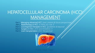
  • 1. HEPATOCELLULAR CARCINOMA (HCC): MANAGEMENT Pratap Sagar Tiwari Part 1: Managing Advanced HCC (past, present & future perspectives) Part 2: Radiotherapy in HCC (is there any role ?) Part 3: Locoregional Therapies in HCC (guidelines & beyond) Part 4: Surgery in HCC (limit and limitations ?) Part 5: Liver transplantation (HCC Perspective)
  • 2. PART 3 LOCOREGIONAL THERAPIES IN HCC (GUIDELINES & BEYOND) Clipart :http://www.scientificanimations.com/wiki-images/
  • 3. HCC MX: OVERVIEW Meets criteria for LT ? Resectable ? Locally Advanced? Advance HCC ? End stage HCC? LT Surgery Locoregional Therapies Chemotherapy BSC
  • 4. LOCOREGIONAL THERAPY MODALITIES Transarterial Chemoembolisation (TACE)Per cutaneous Ethanol Injection (PEI) Ablation therapies Vascular Intervention Therapies Combination therapies/ Future DirectivesIrreversible Electroporation (IRE) Microwave Ablation (MWA) Radiofrequency ablation(RFA) High-intensity focused ultrasound (HIFU) Hepatic artery infusion chemotherapy (HAIC) Transarterial Radioembolisation (TARE) • Laser-induced thermotherapy • Cryoablation • Percutaneous acetic acid injection
  • 5. LOCOREGIONAL THERAPY MODALITIES Per cutaneous Ethanol Injection (PEI) Ablation therapies
  • 6. PERCUTANEOUS ETHANOL ABLATION/INJECTION(PEI)  PEI for treatment of hepatic tumors was first described by Sugiura et al. [1] in 1983.  PEI was recommended as the standard percutaneous treatment for early-stage, non-surgical HCC in the 2001 guidelines of the EASL [2].  PEI has been reported to induce chemical coagulative necrosis in 70-80% of HCC lesions ≤30 mm in diameter [3-5], 95% of tumours <2 cm[6–8], but 50% when of 3–5 cm diameter [6–8], with survival of >50% at 5 years [9]. The best candidates are in Child-Pugh A stage with small HCC [10].  The ablation is limited to the lesion itself and may be suboptimal for the presence of internal septa: residual nests of vital neoplasia inside or just close to the pseudocapsule are hence detected in about one-third of cases during follow-up [11]. References are at the end of the slides.
  • 7. PEI: THE PROCEDURE  The procedure is discussed with the pt/patient’s family. Alternative treatments are discussed with the pt so an informed choice can be made. The pt is instructed to fast for 8 hr before the procedure, and a coagulation profile is checked. A combination of IV midazolam and fentanyl is used for sedation and analgesia.  The lesion or lesions are visualized via USG or CT . A single 20- or 22-gauge needle is inserted into the deep portion of the lesion, or more than one needle is inserted if the lesion is >3 cm. It is important to make as few passes as possible into the lesion because of possible reflux of alcohol back through excessive needle tracks . Lee MJ, et al. Percutaneous Ethanol Injection for the Treatment of Hepatic Tumors: Indications, Mechanism of Action, Technique, and Efficacy.AJR 1995;164:215-220
  • 8. PEI: THE PROCEDURE  Usually, most hepatologists inject approx 8-10 ml of alcohol per treatment session per lesion. However, larger volumes of alcohol have been injected, depending on the pt’s tolerance.  First, alcohol is injected in the deep part of the lesion, and then the needle is slowly withdrawn to the more superficial part while alcohol continues to be injected. In this way, it is hoped that ethanol will be distributed evenly through the lesion.  After alcohol injection, the needle(s) are left in place for 2-3 min so reflux of alcohol along the track is reduced. Reflux of alcohol along the needle track can lead to significant pain from penitoneal irritation.  Alternatively, gelatin sponge can be injected as the needle is withdrawn to prevent reflux of alcohol Lee MJ, et al. Percutaneous Ethanol Injection for the Treatment of Hepatic Tumors: Indications, Mechanism of Action, Technique, and Efficacy.AJR 1995;164:215-220
  • 9. PEI: THE PROCEDURE/RESPONSE  The injection is repeated once or twice a week for up to six to eight sessions, depending on the tumor size.  The therapeutic effect of PEI can be evaluated by contrast CT scan.  A CT scan was performed 1–3 days after the procedure to evaluate technique effectiveness (14). Complete ablation was defined as hypoattenuation of the entire tumour without contrast enhancement[1]. When the presence of unablated tumour portions was suspected, a few more procedures were performed.  Color Doppler USG and MRI are alternative imaging techniques for assessing the response to PEI.[2,3](done every 4 months in a largest study[4] 1. Ebara M, Kita K, Sugiura N, et al. Therapeutic effect of percutaneous ethanol injection on small hepatocellular carcinoma: evaluation with CT. Radiology 1995; 195:371–377. 2. Lencioni R, Caramella D, Bartolozzi C. Hepatocellular carcinoma: use of color Doppler US to evaluate response to treatment with percutaneous ethanol injection. Radiology 1995; 194:113–118. 3. Bartolozzi C, Lencioni R, Caramella D, et al. Treatment of hepatocellular carcinoma with percutaneous ethanol injection: evaluation with contrast-enhanced MR imaging. AJR Am J Roentgenol 1994; 162:827–831. 4. Shiina S, et al. Percutaneous ethanol injection for hepatocellular carcinoma: 20-year outcome and prognostic factors. Liver Int. 2012 Oct; 32(9): 1434–1442.
  • 10. PEI: INDICATIONS/CONTRAINDICATIONS  It is generally agreed that pts with HCCs 3 cm or smaller and three or fewer in number are the best candidates for PEI, although many centers perform PEI for HCCs up to 5 cm.[1-3]  PEI is CI in the presence of gross ascites, severe thrombocytopenia, or coagulopathy (platelet count <40,000/mm3, INR >1.75, in a study[5])because of a high risk of bleeding.  Large infiltrative tumors, thrombosis in the main portal or hepatic vein, and extrahepatic metastasis are also considered CIs in most centers.  Pts with tumors on the surface of the liver are not favorable candidates for PEI because the injected ethanol can leak back into the peritoneal cavity, and there is also a higher risk of tumor implantation into the peritoneal cavity.[4] 1. Ebara M, Ohto M, Sugiura N, et al. Percutaneous ethanol injection for treatment of small hepatocellular carcinoma. Study of 95 patients. J Gastroenterol Hepatol 1990; 5:616–626. 2. Livraghi T, Bolondi L, Lazzaroni S, et al. Percutaneous ethanol injection in the treatment of hepatocellular carcinoma in cirrhosis: A study on 207 patients. Cancer 1992; 69:925–929. 3. Di Stasi M, Buscarini L, Livraghi T, et al. Percutaneous ethanol injection in the treatment of hepatocellular carcinoma. A multicenter survey of evaluation practices and complication rates. Scand J Gastroenterol 1997; 32:1168–1173. 4. Shina S, Tagawa K, Unuma T, et al. Percutaneous ethanol injection therapy for hepatocellular carcinoma. A histopathologic study. Cancer 1991; 68:1524–1530. 5. brunello F, et al. Radiofrequency ablation versus ethanol injection for early hepatocellular carcinoma: A randomized controlled trial. Scandinavian Journal of Gastroenterology, 2008; 43: 727735
  • 11. PEI: COMPLICATIONS  The MC AES include fever, pain, and a feeling of alcohol intoxication. These are generally transient and may only require symptomatic therapy.  Depending on the site of the hepatic lesion, serious complications may occur.  When a lesion is subdiaphragmatic, the pleura may be transgressed, with possible resulting complications such as pneumothorax, pleural effusion, or hemothorax.  Intrapenitoneal hemorrhage has been reported in three of 190 pts by Shiina et al. [1].  Frequency of tumor seeding should theoretically be low because of the cytotoxic effect of ethanol on any tumor cells that may find their way into the percutaneous track [2]. 1. Shiina S, Niwa Y, Omata M. Percutaneous ethanol injection therapy for liver neoplasms. Semin Interient Radiol l993;1O(2):57-68 2. Zerbey AL, Mueller PA, Dawson SL, Hoover HC Jr. Pleural seeding from hepatocellular carcinoma: a complication of percutaneous alcohol ablation. Radiology l994
  • 12. PEI: COMPLICATIONS 1. Shiina S, Niwa Y, Omata M. Percutaneous ethanol injection therapy for liver neoplasms. Semin Interient Radiol l993;1O(2):57-68 2. Zerbey AL, Mueller PA, Dawson SL, Hoover HC Jr. Pleural seeding from hepatocellular carcinoma: a complication of percutaneous alcohol ablation. Radiology l994 • PEI is a minimally invasive therapy with a good safety record. • In a study of 746 pts with HCC treated by PEI, the treatment-related death rate was only 0.1%, and the rate of severe complications was 1.7%.[1] • A similar death rate (0.09%) and complication rate (3.2%) have been reported in a multicenter survey of 1,066 pts after PEI for HCC.[2] • Tumor seeding along the needle track after PEI for HCC has been reported, with an incidence of 1% in a study of 348 pts.[3] 1. Livraghi T, Giorgio A, Marin G, et al. Hepatocellular carcinoma and cirrhosis in 746 patients: long-term results of percutaneous ethanol injection. Radiology 1995; 197:101–108. 2. Di Stasi M, Buscarini L, Livraghi T, et al. Percutaneous ethanol injection in the treatment of hepatocellular carcinoma. A multicenter survey of evaluation practices and complication rates. Scand J Gastroenterol 1997; 32:1168–1173. 3. Ishii H, Okada S, Okusaka T, et al. Needle tract implantation of hepatocellular carcinoma after percutaneous ethanol injection. Cancer 1998; 82:1638–1642.
  • 13. LOCOREGIONAL THERAPY MODALITIES Per cutaneous Ethanol Injection (PEI) Ablation therapies Outcome of PEI ? PEI Vs LR ? PEI Vs RFA ? Outcome of RFA ? RFA Vs LR ? Radiofrequency Ablation (RFA)
  • 14. RADIOFREQUENCY ABLATION (RFA)  In the mid-1990s, RFA was proposed for the treatment of small HCC [1,2].  RFA is based on heat-generated coagulative necrosis that induces the ablation of the tumour itself and may involve small portions of surrounding liver tissue as well.  In RFA, an electrical current is delivered through a needle electrode under imaging or surgical guidance, producing heat- based thermal cytotoxicity[3].  Temperatures range between 60 °C to 100 °C and result in almost instant coagulation necrosis[4]. 1. Rossi S, Di Stasi M, Buscarini E, Quaretti P, Garbagnati F, Squassante L, et al. Percutaneous RF interstitial thermal ablation in the treatment of hepatic cancer. Am J Roengtenol 1996;167:75968. 2. De Baere T, Risse O, Kuoch V, Dromain C, Sengel C, Smayra T, et al. Adverse events during radiofrequency treatment of 582 hepatic tumors. Am J Roengtenol 2003;181:695700. 3. Goldberg SN, Gazelle GS, Mueller PR. Thermal ablation therapy for focal malignancy: a unified approach to underlying principles, techniques, and diagnostic imaging guidance. AJR Am J Roentgenol. 2000;174:323–331. 4. Gervais DA, Arellano RS. Percutaneous tumor ablation for hepatocellular carcinoma. AJR Am J Roentgenol. 2011;197:789–794. Clipart from: https://www.alamy.com/radiofrequency-ablation-image66575282.html
  • 15. RADIOFREQUENCY ABLATION (RFA) • RFA is moderated by the heat-sink effect, a phenomenon that occurs when thermal energy is dispersed from the target lesion due to blood flow in the vessels adjacent to it[1]. • Consequently, the shape and size of the ablation zone may be unpredictable and the efficacy of RFA may be restricted as multiple sessions are necessary for complete tumour eradication[2]. • Caution is needed in treating lesions close to hollow viscera owing to the risk of perforation. • Deep sedation is recommended. 1. Lu DS, Raman SS, Limanond P, Aziz D, Economou J, Busuttil R, Sayre J. Influence of large peritumoral vessels on outcome of radiofrequency ablation of liver tumors. J Vasc Interv Radiol. 2003;14:1267–1274. 2. Mulier S, Ni Y, Jamart J, Ruers T, Marchal G, Michel L. Local recurrence after hepatic radiofrequency coagulation: multivariate meta-analysis and review of contributing factors. Ann Surg. 2005;242:158–171.
  • 16. RADIOFREQUENCY ABLATION (RFA): CI/COMPLICATIONS  The ablation success rate for lesions <2 cm reaches 90% with a local recurrence rate of 1%[1]. For this reason, RFA is considered effective for tumours < 3 cm; combined LRT should be considered for lesions > 3 cm[2].  The main CI are severe bleeding diathesis (platelet count <50000/μL), ascites, jaundice and presence of metallic devices such as pacemakers.  RFA should also be avoided for tumours within 1 cm proximity to the hepatic portal tract.  Major complications include liver failure, bleeding, infection, abscesses, intercostal nerve injury, organ injury, tumour lysis syndrome and pneumothorax[3]. 1. Livraghi T, Meloni F, Di Stasi M, Rolle E, Solbiati L, Tinelli C, Rossi S. Sustained complete response and complications rates after radiofrequency ablation of very early hepatocellular carcinoma in cirrhosis: Is resection still the treatment of choice? Hepatology. 2008;47:82–89. 2. Peng ZW, Zhang YJ, Chen MS, Xu L, Liang HH, Lin XJ, Guo RP, Zhang YQ, Lau WY. Radiofrequency ablation with or without transcatheter arterial chemoembolization in the treatment of hepatocellular carcinoma: a prospective randomized trial. J Clin Oncol. 2013;31:426–432. 3. Georgiades CS, Hong K, Geschwind JF. Radiofrequency ablation and chemoembolization for hepatocellular carcinoma. Cancer J. 2008;14:117–122. 4. Livraghi T, Solbiati L, Meloni MF, Gazelle GS, Halpern EF, Goldberg SN. Treatment of focal liver tumors with percutaneous radio-frequency ablation: complications encountered in a multicenter study. Radiology. 2003;226:441–451. In a multicenter study of RFA for malignant liver tumours in 2320 pts, the rate of major complications reached 2.2% [4].
  • 17. ASSESSING RESPONSE  Typically the efficacy of percutaneous ablation therapy is assessed by dynamic cross-sectional imaging (CT or MRI) one month after therapy.  Standard methods to assess treatment response (eg, unidimensional Response Evaluation Criteria in Solid Tumors [RECIST] criteria ) involve measurement of tumor dimensions before and after treatment, but, methods such as these disregard the extent of necrosis, which is the end result of locoregional ablative therapies [1].  Others have proposed guidelines for assessment of response to locoregional ablative therapy that are based upon reduction in viable tumor burden. Although not entirely reliable, the absence of contrast uptake within the tumor is thought to reflect tumor necrosis, while the persistence of contrast uptake indicates persistent disease. Recurrence of tumor in the treated area (or elsewhere) is signaled by the reappearance of vascular enhancement. 1. Bruix J, Reig M, Rimola J, et al. Clinical decision making and research in hepatocellular carcinoma: pivotal role of imaging techniques. Hepatology 2011; 54:2238.
  • 18. IMAGING RESPONSE CRITERIA USED IN EVALUATION OF HCC AFTER TREATMENT 1. Modified from: WHO handbook for reporting results of cancer treatment. World Health Organization, Geneva, 1979. 2. Yaghmai V, Besa C, Kim E, et al. Imaging assessment of hepatocellular carcinoma response to locoregional and systemic therapy. AJR Am J Roentgenol 2013; 201:80. • A partially necrotic (black) HCC with an arterially- enhancing (viable) component (white) is shown. The arrows illustrate different measurements obtained for assessment of response according to the various criteria.  WHO measures two perpendicular dimensions of the entire lesion;  RECIST measures the single largest dimension of the entire lesion;  EASL measures perpendicular dimensions of just the enhancing (viable) tumor;  modified RECIST measures one single largest dimension of the enchancing tumor. • Both EASL criteria and modified RECIST are based upon measurements obtained using cross sectional imaging (CT or MRI) and contrast-enhanced arterial phase images.
  • 19. MODIFIED RECIST FOR HEPATOCELLULAR CARCINOMA
  • 21. PEI ; OUTCOME? Shiina S, et al. Percutaneous ethanol injection for hepatocellular carcinoma: 20-year outcome and prognostic factors. Liver Int. 2012 Oct; 32(9): 1434–1442. • Background: PEI is the best-known image-guided percutaneous ablation for HCC and a well-tolerated, inexpensive procedure with few adverse effects. However, there have been few reports on its long-term results. • Aims: To report a 20-year consecutive case series at a tertiary referral centre. • Methods: performed 2147 ethanol injection treatments on 685 primary HCC pts and analysed a collected database.
  • 22. BASELINE CHARACTERISTICS OF THE 685 PTS UNDERGOING PEI FOR PRIMARY HCC Shiina S, et al. Percutaneous ethanol injection for hepatocellular carcinoma: 20-year outcome and prognostic factors. Liver Int. 2012 Oct; 32(9): 1434–1442. *Anti-HCV was not tested in 12 pts. †Serum DCP level was not measured in 168 pts. ‡Serum AFP-L3 level was not measured in 461 pts. HBs-Ag, hepatitis B surface antigen; HCV, hepatitis C virus; AFP, α-fetoprotein; DCP, des-gamma-carboxy- prothrombin; AFP-L3, lectin-reactive α-fetoprotein. Data are expressed as mean ± standard deviation.
  • 23. PEI ; OUTCOME? Shiina S, et al. Percutaneous ethanol injection for hepatocellular carcinoma: 20-year outcome and prognostic factors. Liver Int. 2012 Oct; 32(9): 1434–1442. • With a median follow-up of 51.6 months, 5-, 10- and 20-year survival rates were 49.0% [95% CI= 45.3–53.0%], 17.9% (95% CI = 15.0–21.2%) and 7.2% (95% CI = 4..5–11.5%) respectively.
  • 24. COMPLICATIONS IN 2147 TREATMENTS OF ETHANOL INJECTION FOR HCC Shiina S, et al. Percutaneous ethanol injection for hepatocellular carcinoma: 20-year outcome and prognostic factors. Liver Int. 2012 Oct; 32(9): 1434–1442.
  • 25. PEI ; OUTCOME? Shiina S, et al. Percutaneous ethanol injection for hepatocellular carcinoma: 20-year outcome and prognostic factors. Liver Int. 2012 Oct; 32(9): 1434–1442. Results • Final CT demonstrated complete ablation of treated tumours in 2108 (98.2%) of 2147 RX. • With a median follow-up of 51.6 months, 5-, 10- and 20-year survival rates were 49.0% [95% CI= 45.3–53.0%], 17.9% (95% CI = 15.0–21.2%) and 7.2% (95% CI = 4..5–11.5%) respectively. • Multivariate analysis demonstrated that age, CP class, tumour size, tumour number and serum AFP were significant prognostic factors for survival. • Five-, 10- and 20-year local tumour progression rates were 18.2% (95% CI = 15.0–21.4%), 18.4% (95% CI = 15.2–21.6%) and 18.4% (95% CI = 15.2–21.6%) respectively. • There were 45 complications (2.1%) and two deaths (0.09%). Conclusions: • Ethanol injection was potentially curative for HCC, resulting in survival for more than 20 years. • This study suggests that new ablation therapies will achieve similar or even better long-term results in HCC.
  • 26. SURVIVAL RESULTS AFTER PEI THERAPY FOR HCC Poon RTP, et al. Locoregional Therapies for Hepatocellular Carcinoma: A Critical Review From the Surgeon’s Perspective. ANNALS OF SURGERY Vol. 235, No. 4, 466– 486 References are included at the end of slides Study/Year No. of Patients Tumor Size Survival % 1 year 3 year 5 year Ebara et al,[1] 1990 95 <3 cm 93 65 28 Livraghi et al,[2] 1992 162 <5 cm 90 63 - Castells et al,[3] 1993 30 ≤4 cm 83 55 - Shiina et al,[4] 1993 146 1–6.5 cm 79 46 38 Isobe et al,[5] 1994 37 ≤2 cm 95 70 - Lencioni et al,[6] 1995 105 <5 cm 96 68 32 Livraghi et al,[7] 1995 246 224 ≤3 cm 3–5 cm 97 94 68 57 40 37 Orlando et al,[8] 1997 35 <4 cm 86 33 - Castellano et al,[9] 1997 71 ≤5 cm 89 54 24 Lin et al,[10] 1999 47 ≤5 cm 85 61 -
  • 27. SURVIVAL RESULTS AFTER PEI THERAPY FOR HCC  BACKGROUND: The objective of this study was to evaluate the indications for PEI performed in a single session under general anesthesia for treating pts with LC and large (tumors > 5 cm) HCC, and relevant survival curves.  METHODS: Between November 1991 and November 1996, 108 pts were treated (a total of 128 procedures). They fell into 3 groups:  GROUP A: 24 pts with single, encapsulated HCC measuring from 5-8.5 cm  GROUP B: 63 pts with single, infiltrating HCC measuring from 5-10 cm or multiple HCC  GROUP C: 21 pts with advanced disease, either hepatic (Child's Class C) or HCC with PVT.  The mean amount of ethanol injected was 62 mL. The average hospital stay was 3.8 days. The mean follow-up time was 40 months. 1. Livraghi T, Benedini V, Lazzaroni S, et al. Long-term results of single-session percutaneous ethanol injection in patients with large hepatocellular carcinoma. Cancer 1998; 83:48–57.
  • 28. SURVIVAL RESULTS AFTER PEI THERAPY FOR HCC RESULTS:  The 1-, 2-, 3-, and 4-year survival rates were: 72%, 65%, 57%, and 44%, respectively, for Group A; 73%, 60%, 42%, and 18%, respectively, for Group B; and 46%, 25%, and 0%, respectively, for Group C.  Mortality was 0.7% (bleeding from esophageal varices in a Child's Class C patient).  The rate of major complications was 4.6% (1 case of peritoneal hemorrhage, 1 case of severe liver failure, 1 case of transient renal insufficiency, 1 case of peritoneal seeding, and 2 cases of infarctions of a segment adjacent to the tumor). 1. Livraghi T, Benedini V, Lazzaroni S, et al. Long-term results of single-session percutaneous ethanol injection in patients with large hepatocellular carcinoma. Cancer 1998; 83:48–57.
  • 29. PERCUTANEOUS ETHANOL ABLATION/INJECTION(PEI) OUTCOME? HAVE A DEFINITE ROLE IN EARLY STAGE OF HCC, GENERALLY ≤3CM 1 YR SURVIVAL: 90-95 % 3 YR SURVIVAL: 60-65 % 5 YR SURVIVAL: 30-40 %
  • 30. PEI VS LIVER RESECTION ?
  • 31. PEI VS LIVER RESECTION ?  OBJECTIVE: To compare disease recurrence and survival.  METHODS: A total of 76 pts were randomly assigned to 2 groups based on treatment; all had one or 2 tumors with diameter </=3 cm, with hepatitis without cirrhosis or Child class A or B cirrhosis without evident ascites or bleeding tendency. 1. Huang GT, Lee PH, Tsang YM, Lai MY, Yang PM, Hu RH, Chen PJ, Kao JH, Sheu JC, Lee CZ, et al. Percutaneous ethanol injection versus surgical resection for the treatment of small hepatocellular carcinoma: a prospective study. Ann Surg. 2005;242:36–42.
  • 32. PEI VS LIVER RESECTION ?  RESULTS: Follow-up ranged from 12 to 59 months.  Among PEI pts, 18 had recurrence 1 to 37 months . Three injection therapy patients died of 25, 37, and 57 months after treatment.  For the LR group, 15 had recurrence 2 to 54 months after treatment. Five resection patients died of cancer at 11, 20, 23, 26, and 52 months, respectively.  By Cox regression model and Kaplan-Meier survival analysis, there is no statistical significance for recurrence and survival between treatment groups.  However, tumor size larger than 2 cm and alpha-fetoprotein over 200 ng/mL correlated with higher recurrence rate, and Child class B liver cirrhosis correlated with shorter survival. 1. Huang GT, Lee PH, Tsang YM, Lai MY, Yang PM, Hu RH, Chen PJ, Kao JH, Sheu JC, Lee CZ, et al. Percutaneous ethanol injection versus surgical resection for the treatment of small hepatocellular carcinoma: a prospective study. Ann Surg. 2005;242:36–42. CONCLUSIONS: Percutaneous ethanol injection therapy appears to be as safe and effective as resection, and both treatments can be considered first-line options for small hepatocellular carcinoma.
  • 33. PEI VS LIVER RESECTION ?  On the other hand, Cho et al[1] reported that LR(n=113) had a trend towards better survival profile as compared to PEI (n=1160) in HCC patients (5-year overall survival 65% vs 49%) (P = 0.059). 1. Cho YB, Lee KU, Suh KS, Kim YJ, Yoon JH, Lee HS, Hahn S, Park BJ. Hepatic resection compared to percutaneous ethanol injection for small hepatocellular carcinoma using propensity score matching. J Gastroenterol Hepatol. 2007;22:1643–1649.
  • 34. PEI IN LIVER METASTASES ?  The effectiveness of PEI for the treatment of CLM is unclear. It was reported that in more than 50% of cases of liver metastases < 4 cm, complete necrosis can be obtained by means of PEI[1].  Giorgio et al[2] (n=33) pts with 62 large (> 3.5 cm) reported that the survival rates of pts with liver metastases who underwent PEI were 94%, 80%, 80%, and 44% at 12 mo, 24 mo, 36 mo, and 44 mo, respectively. 1. Giovannini M. Percutaneous alcohol ablation for liver metastasis. Semin Oncol. 2002;29:192–195 2. Giorgio A, Tarantino L, Mariniello N, De Stefano G, Perrotta A, Aloisio V, Del Viscovo L, Alaia A. [Ultrasonography-guided percutaneous ethanol injection in large an/or multiple liver metastasis] Radiol Med. 1998;96:238–242
  • 35. PEI; RECURRENCE RATE?  One major concern of PEI for HCC is the high incidence of recurrence.  The cumulative intrahepatic recurrence rates at 1, 3, and 5 years after PEI for small HCCs (5 cm) were in the range of 26-32%, 51-81%, and 60- 83%, respectively, in reported series.[1-6]  The majority of recurrences are new lesions at different portions of the liver, but local recurrence at the site of the initial lesion treated by PEI accounted for 16-38% .[1,4,6]  The substantial local recurrence rate may be a result of inhomogeneous diffusion of ethanol in the tumor. 1. Shiina S, Tagawa K, Niwa Y, et al. Percutaneous ethanol injection therapy for hepatocellular carcinoma: results in 146 patients. AJR Am J Roentgenol 1993; 160:1023–1028. 2. Castellano L, Calandra M, Del Vecchio Blanco C, de Sio I. Predictive factors of survival and intrahepatic recurrence of hepatocellular carcinoma in cirrhosis after percutaneous ethanol injection: analysis of 71 patients. J Hepatol 1997; 27:862– 870. 3. Pompili M, Rapaccini GL, de Luca F, et al. Risk factors for intrahepatic recurrence of hepatocellular carcinoma in cirrhotic patients treated by percutaneous ethanol injection. Cancer 1997; 79:1501–1508. 4. Hasegawa S, Yamasaki N, Hiwaki T, et al. Factors that predict intrahepatic recurrence of hepatocellular carcinoma in 81 patients initially treated by percutaneous ethanol injection. Cancer 1999;861682–1690. 5. Khan KN, Yatsuhashi H, Yamasaki K, et al. Prospective analysis of risk factors for early intrahepatic recurrence of hepatocellular carcinoma following ethanol injection. J Hepatol 2000; 32:269–278. 6. Koda M, Murawaki Y, Mitsuda A, et al. Predictive factors for intrahepatic recurrence after percutaneous ethanol injection therapy for small hepatocellular carcinoma. Cancer 2000; 88:529–537.
  • 36. PEI; RECURRENCE RATE?  A high intrahepatic recurrence rate is also a major problem after LR of HCC.[1] • Few studies have compared the recurrence rate after PEI and LR for small HCCs. • In a retrospective study, Okuda[2] found similar rates of new lesions after PEI and LR in two comparable groups of pts. • However, In a study comparing two cohorts of pts with solitary HCCs ≤ 4 cm treated by PEI and LR, the 2-year recurrence rates after PEI and LR were 66% and 45%, respectively, and the difference was most obvious among pts with tumors 3 to 4 cm in size.[3] • One advantage of PEI is its easy repeatability, which allows further treatment of both local and distant intrahepatic recurrences. • Thus, PEI can be used for treating recurrence in the liver remnant after resection of HCC.[1] 1. Poon RTP, Fan ST, Wong J. Risk factors, prevention and management of postoperative recurrence after resection of hepatocellular carcinoma. Ann Surg 2000; 232:10–24. 2. Okuda K. Intratumor ethanol injection. J Surg Oncol 1993(suppl); 3:97–99. 3. Castells A, Bruix J, Bru C, et al. Treatment of small hepatocellular carcinoma in cirrhotic patients: a cohort study comparing surgical resection and percutaneous ethanol injection. Hepatology 1993; 18:1121–1126.
  • 37. PEI VS LIVER RESECTION ? • NOT ADEQUATE EVIDENCES TO DRAW A CONCLUSION • HIGH INTRAHEPATIC RECURRENCE RATES WITH PEI • PEI IN LIVER METS: INCONCLUSIVE • NOT BENEFICIAL IN LARGER LESIONS (>3 CM) UNLIKE SURGERY • ADVANTAGES: EASY, CHEAP AND REPEATABILITY & CAN BE USED FOR TREATING RECURRENCE IN THE LIVER REMNANT AFTER RESECTION OF HCC.
  • 38. LOCOREGIONAL THERAPY MODALITIES Per cutaneous Ethanol Injection (PEI) Ablation therapies Outcome of PEI ? PEI Vs LR ? PEI Vs RFA ? Outcome of RFA ? RFA Vs LR ? Radiofrequency Ablation (RFA)
  • 39. RADIOFREQUENCY ABLATION: OUTCOME?  Reported 3- and 5-year survival rates following RFA for pts fulfilling the Milan transplantation criteria (single tumor <5 cm, or three or fewer tumors, each ≤3 cm) range from 70-91, and 48-77 %, respectively [1-3].  Long-term outcomes are better in pts with smaller lesions [4-6]. In one series of 302 pts, the 3- year survival rates for lesions >5 cm, 2.1 to 5 cm, and ≤2 cm were 59, 74, and 91 %, respectively [4]. 1. Lencioni R, Cioni D, Crocetti L, et al. Early-stage hepatocellular carcinoma in patients with cirrhosis: long-term results of percutaneous image-guided radiofrequency ablation. Radiology 2005; 234:961. 2. Takahashi S, Kudo M, Chung H, et al. Initial treatment response is essential to improve survival in patients with hepatocellular carcinoma who underwent curative radiofrequency ablation therapy. Oncology 2007; 72 Suppl 1:98. 3. Choi D, Lim HK, Rhim H, et al. Percutaneous radiofrequency ablation for early-stage hepatocellular carcinoma as a first-line treatment: long-term results and prognostic factors in a large single- institution series. Eur Radiol 2007; 17:684. 4. Tateishi R, Shiina S, Teratani T, et al. Percutaneous radiofrequency ablation for hepatocellular carcinoma. An analysis of 1000 cases. Cancer 2005; 103:1201. 5. Sala M, Llovet JM, Vilana R, et al. Initial response to percutaneous ablation predicts survival in patients with hepatocellular carcinoma. Hepatology 2004; 40:1352. 6. Livraghi T, Meloni F, Di Stasi M, et al. Sustained complete response and complications rates after radiofrequency ablation of very early hepatocellular carcinoma in cirrhosis: Is resection still the treatment of choice? Hepatology 2008; 47:82.
  • 40. RADIOFREQUENCY ABLATION: OUTCOME?  In an analysis of five and 10-year survival following RFA in a Japanese series of 1170 pts with HCC, the majority of whom fulfilled the Milan criteria [1]. Five and 10-year survival rates were 60 and 27 %, respectively. While the five and 10-year local progression rates were only 3 % each, the distant recurrence rates were 75 and 81 %, respectively.  Local recurrence rates are variable, ranging from 2 to 36 percent in other studies [2-6]. 1. Shiina S, Tateishi R, Arano T, et al. Radiofrequency ablation for hepatocellular carcinoma: 10-year outcome and prognostic factors. Am J Gastroenterol 2012; 107:569. 2. Curley SA, Izzo F, Ellis LM, et al. Radiofrequency ablation of hepatocellular cancer in 110 patients with cirrhosis. Ann Surg 2000; 232:381. 3. Lencioni RA, Allgaier HP, Cioni D, et al. Small hepatocellular carcinoma in cirrhosis: randomized comparison of radio-frequency thermal ablation versus percutaneous ethanol injection. Radiology 2003; 228:235. 4. Horiike N, Iuchi H, Ninomiya T, et al. Influencing factors for recurrence of hepatocellular carcinoma treated with radiofrequency ablation. Oncol Rep 2002; 9:1059. 5. Hori T, Nagata K, Hasuike S, et al. Risk factors for the local recurrence of hepatocellular carcinoma after a single session of percutaneous radiofrequency ablation. J Gastroenterol 2003; 38:977. 6. Harrison LE, Koneru B, Baramipour P, et al. Locoregional recurrences are frequent after radiofrequency ablation for hepatocellular carcinoma. J Am Coll Surg 2003; 197:759.
  • 41. FOLLOW-UP RESULTS AFTER RFA THERAPY FOR HCC Study No. Tumor Size (cm) Route Mean FU (mnths) CN % RR % Survival Rossi et al,[1] 1996 39 ≤3 P 22.6 95 41 1-yr, 94%, 3-yr, 68% 5-yr, 40% Rossi et al,[2] 1998 23 ≤3.5 P 10 100 28 - Allgaier et al,[3] 1999 12 - P 5 100 0 - Francica & Marone,[4] 1999 15 1–4.3 P 15* 90 33 - Curley et al,[5] 1999 48 - P (26), I (22) 15* 100 2.1 - Nicoli et al,[6] 2000 47 1–6 P (33), I (14) 11.8 100 - 2-yr, 83% Curley et al,[7] 2000 110 Mean 3.4 P (76), L (31), I (3) 19* 95 49 - Poggi et al,[8] 2001 15 1.5–6.2 P 9.2 88 20 - Buscarini et al,[9] 2001 88 ≤3.5 P 34 93 39 1-year, 89% 3-year, 62% 5-year, 33%
  • 43. RFA VS LIVER RESECTION  Two systematic reviews[1,2] showed no significant differences in OS between RFA and LR for HCC within the Milan criteria, although RFA was a/with higher recurrence rates. (note: in study by Cho YK (inc 2 RCT/550/550)stated as: OS for very early stage HCC, defined as an asymptomatic solitary small HCC ≤ 2 cm, showed that primary RFA with a 9% local recurrence rate is comparable to LR with a 3% operative mortality rate.)  Zhou et al[3] performed a meta-analysis of 1 RCT and 9 controlled studies comparing RFA(n=774) and LR(n=667) for HCC. The 5-year OS rate was borderline significant [OR 0.60 (0.36-1.01)]. RFA was a/with higher local recurrence [OR 4.50 (2.45-8.27)] and the disease-free survival rates were significantly better for LR pts. 1. Cho YK, Rhim H, Noh S. Radiofrequency ablation versus surgical resection as primary treatment of hepatocellular carcinoma meeting the Milan criteria: a systematic review. J Gastroenterol Hepatol. 2011;26:1354–1360. 2. Zhou Y, Zhao Y, Li B, Xu D, Yin Z, Xie F, Yang J. Meta-analysis of radiofrequency ablation versus hepatic resection for small hepatocellular carcinoma. BMC Gastroenterol. 2010;10:78. RFA VS LR  OS: NO SIGNIFICANT DIFFERENCE  RECURRENCE RATE: HIGHER IN RFA  DISEASE FREE SURVIVAL: HIGHER IN LR
  • 44. SUMMARY OF META-ANALYSES OF RFA VS LR IN HCC Author RX N 5 YR Local Recurrence (OR) 95% CI 5 YR Overall survival (OR) 95% CI Changyong et al [1] (inc 3 RCT) LR RFA 2313 2419 0.57 (0.50–0.65) P < 0.00001 Xu G et al [2] (Inc 2 RCT) LR RFA 1233 1302 1.68 (1.21 to 2.34) 0.60 (0.43 to 0.84) 1. Chamhgyong E, et al. Efficacy comparison of radiofrequency ablation and hepatic resection for hepatocellular carcinoma: A meta-analysis. JCRT 2017;13 (4): 625-30 2. Xu gang, et al. Meta-analysis of surgical resection and radiofrequency ablation for early hepatocellular carcinoma. World Journal of Surgical Oncology 2012;10:163 XU G et al: The complication of LR was higher than RFA (OR, 6.25 (95%CI, 3.12 to 12.52); P = 0.000).
  • 45. COMPARISON OF LR AND RFA FOR SMALL HCC: A META- ANALYSIS OF 16,103 PTS OS No of studies pts RFA (%) LR (%) OR [95% CI] P value ≤5 cm 3 YR 31 16,103 78.6 83.9 0.65 [0.53, 0.80] <0.001 5 YR 30 14,655 60.8 71.4 0.57 [0.48, 0.67] <0.001 ≤3 cm 3 YR 19 13,298 81.2 85.7 0.62 [0.43, 0.89] 0.009 5 YR 16 13,075 61.8 71.9 0.55 [0.42, 0.72] <0.001 ≤2 cm 3 YR 4 442 80.6 83.7 0.54 [0.12, 2.37] 0.41 5 YR 4 442 69.0 74.2 0.65 [0.27, 1.55] 0.33 Xu Q, et al. Comparison of Hepatic Resection and Radiofrequency Ablation for Small Hepatocellular Carcinoma: A Meta-Analysis of 16,103 Patients. Nature 2014
  • 46. COMPARISON OF LR AND RFA FOR SMALL HCC: A META- ANALYSIS OF 16,103 PTS • The analysis showed that although LR was a/ with higher complication rate and longer hospital-stay, LR is proposed as the first-line treatment rather than RFA for pts with HCCs larger than 2 cm. • For pts with HCCs of 2 cm or less, RFA may be an alternative to LR because of their comparable long-term efficacy. Xu Q, et al. Comparison of Hepatic Resection and Radiofrequency Ablation for Small Hepatocellular Carcinoma: A Meta-Analysis of 16,103 Patients. Nature 2014
  • 47. RFA VS LIVER RESECTION IN CLM  Wu et al[1](non RCT) metanalysis that compared RFA and LR for solitary CLM showed that LR was better than RFA in terms of 5-year overall survival, as well as local control of the disease.  Amerongen et al [2] also showed similar results. 1. Wu YZ, Li B, Wang T, Wang SJ, Zhou YM. Radiofrequency ablation vs hepatic resection for solitary colorectal liver metastasis: a meta-analysis. World J Gastroenterol. 2011;17:4143–4148. 2. Amerongen MJ,et al. Radiofrequency ablation compared to surgical resection for curative treatment of patients with colorectal liver metastases – a meta-analysis. HPBONLINE September 2017Volume 19, Issue 9, Pages 749–756 Author DX RX N Local Recurrence (OR) Overall survival (OR) Wu et al[1] CLM LR RFA 273 574 - 4.89, 95% CI: 1.73- 13.87, P = 0.003 0.41, 95% CI: 0.22- 0.90, P = 0.008(5 yr) - Amerongen et al [2] CLM RFA LR 1060 1817 1.66, 95% CI = 1.15–2.40, P = 0.007 2.35, 95% CI = 1.49–3.69, P = 0.001(5 yr)
  • 48. RFA VS LIVER RESECTION IN CLM • In conclusion, RFA is a minimally invasive modality for the treatment of CLM which has lower complication rates as compared to LR. • However, RFA is also currently a/with increased recurrence rates and lower disease-free and overall survival and should therefore only be used for pts who are not optimal candidates for surgery. • Furthur RCT is needed for definite summary.
  • 49. RFA LR Less Invasive Invasive Less Complications More Complications Short hospital stay Longer hospital stay Cost effective Cost ineffective Above benefit more on tumor <3 cm (limited ablation volume) Unrelated to size, provided adequate FHR High recurrence (Heat sink effect/needle tract seedling) Less recurrence Not suitable for specially located tumors, such as underneath the liver capsule, the adjacent Gb, or diaphragm. (Technically infeasible tumors) Allows the identification of histological parameters that predict recurrence risk. Similar efficacy to LR in CP-A , HCC<3 CM If LT is not feasible, ablation could be considered as the first-line option for pts with very early-stage HCC, and LR could be second line for pts in whom ablation cannot be done or fails.[1] 1. Forner A,et al. Hepatocellular carcinoma. Lancet 2018; 391: 1301–14
  • 51. PEI VS RFA ? Lencioni RA. Small hepatocellular carcinoma in cirrhosis: randomized comparison of radio-frequency thermal ablation versus percutaneous ethanol injection. Radiology. 2003 Jul;228(1):235-40. Epub 2003 May 20. PURPOSE: To compare the effectiveness of RFA with that of PEI for the RX of small HCC in pts with cirrhosis. MATERIALS AND METHODS: A series of 102 pts with LC and either single HCC 5 cm in diameter or smaller or as many as three HCCs each 3 cm or smaller (overall number of lesions, 142) randomly received either RFA (n = 52) or PEI (n = 50) as the sole first-line anticancer treatment. Mean follow-up was 22.9 months +/- 9.4 (SD) in the RF group and 22.4 months +/- 8.6 in the PEI group.
  • 52. PEI VS RFA ? Lencioni RA. Small hepatocellular carcinoma in cirrhosis: randomized comparison of radio-frequency thermal ablation versus percutaneous ethanol injection. Radiology. 2003 Jul;228(1):235-40. Epub 2003 May 20.
  • 53. PEI VS RFA ? • One- and 2-year local recurrence-free survival rates were 98% and 96% in the RF group and 83% and 62% in the PEI group, respectively (RR = 0.17; 95% CI: 0.06, 0.51; P =.002).
  • 54. PEI VS RFA ? Lencioni RA. Small hepatocellular carcinoma in cirrhosis: randomized comparison of radio-frequency thermal ablation versus percutaneous ethanol injection. Radiology. 2003 Jul;228(1):235-40. Epub 2003 May 20. • RESULTS: One- and 2-year survival rates were 100% and 98% in the RF group and 96% and 88% in the PEI group, respectively ( RR = 0.20; 95% CI: 0.02, 1.69; P =.138). • One- and 2-year local recurrence-free survival rates were 98% and 96% in the RF group and 83% and 62% in the PEI group, respectively (RR = 0.17; 95% CI: 0.06, 0.51; P =.002). • One- and 2-year event-free survival rates were 86% and 64% for the RF group and 77% and 43% for the PEI group, respectively (RR = 0.48; 95% CI: 0.27, 0.85; P =.012). CONCLUSION: RFA is superior to PEI with respect to local recurrence-free survival rates.
  • 55.  To compare PEI, the standard approach which has been used for many years to treat early non- surgical HCC in cirrhotic pts, and RFA, which has become an interesting alternative.  MATERIAL AND METHODS: A randomized trial was carried out on 139 cirrhotic pts in C-P A/B with 1-3 nodes of HCC (diameter 15-30 mm), for a total of 177 lesions.  Pts were randomized to receive RFA (n=70) or PEI (n=69).  The pEP was complete response (CR) 1 year after the percutaneous ablation of all HCC nodes identified at baseline.  sEP : early (30-50 days) CR, complications, survival and costs.
  • 56.
  • 57.
  • 58. The OS rate was not significantly different in the RFA versus PEI arm (HR=0.88, 95% CI: 0.50-1.53).
  • 59.  Twenty-three pts in the PEI arm needed a second cycle of treatment, whereas just 9 of the RFA arm underwent a second cycle.  Treatment failures were 20 (2 patients died) in the PEI arm and 3 in the RFA arm.  Complications occurred in 10 (14.3%) of the 70 pts treated by RFA and in 12 (17.4%) of the 69 treated by PEI (p0.616).  There were 4 major complications: 2 in the RFA arm (1 haemoperitoneum; 1 right haemothorax that needed urgent thoracotomy) and 2 in the PEI arm (1 haemoperitoneum ; 1 death).  The fatality occurred in a man, aged 78 years, who was treated by PEI for two lesions and developed thrombosis of the left portal branch; the patient died 10 days after the procedure with a clinical syndrome suggestive of bowel infarction, and the necropsy showed diffuse ischaemic damage to the small bowel. The fatality was considered possibly to be related to PEI.  No case of seeding was observed.
  • 60.  RESULTS: In an intention-to-treat analysis, 1-year CR was achieved in 46/70 (65.7%) and in 25/69 (36.2%) patients treated by RFA and PEI, respectively (p=0.0005).  For lesions >20 mm in diameter, there was a larger CR rate in the RFA group (68.1% versus 26.3%).  An early CR was obtained in 67/70 (95.7%) pts treated by RFA compared with 42/64 (65.6%) pts treated by PEI (p=0.0001).  Complications occurred in 10 and 12 pts treated by RFA and PEI, respectively.  The OS rate was not significantly different in the RFA versus PEI arm (adjusted HR=0.88, 95% CI: 0.50-1.53).  There was an incremental health-care cost of 8286 euro for each additional pt successfully treated by RFA. CONCLUSIONS: The 1-year CR rate after percutaneous treatment of early HCC was significantly better with RFA than with PEI but did not provide a clear survival advantage in cirrhotic pts.
  • 61. SUMMARY OF META-ANALYSES OF RFA VS PEI Author DX RX N Local Recurrence (RR) Overall survival (RR) Bouza et al[1] HCC RFA PEI 396 391 0.37 (0.23-0.59) 1.24 (1.05-1.48) (4 yr) 1. Bouza C, López-Cuadrado T, Alcázar R, Saz-Parkinson Z,Amate JM. Meta-analysis of percutaneous radiofrequency ablation versus ethanol injection in hepatocellular carcinoma. BMC Gastroenterol 2009; 9: 31 • Six studies were eligible. (5 RCT ,1 qRCT ). In general, subjects were in CP-A (74%) and had unresectable HCC (mean size 2.5 cm). Mean follow-up was 25 +/- 11 months. • The survival rate showed a significant benefit for RFA over PEI at one, two, three and four years. The advantage in survival increased with time with Relative Risk values of: 1.28 (95%CI:1.12-1.45) and 1.24 (95%CI:1.05-1.48) for RFA versus PEI at 3- and 4-years respectively. • Likewise, RFA achieved significantly lower rates of local recurrence (RR: 0.37, 95%CI: 0.23-0.59). • The overall rate of AEs was higher with RFA (RR:2.55, 95%CI: 1.8-3.6) yet no significant differences were found .
  • 62. PERCUTANEOUS ACETIC ACID INJECTION  The efficacy of PEI is limited by the presence of septa in the tumor nodule, which prevents uniform diffusion of ethanol and necessitates repeated treatment sessions. Further, capsular invasion cannot be ablated because ethanol cannot dissolve the fibrous capsule. Percutaneous acetic acid injection (PAI) has been used as an alternative.[1]  Acetic acid has a stronger necrotizing power than ethanol because it can dissolve lipids and extract collagen. Its low pH induces swelling of the fibers and promotes dissociation of intermolecular collagen cross-links. 1. Ohnishi K, Ohyama N, Ito S, Fujiwara K. Small hepatocellular carcinoma: treatment with US-guided intratumoral injection of acetic acid. Radiology 1994; 193:747– 752.
  • 63. PERCUTANEOUS ACETIC ACID INJECTION  One randomized controlled trial conducted in 60 pts with HCCs smaller than 3 cm showed that the survival of pts treated with PAI was significantly better than those treated with PEI (2-year survival rate, 92% vs. 63%).[1]  The local recurrence rate was also significantly lower in the PAI group compared with the PEI group (2-year local recurrence rate, 10% vs.44%).  In that study, major treatment-related complications were rare with both modalities. However, no further data are available to confirm the advantages of PAI over PEI. 1. Ohnishi K, Yoshioka H, Ito S, Fujiwara K. Prospective randomized controlled trial comparing percutaneous acetic acid injection and percutaneous ethanol injection for small hepatocellular carcinoma. Hepatology 1998; 27:67–72.
  • 64. PEI VS PAI VS RFA ?  BACKGROUND & AIMS: To evaluate the evidence comparing RFA, PEI and PAI using meta- analytical techniques.  METHODS: Literature search was undertaken until December 2008 to identify comparative studies evaluating survival, recurrence, complete necrosis of tumour and complications. Only randomized clinical trials and quasi-randomized studies were included. 1. Germani G, Pleguezuelo M, Gurusamy K, Meyer T, Isgrò G, Burroughs AK. Clinical outcomes of radiofrequency ablation, percutaneous alcohol and acetic acid injection for hepatocelullar carcinoma: a meta-analysis. J Hepatol. 2010 Mar;52(3):380-8. doi: 10.1016/j.jhep.2009.12.004. Epub 2010 Jan 17.
  • 65. PEI VS PAI VS RFA ?  RESULTS: Eight studies were identified: RFA vs. PEI (n=5), PAI vs. PEI (n=2) and RFA vs. PAI vs. PEI (n=1) including 1035 pts.  PAI did not differ significantly from PEI for survival (OR 0.55; 95% CI 0.23-1.33; p=0.18), and local recurrence but required less sessions.  PAI had similar outcomes, except local recurrence, to RFA in the direct and indirect comparison.  RFA was superior to PEI for survival (OR 0.52; 95% CI 0.35-0.78; p=0.001), complete necrosis of tumour and local recurrence.  For tumours ≤2 cm RFA was not significantly better than PEI. 1. Germani G, Pleguezuelo M, Gurusamy K, Meyer T, Isgrò G, Burroughs AK. Clinical outcomes of radiofrequency ablation, percutaneous alcohol and acetic acid injection for hepatocelullar carcinoma: a meta-analysis. J Hepatol. 2010 Mar;52(3):380-8. doi: 10.1016/j.jhep.2009.12.004. Epub 2010 Jan 17. CONCLUSIONS: • RFA seems to be a superior ablative therapy than PEI for HCC, particularly for tumours >2 cm. • For tumours ≤2 cm outcome benefits comparing RFA and PEI are similar. • PAI did not differ significantly from PEI for all the outcomes evaluated. • RFA and PAI have similar survival rates. • PAI needs re-evaluation versus both PEI and RFA for tumours ≤2 cm.
  • 66. LOCOREGIONAL THERAPY MODALITIES Per cutaneous Ethanol Injection (PEI) Ablation therapies Outcome of PEI ? PEI Vs LR ? PEI Vs RFA ? Outcome of RFA ? RFA Vs LR ? Radiofrequency Ablation (RFA) • The meta-analysis of several studies shows conclusively that RFA is superior to PEI in terms of proportion dead, complete necrosis of targeted nodule, incidence of local recurrence, and number of treatment sessions. • Moreover time to death and time to recurrence were significantly better in pts treated with RFA. Winner: RFA
  • 67. MICROWAVE ABLATION (MWA): INTRO  MWA Vs RFA: They differ in their MOA: RFA uses electric current, whereas MWA uses electromagnetic energy.  However, the principle underlying these two treatment methods is similar, in that high temperatures are induced in localized areas to coagulate tissue.  MWA has several advantages over RFA. The heat-sink effect, is an important disadvantage of RFA. Consequently, the shape and size of the ablation zone in RFA may be unpredictable, and such limitations can lead to an inadequate ablation zone.  For MWA, the time needed for ablation is less than that required for RFA as MWA can deliver higher temperatures in the ablation zone.  These features of MWA lead to a more predictable ablation zone.  Therefore, the apparent superiority of MWA over RFA has made it an alternative method to RFA, and it may potentially be a more powerful technique than RFA.
  • 68. RFA VS MWA RFA MWA Electric current Electromagnetic energy Grounding pads (risk of burns due to ground pads) No grounding pads (no risk of burns) Lower intratumoral temperatures Higher intratumoral temperatures More peri-procedural pain Less peri-procedural pain Unpredictable ablation zone More predictable ablation zone Heat-sink effect Less susceptible to heat-sink effect Single lesion can be treated Simultaneous treatment of multiple lesions More procedural time Shorter procedural time Less ablation volume Larger ablation volume Similar complications and complication rate Surgical clips or pacemaker are contraindications Surgical clips or a pacemaker not a contraindication Modified from: Poulou LS,et al. Percutaneous microwave ablation vs radiofrequency ablation in the treatment of hepatocellular carcinoma. World J Hepatol. 2015 May 18; 7(8): 1054–1063.
  • 69. MICROWAVE ABLATION (MWA)  Indications and CIs for MWA are the same as those for RFA, apart from the size of a lesion that can be ablated; according to most studies, MWA can treat 5-8 cm tumours[1].  Furthermore, MWA allows simultaneous ablation of multiple tumours or even combined resection and ablation. 1. Liang P, Yu J, Lu MD, Dong BW, Yu XL, Zhou XD, Hu B, Xie MX, Cheng W, He W, et al. Practice guidelines for ultrasound-guided percutaneous microwave ablation for hepatic malignancy. World J Gastroenterol. 2013;19:5430–5438.
  • 70. MWA: COMPLICATIONS  In a multicentre study, 736 pts with hepatic lesions underwent MWA; the reported rate of major complications was 2.9%.  MWA was not proven to increase the risk of damage of vascular structures and/or bleeding.  Minor complications included pain, post-ablation syndrome, and asymptomatic pleural effusions, which are usually self-limiting and do not require any further treatment.  With the peri-procedural mortality rate being reported to be as low as < 0.01%, the safety of MWA was established[1]. 1. Livraghi T, Meloni F, Solbiati L, Zanus G; Collaborative Italian Group using AMICA system. Complications of microwave ablation for liver tumors: results of a multicenter study. Cardiovasc Intervent Radiol. 2012;35:868–874.
  • 71. MWA: TUMOR RECURRENCE  MWA shares a high rate of local recurrence in HCC with all other ablation modalities.  Lee et al[1](n=26) studied MWA in tumours of 2-6 cm in diameter. All early postoperative CT imaging showed no residual lesions; however, after a median follow-up of 14 (range, 4-26) months. , 42% of pts experienced local tumour progression.  As Lee et al[1] noted, high local tumour progression is a drawback of MWA and can be attributed to the use of a large applicator (5 mm in diameter), which increases the risk of tumour puncture and subsequent tumour seeding. 1. Lee KF, Hui JW, Cheung YS, Wong JS, Chong CN, Wong J, Yu SC, Lai PB. Surgical ablation of hepatocellular carcinoma with 2.45-GHz microwave: a critical appraisal of treatment outcomes. Hong Kong Med J. 2012;18:85–91.
  • 72. MWA: OUTCOME  One of the largest series reporting long-term outcomes from MWA involved 288 pts treated at a single institution over an eight year period [1].  The mean maximum tumor diameter was 3.75 cm, and 63 % had a single lesion.  At a mean follow-up of 31 months, only 24 pts (8 %) had local regrowth of a treated lesion, while 76 (26 %) developed new tumors elsewhere.  Cumulative survival rates at one, three, and five years were 93, 72, and 52 percent, respectively, and were significantly better for pts with maximum tumor size ≤4 cm as compared to larger tumors. 1. Liang P, Dong B, Yu X, et al. Prognostic factors for survival in patients with hepatocellular carcinoma after percutaneous microwave ablation. Radiology 2005; 235:299.
  • 74. MICROWAVE ABLATION (MWA)  BACKGROUND: MWA has several advantages over RFA for the treatment of HCC. Aimed to compare the efficacy and safety of MWA with those of RFA for HCC from the perspectives of percutaneous and laparoscopic approaches.  METHODS: PubMed/MEDLINE, Embase, the Cochrane library, and China Biology Medicine databases were searched. Studies comparing the efficacy and safety of MWA with those of RFA in patients with HCC were considered eligible. Complete ablation (CA), local recurrence (LR), disease-free survival (DFS), overall survival (OS), and the major complication rate were compared between MWA and RFA.
  • 75. CHARACTERISTICS OF THE INCLUDED STUDIES
  • 76. MWA can produce more uniform and larger tumor necrosis. However, these characteristics, in turn, are associated with a theoretically increased risk of damaging neighboring organs, especially vascular and biliary structures. A larger ablation area might account for a higher complication rate MICROWAVE ABLATION (MWA)  RESULTS: Four RCTs and 10 cohort studies were included.  For percutaneous ablation, no significant difference was found between MWA and RFA regarding CA, LR, DFS, OS, and the major complication rate.  A subgroup analysis of tumors measuring ≥3 cm revealed no difference in CA and LR for percutaneous ablation.  For laparoscopic ablation, a significantly lower LR rate and a non-significant trend toward a higher major complication rate were observed for the MWA group (OR 2.16, 95% CI 1.16-4.02, p = .01 for LR; OR 0.21, 95% CI 0.04-1.03, p = .05 for major complication rate).  CA, DFS, and OS were similar between the two groups.
  • 77. CONCLUSIONS: Percutaneous (P)-MWA had similar therapeutic effects compared with P-RFA for HCC. Pts undergoing laparoscopic MWA had a lower LR rate; however, their major complication rate appeared to be higher. The superiority of MWA over RFA remains unclear and needs to be confirmed by high-quality evidence.
  • 78. OTHER TREATMENTS Irreversible Electroporation (IRE) Laser-induced thermotherapy (LA) Cryoablation (CA) High-intensity focused ultrasound (HIFU)
  • 79. LASER ABLATION THERAPY (LA)  It causes tissue destruction by hyperthermic coagulative necrosis.  Using a single conventional neodymium-yttrium-aluminum-garnet (Nd-YAG) laser fiber, the maximum diameter of the ablated lesion is 2 cm, although larger lesions can be ablated by laser splitting with simultaneous heating of multiple probes or diffuse-tip fibers that emit the laser light in a more diffuse fashion.[1]  Even with these newly designed laser fibers, the limiting diameter for adequate tissue destruction using laser currently is approx 5 cm.[2]  The cooling effect of blood flow in nearby vessels is also a problem with laser ablation, but the size of ablation can be significantly ↑ by eliminating portal flow using the Pringle maneuver.[1] 1. Heisterkamp J, van Hillegersberg R, Mulder PG, et al. Importance of eliminating portal flow to produce larger intrahepatic lesions with interstitial laser coagulation. Br J Surg 1997; 84:1245–1248. 2. Christophi C, Muralidharan V. Treatment of hepatocellular carcinoma by percutaneous laser hyperthermia. J Gastroenterol Hepatol 2001; 16:548–552.
  • 80. LA  Laser ablation can be performed percutaneously, laparoscopically, or during surgery.  A study of percutaneous laser ablation of 676 pts with liver secondaries or HCC revealed no clinically relevant complications.[1]  However, careful pt selection in terms of liver function is important, because liver failure and death after laser ablation of HCC have been reported in pts with Child C cirrhosis.[2] 1. Vogl TJ, Eichler K, Straub R, et al. Laser-induced thermotherapy of malignant liver tumors: general principles, equipment, procedure, side effects, complications and results. Eur J Ultrasound 2001; 13: 117–127. 2. Giorgio A, Tarantino L, de Stefano G, et al. Interstitial laser photocoagulation under ultrasound guidance of liver tumors: results in 104 treated patients. Eur J Ultrasound 2000; 11:181–188.
  • 81. LA: OUTCOME  Most of the studies on laser therapy for liver tumors involved pts with metastatic disease, and few data exist on the clinical efficacy of laser ablation for HCC.  A study showed that PCLA induced CN in 70 (82%) of 85 HCC nodules,[1] but there were no long-term data on survival or recurrence.  In a small series of 8 pts with HCC treated by PCLT ,follow-up CT scan showed CN in all pts with HCCs <4 cm but not in tumors >5 cm despite repeated treatments, and histologic examination of two large HCCs revealed a peripheral rim of viable cells after laser therapy.[2] 1. Giorgio A, Tarantino L, de Stefano G, et al. Interstitial laser photocoagulation under ultrasound guidance of liver tumors: results in 104 treated patients. Eur J Ultrasound 2000; 11:181–188. 2. Christophi C, Muralidharan V. Treatment of hepatocellular carcinoma by percutaneous laser hyperthermia. J Gastroenterol Hepatol 2001; 16:548–552.
  • 82. LASER THERAPY: OUTCOME ?  PURPOSE: Long-term outcome of LC pts with early HCC treated with USG-guided percutaneous LA  PATIENTS AND METHODS: retrospectively analyzed treatment and survival parameters of 432 cirrhotic pts with nonsurgical early HCC (single nodule < or = 4 cm or three nodules < or = 3 cm each) who had received PLA in nine Italian centers. 1. Pacella CM, Francica G, Di Lascio FM, Arienti V, Antico E, Caspani B, Magnolfi F, Megna AS, Pretolani S, Regine R, Sponza M, Stasi R. Long-term outcome of cirrhotic patients with early hepatocellular carcinoma treated with ultrasound-guided percutaneous laser ablation: a retrospective analysis. J Clin Oncol. 2009 Jun 1;27(16):2615-21.
  • 83. LASER THERAPY: OUTCOME ?  RESULTS: Single tumors were seen in 344 (80%) of 432 pts, and two to three nodules were seen in 88 pts (20%), for a total of 548 tumors.  An initial complete response after PLA was observed in 338 pts (78%).  Median OS was 47 months (95% CI, 41 to 53 months).  The 3- and 5-year cumulative survival rates were 61% and 34%, respectively.  In multivariate analysis, independent predictors of survival were serum albumin >3.5 g/dL (P = .002; RR = 0.580; 95% CI, 0.409 to 0.821), the achievement of a CA (P = .001; RR = 0.517; 95% CI, 0.346 to 0.771), and age <73 years (P < .001; RR = 0.466; 95% CI, 0.320 to 0.681).  C-P A pts had a 5-year cumulative survival rate of 41%; this figure increased up to 60% with a median survival time of 63 mnths (95% CI, 48- 78 months) in pts with tumors < or = 2.0 cm. 1. Pacella CM, Francica G, Di Lascio FM, Arienti V, Antico E, Caspani B, Magnolfi F, Megna AS, Pretolani S, Regine R, Sponza M, Stasi R. Long-term outcome of cirrhotic patients with early hepatocellular carcinoma treated with ultrasound-guided percutaneous laser ablation: a retrospective analysis. J Clin Oncol. 2009 Jun 1;27(16):2615-21. CONCLUSION: This analysis confirms that a complete tumor ablation results in improved survival in patients with nonsurgical HCC. Ideal candidates for PLA are younger pts with normal serum albumin levels and tumor size < or = 2 cm.
  • 84. LA VS RFA ; WHICH ONE IS SUPERIOR ?
  • 85. LA VS RFA  Aim: In pts with cirrhosis and small HCC, thermal ablation is currently recognized as an effective local treatment. Among thermal procedures, RFA is the most diffusely used and is the standard against which any new treatment should be compared.  In retrospective studies, laser ablation (LA) resulted as safe and effective as RFA. Therefore, performed a non-inferiority RCT comparing RFA with LA in pts with cirrhosis and HCC within Milan criteria.  Methods: Overall, 140 pts with 157 HCC nodules were randomly assigned to receive RFA(70) or LA(70).  The pEP was the proportion of complete tumor ablation (CTA).  sEP were time to local progression (TTLP) and overall survival (OS). 1. Pacella CM, et al. Radiofrequency Ablation versus Laser Ablation for the Treatment of Small Hepatocellular Carcinoma in Cirrhosis: a Randomized Trial.Journal of Gastroenterology and Hepatology 2015 30(3):559-565
  • 86. The design of the study scheduled up to 3 ablation procedures during a 6-month period. Primary outcome measure was CTA defined as absence of any contrast enhancement within or at the periphery of the HCC nodule. Treatment failure was defined when imaging showed a residual nodule activity after the 3 scheduled ablations. For these pts, TACE was recommended. LA VS RFA  Results: Per pt CTA rates after RFA and LA were 97.4% (95% CI, 91.0–99.3) and 95.7% (88.1–98.5), respectively (difference =1.4%, 95% CI from −6.0% to +9.0%).  Per nodule CTA rates for RFA and LA were 97.4% (91.0–99.3) and 96.3% (89.6–98.7), respectively (difference =1.1%, from −5.7% to +8.1%).  The mean TTP was comparable between RFA group (42.0 months; 95% CI, 36.83–47.3) and LA group (46.7 months; 95% CI, 41.5–51.9) (P=.591).  The mean OS was 42 months in both groups and survival probability at 1 and 3 years was 94% and 89% in RFA group, and 94% and 80% in LA group. 1. Pacella CM, et al. Radiofrequency Ablation versus Laser Ablation for the Treatment of Small Hepatocellular Carcinoma in Cirrhosis: a Randomized Trial.Journal of Gastroenterology and Hepatology 2015;30(3):559-565
  • 87. LA VS RFA Conclusion: LA resulted not inferior to RFA in inducing the CP-A of HCC nodules and therefore it should be considered as an evaluable alternative for thermal ablation of small HCC in cirrhotic patients
  • 88. DIFFERENCES BETWEEN LA VS RFA 1. Pacella CM, et al. Radiofrequency Ablation versus Laser Ablation for the Treatment of Small Hepatocellular Carcinoma in Cirrhosis: a Randomized Trial.Journal of Gastroenterology and Hepatology 30(3):559-565 RFA LA Validation Yes ; recommended by internationally endorsed guidelines No Needle Size 17 G (1.5mm) 21 G (0.7mm) Thin and flexible needles are preferable to reach tumors more easily and safely No of needles 1/ less time required for the positioning 1-4 /May be challenging for less experienced operators Ablation time 10-12 minutes 4-6 Minutes The time factor is relevant when multiple ablations for large or multifocal HCC are required: in this case the procedural time becomes considerably longer with the use of RFA Cost/nodule 2,000 Euros 600-1200 Euros
  • 89. CRYOABLATION THERAPY (CA)  CA has been used for the RX of liver tumors since the 1980s, with the initial experience mainly in pts with metastatic malignancies.[1]  Rapid freezing to subzero temperature leads to ice formation causing cellular damage by dehydration and destruction.[1]  The procedure is usually performed during surgery with insertion of a cryoprobe cooled with liquid nitrogen or liquid argon into the tumor mass .Intraoperative USG provides guidance to the placement of the cryoprobe in the liver mass, and it also provides a means of monitoring the growth of the freeze zone to ensure an adequate margin. The growing ice ball can be seen as an expanding hyperechoic lesion, and a margin of at least 1 cm of liver tissue around the tumor should be frozen to ensure CTA.[2] 1. Ravikumar TS, Steele GD Jr. Hepatic cryosurgery. Surg Clin North Am 1989; 69:433–440. 2. Cuschieri A, Crosthwaite G, Shimi S, et al. Hepatic cryotherapy for liver tumors. Development and clinical evaluation of a high-efficiency insulated multineedle probe system for open and laparoscopic use. Surg Endosc 1995; 9:483–489.
  • 90. CRYOABLATION (CA)  The ability to monitor the ablation process precisely by ultrasound is an advantage of CA over the other currently available local ablative therapies. Although CA for liver tumor is usually performed through laparotomy, percutaneous or laparoscopic CA has also been reported.[1-3]  CA is most effective for tumors <5 cm, although larger tumors can be treated by multiple probes inserted simultaneously.[1]  The main concern of cryotherapy is its associated complications that ranged from 8% to 41%, and the death rate ranged from 0% to 17%.[4-10]  Specific complications of cryoablation include bleeding from cracking of liver parenchyma, freezing injury to adjacent structures such as the colon, intrahepatic abscess, and bile duct damage resulting in the formation of biloma or biliary fistula. [4-10] 1. References are at the end of the slides
  • 91. CRYOABLATION  A syndrome of thrombocytopenia, DIC, ARDS, and AKI after CA has been described and termed “cryoshock phenomenon”; this has been observed most frequently after ablation of large tumors.[1]  Although the exact cause of this phenomenon is not certain, it may be related to the release of cytokines such as IL-6 and TNF-alpha after large volume freezing or repeated cycles of freezing and thawing. [1]  Cryoshock phenomenon occurred in only 1% of patients, but it was a/with a 28% risk of death.[1] 1. Seifert JK, Morris DL. World survey on the complications of hepatic and prostate cryotherapy. World J Surg 1999; 23:109–114.
  • 92. CRYOABLATION: OUTCOME  Only limited data are available on the survival results after cryotherapy for HCC, mostly from small series of 8 to 12 pts with a short duration of follow-up.[1-5]  The reported 2-year survival rate after CA of HCC was 30% to 60%.[2,4,5,6]  The largest series of cryotherapy for HCC was reported by a Chinese group, who found a 5-year survival rate of 37.9% among 191 pts, and a 5-year survival rate of 53.1% in a subgroup of 56 pts with tumors <5 cm.[7] These survival results appear to be comparable with that after hepatic resection.  So far no study has directly compared the results of CA and LR for HCC.  Pearson et al[8] reported local recurrence in 12 (13.6%) of 88 primary or metastatic liver tumors treated by CA after a median follow-up of 15 months. 1. Wren SM, Coburn MM, Tan M, et al. Is cryosurgical ablation appropriate for treating hepatocellular cancer? Arch Surg 1997; 132: 599–603. 2. Adam R, Akpinar E, Johann M, et al. Place of cryosurgery in the treatment of malignant liver tumors. Ann Surg 1997; 225:39–48. 3. Crews KA, Kuhn JA, McCarty TM, et al. Cryosurgical ablation of hepatic tumors. Am J Surg 1997; 174:614–617. 4. Wong WS, Patel SC, Cruz FS, et al. Cryosurgery as a treatment for advanced-stage hepatocellular carcinoma: results, complications, and alcohol ablation. Cancer 1998; 82:1268–1278. 5. Cha C, Lee FT, Rikkers LF, et al. Rationale for the combination of cryoablation with surgical resection of hepatic tumors. J Gastrointest Surg 2001; 5:206–213. 6. Zhou XD, Tang ZY, Yu YQ, et al. Clinical evaluation of cryosurgery in the treatment of primary liver cancer: report of 60 cases. Cancer 1988; 61:1889–1892. 7. Zhou XD, Tang ZY. Management of hepatocellular carcinoma: longterm outcome in 2639 cases. Gan To Kagaku Ryoho 1997; 24(suppl 1):9–16. 8. Pearson AS, Izzo F, Fleming RY, et al. Intraoperative radiofrequency ablation or cryoablation for hepatic malignancies. Am J Surg 1999; 178:592–599.
  • 93. CRYOABLATION  So far one RCT has compared cryoablation with RFA in 360 pts with HCC. No differences were observed between the two techniques concerning OS, and tumour-free survival. Local tumour progression was significantly lower in the cryoablation group than the RFA group.[1] 1. Wang C, Wang H, Yang W, Hu K, Xie H, Hu K-Q, et al. Multicenter randomized controlled trial of percutaneous cryoablation vs. radiofrequency ablation in hepatocellular carcinoma. Hepatology 2015;61: 1579–1590. SUMMARY: Overall, current data suggest that cryotherapy is an effective local ablative therapy for unresectable HCC, although it is associated with a relatively high complication rate.
  • 94. IRREVERSIBLE ELECTROPORATION (IRE)  IRE is a recently introduced ablation technique. It acts at the level of the cell membrane and achieves apoptotic cell death by creating irreversible pores [1]. This effect is achieved via delivery of ultrashort pulses of high voltage and high-intensity current [2].  The connective tissue framework and the adjacent vital structures are not damaged & it is not affected by the heat sink effect.  These distinct advantages have allowed IR to use IRE for treatment of HCCs that are otherwise candidates for curative treatment but the location renders them unfit for other ablative methods.  A number of studies with small cohorts have already explored the safety and feasibility of IRE in the management of HCC [3-10]. Note: References are at the end of slides
  • 95. HIGH-INTENSITY FOCUSED ULTRASOUND (HIFU)ABLATION  HIFU uses externally generated sonic waves to create a sharply delineated area of thermal energy that destroys the target tissue. Devices for HIFU (Ablatherm, Sonablate) are approved in Europe and elsewhere, but are investigational in the United States.  The efficacy and safety of HIFU for primary [1-5] and recurrent HCC [6] has been predominantly shown by one group in Hong Kong.  The pt characteristics that are a/with good outcomes are similar to those that suggest a good outcome from RFA and other thermal ablation methods [5,7].  There are no comparative trials versus other forms of local ablation, and the place of HIFU in the overall treatment schema of HCC is currently undefined. 1. Cheung TT, Fan ST, Chu FS, et al. Survival analysis of high-intensity focused ultrasound ablation in patients with small hepatocellular carcinoma. HPB (Oxford) 2013; 15:567. 2. Cheung TT, Chu FS, Jenkins CR, et al. Tolerance of high-intensity focused ultrasound ablation in patients with hepatocellular carcinoma. World J Surg 2012; 36:2420. 3. Xu G, Luo G, He L, et al. Follow-up of high-intensity focused ultrasound treatment for patients with hepatocellular carcinoma. Ultrasound Med Biol 2011; 37:1993. 4. Fukuda H, Ito R, Ohto M, et al. Treatment of small hepatocellular carcinomas with US-guided high-intensity focused ultrasound. Ultrasound Med Biol 2011; 37:1222. 5. Ng KK, Poon RT, Chan SC, et al. High-intensity focused ultrasound for hepatocellular carcinoma: a single-center experience. Ann Surg 2011; 253:981. 6. Chan AC, Cheung TT, Fan ST, et al. Survival analysis of high-intensity focused ultrasound therapy versus radiofrequency ablation in the treatment of recurrent hepatocellular carcinoma. Ann Surg 2013; 257:686. 7. Shen HP, Gong JP, Zuo GQ. Role of high-intensity focused ultrasound in treatment of hepatocellular carcinoma. Am Surg 2011; 77:1496.
  • 96. WHAT DO THE GUIDELINE SAY ? clipart from: https://www.canstockphoto.com/illustration/confused.html
  • 97.
  • 98. MODIFIED BCLC STAGING SYSTEM AND TREATMENT STRATEGY.
  • 99. NOTES FROM: MODIFIED BCLC STAGING SYSTEM AND TREATMENT STRATEGY.  1.‘‘Preserved liver function” refers to Child-Pugh A without any ascites, considered conditions to obtain optimal outcomes. This prerequisite applies to all treatment options apart from transplantation, that is instead addressed primarily to patients with decompensated or end-stage liver function.  2. PS 1 refers to tumour induced (as per physician opinion) modification of performance capacity.  3. Optimal surgical candidacy is based on a multiparametric evaluation including compensated Child-Pugh class A liver function with MELD score <10, to be matched with grade of portal hypertension, acceptable amount of remaining parenchyma and possibility to adopt a laparoscopic/minimally invasive approach. The combination of the previous factors should lead to an expected perioperative mortality <3% and morbidity <20% including a postsurgical severe liver failure incidence <5%.
  • 100. RECOMMENDATIONS FROM EASL 18  Thermal ablation with radiofrequency is the standard of care for pts with BCLC 0 and A tumours not suitable for surgery (evidence high; recommendation strong).  Thermal ablation in single tumours 2 to 3 cm in size is an alternative to surgical resection based on technical factors (location of the tumour), hepatic and extrahepatic pt conditions.  In pts with very early stage HCC (BCLC-0) radiofrequency ablation in favourable locations can be adopted as first-line therapy even in surgical pts (evidence moderate; recommendation strong).  Microwave ablation showed promising results for local control and survival (evidence low). Other ablative therapies are under investigation.  Ethanol injection is an option in some cases where thermal ablation is not technically feasible, especially in tumours <2 cm (evidence high; recommendation strong)
  • 101.  TOPICS NOT COVERED .ROLE OF LRT AS BRIDGING/DOWNSTAGING THERAPY/ADJUVANT THERAPY .COMBINATION THERAPIES WITH LRT .TACE END OF SLIDES
  • 102. REFERENCES: CRYOABLATION (CA) 1. Cuschieri A, Crosthwaite G, Shimi S, et al. Hepatic cryotherapy for liver tumors. Development and clinical evaluation of a high-efficiency insulated multineedle probe system for open and laparoscopic use. Surg Endosc 1995; 9:483–489. 2. Lezoche E, Paganini AM, Feliciotti F, et al. Ultrasound-guided laparoscopic cryoablation of hepatic tumors: preliminary report. World J Surg 1998; 22:829–836. 3. Silverman SG, Tuncali K, Adams DF, et al. MR imaging-guided percutaneous cryotherapy of liver tumors: initial experience. Radiology 2000; 217:657–664. 4. Wren SM, Coburn MM, Tan M, et al. Is cryosurgical ablation appropriate for treating hepatocellular cancer? Arch Surg 1997; 132: 599–603. 5. Adam R, Akpinar E, Johann M, et al. Place of cryosurgery in the treatment of malignant liver tumors. Ann Surg 1997; 225:39–48. 6. Crews KA, Kuhn JA, McCarty TM, et al. Cryosurgical ablation of hepatic tumors. Am J Surg 1997; 174:614–617. 7. Zhou XD, Tang ZY. Management of hepatocellular carcinoma: longterm outcome in 2639 cases. Gan To Kagaku Ryoho 1997; 24(suppl 1):9–16. 8. Wong WS, Patel SC, Cruz FS, et al. Cryosurgery as a treatment for advanced-stage hepatocellular carcinoma: results, complications, and alcohol ablation. Cancer 1998; 82:1268–1278. 9. Pearson AS, Izzo F, Fleming RY, et al. Intraoperative radiofrequency ablation or cryoablation for hepatic malignancies. Am J Surg 1999; 178:592–599. 10. Cha C, Lee FT, Rikkers LF, et al. Rationale for the combination of cryoablation with surgical resection of hepatic tumors. J Gastrointest Surg 2001; 5:206–213.
  • 103. REFERENCES TO : PERCUTANEOUS ETHANOL ABLATION/INJECTION(PEI) 1. Sugiura S, Takara K, Ohto M, Okuda K, Hirooka N. Treatment of small hepatocellular carcinoma by percutaneous injection of alcohol into tumor with real-time ultrasound monitoring. Acta Hepatol Jpn 1983;24:920. 2. Bruix J, Sherman M, Llovet JP, Beaugrand M, Lencioni R, Burroughs AK, et al. for the panel of experts on HCC. Clinical management of hepatocellular carcinoma. Conclusions of the Barcelona-2000 EASL Conference. J Hepatol 2001;35:42130. 3. Shiina S, Tagawa K, Niwa Y, Unuma T, Komatsu Y, Yoshiura K, et al. Percutaneous ethanol injection therapy for hepatocellular carcinoma in 146 patients. Am J Roengtenol 1993;160:10238. 4. Shiina S, Tagawa K, Unuma T, Takanashi R, Yoshiura K, Komatsu Y, et al. Percutaneous ethanol injection therapy for hepatocellular carcinoma. A histopathologic study. Cancer 1991;68:152430. 5. Livraghi T, Giorgio A, Marin G, Salmi A, De Sio I, Bolondi L, et al. Hepatocellular carcinoma and cirrhosis in 746 patients: long-term results of percutaneous ethanol injection. Radiology 1995;197:1018. 6. Vilana R, Bruix J, Bru C, Ayuso C, Sole M, Rodes J. Tumor size determines the efficacy of percutaneous ethanol injection for the treatment of small hepatocellular carcinoma. Hepatology 1992;16:353–357. 7. Livraghi T, Giorgio A, Marin G, Salmi A, de Sio I, Bolondi L, et al. Hepatocellular carcinoma and cirrhosis in 746 patients: long-term results of percutaneous ethanol injection. Radiology 1995;197:101–108. 8. Lencioni R, Pinto F, Armillotta N, Bassi AM, Moretti M, Di Giulio M, et al. Long-term results of percutaneous ethanol injection therapy for hepatocellular carcinoma in cirrhosis: a European experience. Eur Radiol 1997;7:514–519. 9. Clark TW. Chemical ablation of liver cancer. Tech Vasc Interv Radiol 2007;10:58–63. 10. Sala M, Llovet JM, Vilana R, Bianchi L, Sole M, Ayuso C, et al. Initial response to percutaneous ablation predicts survival in patients with hepatocellular carcinoma. Hepatology 2004;40:1352–1360. 11. Koda M, Murawaki Y, Mitsuda A, Ohyama K, Horie Y, Suou T, et al. Predictive factors for intrahepatic recurrence after percutaneous ethanol injection therapy for small hepatocellular carcinoma. Cancer 2000:88:52937.
  • 104. REFERENCES: SURVIVAL RESULTS AFTER PERCUTANEOUS ETHANOL THERAPY FOR HEPATOCELLULAR CARCINOMA 1. Ebara M, Ohto M, Sugiura N, et al. Percutaneous ethanol injection for treatment of small hepatocellular carcinoma. Study of 95 patients. J Gastroenterol Hepatol 1990; 5:616–626. 2. Livraghi T, Bolondi L, Lazzaroni S, et al. Percutaneous ethanol injection in the treatment of hepatocellular carcinoma in cirrhosis: A study on 207 patients. Cancer 1992; 69:925–929. 3. Castells A, Bruix J, Bru C, et al. Treatment of small hepatocellular carcinoma in cirrhotic patients: a cohort study comparing surgical resection and percutaneous ethanol injection. Hepatology 1993; 18:1121–1126. 4. Shiina S, Tagawa K, Niwa Y, et al. Percutaneous ethanol injection therapy for hepatocellular carcinoma: results in 146 patients. AJR Am J Roentgenol 1993; 160:1023– 1028. 5. Isobe H, Sakai H, Imari Y, et al. Intratumor ethanol injection therapy for solitary minute hepatocellular carcinoma. A study of 37 patients. J Clin Gastroenterol 1994; 193:747–752. 6. Lencioni R, Bartolozzi C, Caramella D, et al. Treatment of small hepatocellular carcinoma with percutaneous ethanol injection. Analysis of prognostic factors in 105 Western patients. Cancer 1995; 76:1737–1746. 7. Livraghi T, Giorgio A, Marin G, et al. Hepatocellular carcinoma and cirrhosis in 746 patients: long-term results of percutaneous ethanol injection. Radiology 1995; 197:101–108. 8. Orlando A, Cottone M, Virdone R, et al. Treatment of small hepatocellular carcinoma associated with cirrhosis by percutaneous ethanol injection. A trial with a comparison group. Scand J Gastroenterol 1997; 32:598–603. 9. Castellano L, Calandra M, Del Vecchio Blanco C, de Sio I. Predictive factors of survival and intrahepatic recurrence of hepatocellular carcinoma in cirrhosis after percutaneous ethanol injection: analysis of 71 patients. J Hepatol 1997; 27:862–870. 10. Lin SM, Lin DY, Lin CJ. Percutaneous ethanol injection treatment in 47 cirrhotic patients with hepatocellular carcinoma 5 cm or less: a long-term result. Int J Clin Pract 1999; 53:257–262.
  • 105. REFERENCES: FOLLOW-UP RESULTS AFTER RADIOFREQUENCY ABLATION (RFA) THERAPY FOR HCC 1. Rossi S, Di Stasi M, Buscarini E. Percutaneous RF interstitial thermal ablation in the treatment of hepatic cancer. AJR Am J Roentgenol 1996; 167:759–768. 2. Rossi S, Buscarini E, Garbagnati F, et al. Percutaneous treatment of small hepatic tumors by an expandable RF needle electrode. AJR Am J Roentgenol 1998; 170:1015–1028. 3. Allgaier HP, Deibert P, Zuber I, et al. Percutaneous radiofrequency interstitial thermal ablation of small hepatocellular carcinoma. Lancet 1999; 353:1676–1677. 4. Francica G, Marone G. Ultrasound-guided percutaneous treatment of hepatocellular carcinoma by radiofrequency hyperthermia with a “cooled-tip needle.” A preliminary clinical experience. Eur J Ultrasound 1999; 9:145–153. 5. Curley SA, Izzo F, Delrio P, et al. Radiofrequency ablation of unresectable primary and metastatic hepatic malignancies: results in 123 patients. Ann Surg 1999; 230:1–8. 6. Nicoli N, Casaril A, Marchiori L, et al. Intra-operative and percutaneous radiofrequency thermal ablation in the treatment of hepatocellular carcinoma. Chir Ital 2000; 52:29–40. 7. Curley S, Izzo F, Ellis LM, et al. Radiofrequency ablation of hepatocellular carcinoma in 110 patients with cirrhosis. Ann Surg 2000; 232:381–391. 8. Poggi G, Gatti C, Cupella F, et al. Percutaneous US-guided radiofrequency ablation of hepatocellular carcinomas: results in 15 patients. Anticancer Res 2001; 21:739–742. 9. Buscarini L, Buscarini E, Di Stasi M, et al. Percutaneous radiofrequency ablation of small hepatocellular carcinoma: long- term results. Eur Radiol 2001; 11:914–921.
  • 106. REFERENCES: IRREVERSIBLE ELECTROPORATION (IRE) 1. Davalos RV, Mir IL, Rubinsky B. Tissue ablation with irreversible electroporation. Ann Biomed Eng. 2005;33:223–31. 2. Charpentier KP. Irreversible electroporation for the ablation of liver tumors: are we there yet? Arch Surg. 2012;147:1053–61. 3. Cannon R, Ellis S, Hayes D, Narayanan G, Martin RC II. Safety and early efficacy of irreversible electroporation for hepatic tumors in proximity to vital structures. J Surg Oncol. 2013;107:544–9. 4. Cheng RG, Bhattacharya R, Yeh MM, Padia SA. Irreversible electroporation can effectively ablate hepatocellular carcinoma to complete pathologic necrosis. J Vasc Interv Radiol. 2015;26:1184–8. 5. Kingham TP, Karkar AM, D’Angelica MI, et al. Ablation of perivascular hepatic malignant tumors with irreversible electroporation. J Am Coll Surg. 2012;215:379–87. 6. Niessen C, Beyer LP, Pregler B, et al. Percutaneous ablation of hepatic tumors using irreversible electroporation: a prospective safety and midterm efficacy study in 34 patients. J Vasc Interv Radiol. 2016;27:480–6. 7. Niessen C, Igl J, Pregler B, et al. Factors associated with shortterm local recurrence of liver cancer after percutaneous ablation using irreversible electroporation: a prospective single-center study. J Vasc Interv Radiol. 2015;26:694–702. 8. Scheffer HJ, Nielsen K, van Tilborg AA, et al. Ablation of colorectal liver metastases by irreversible electroporation: results of the COLDFIRE-I ablate-and-resect study. Eur Radiol. 2014;24:2467–75. 9. Scheffer HJ, Vroomen LG, Nielsen K, et al. Colorectal liver metastatic disease: efficacy of irreversible electroporation—a single-arm phase II clinical trial (COLDFIRE-2 trial). BMC Cancer. 2015;15:772. 10. Silk MT, Wimmer T, Lee KS, et al. Percutaneous ablation of peribiliary tumors with irreversible electroporation. J Vasc Interv Radiol. 2014;25:112–8.
  • 107. OVERVIEW OF LRT STRATEGIES FOR HCC(A) 1. Ahmed, M.; Brace, C.L.; Lee, F.T., Jr.; Goldberg, S.N. Principles of and advances in percutaneous ablation. Radiology 2011, 258, 351–369. 2. Majumdar, A.; Roccarina, D.; Thorburn, D.; Davidson, B.R.; Tsochatzis, E.; Gurusamy, K.S. Management of people with early- or very early- stage hepatocellular carcinoma: An attempted network meta-analysis. Cochrane Database Syst. Rev. 2017, 3, Cd011650. 3. Crocetti, L.; Bargellini, I.; Cioni, R. Loco-regional treatment of HCC: Current status. Clin. Radiol. 2017, 72, 626–635. 4. Toshimori, J.; Nouso, K.; Nakamura, S.; Wada, N.; Morimoto, Y.; Takeuchi, Y.; Yasunaka, T.; Kuwaki, K.; Ohnishi, H.; Ikeda, F. Local recurrence and complications after percutaneous radiofrequency ablation of hepatocellular carcinoma: A retrospective cohort study focused on tumor location. Acta Med. Okayama 2015, 69, 219–226. 5. Majumdar, A.; Roccarina, D.; Thorburn, D.; Davidson, B.R.; Tsochatzis, E.; Gurusamy, K.S. Management of people with early- or very early- stage hepatocellular carcinoma: An attempted network meta-analysis. Cochrane Database Syst. Rev. 2017, 3, Cd011650. 6. Lencioni, R.; Crocetti, L. Local-regional treatment of hepatocellular carcinoma. Radiology 2012, 262, 43–58. 7. Chinnaratha, M.A.; Chuang, M.Y.; Fraser, R.J.; Woodman, R.J.; Wigg, A.J. Percutaneous thermal ablation for primary hepatocellular carcinoma: A systematic review and meta-analysis. J. Gastroenterol. Hepatol. 2016, 31, 294–301. 8. Liu, Y.; Zheng, Y.; Li, S.; Li, B.; Zhang, Y.; Yuan, Y. Percutaneous microwave ablation of larger hepatocellular carcinoma. Clin. Radiol. 2013, 68, 21–26. 9. Hoffmann, R.; Rempp, H.; Erhard, L.; Blumenstock, G.; Pereira, P.L.; Claussen, C.D.; Clasen, S. Comparison of four microwave ablation devices: An experimental study in ex vivo bovine liver. Radiology 2013, 268, 89–97.
  • 108. REFERENCES:OVERVIEW OF LRT STRATEGIES FOR HCC(B) 1. Lencioni, R.; Crocetti, L. Local-regional treatment of hepatocellular carcinoma. Radiology 2012, 262, 43–58. 2. Distelmaier, M.; Barabasch, A.; Heil, P.; Kraemer, N.A.; Isfort, P.; Keil, S.; Kuhl, C.K.; Bruners, P. Midterm Safety and Efficacy of Irreversible Electroporation of Malignant Liver Tumors Located Close to Major Portal or Hepatic Veins. Radiology 2017, 285, 1023–1031. 3. European Association for the Study of the Liver. EASL Clinical Practice Guidelines: Management of hepatocellular carcinoma. J. Hepatol. 2018, 69, 182–236. 4. Lo, C.M.; Ngan, H.; Tso,W.K.; Liu, C.L.; Lam, C.M.; Poon, R.T.; Fan, S.T.;Wong, J. Randomized controlled trial of transarterial lipiodol chemoembolization for unresectable hepatocellular carcinoma. Hepatology 2002, 35, 1164–1171. 5. Llovet, J.M.; Real, M.I.; Montana, X.; Planas, R.; Coll, S.; Aponte, J.; Ayuso, C.; Sala, M.; Muchart, J.; Sola, R.; et al. Arterial embolisation or chemoembolisation versus symptomatic treatment in patients with unresectable hepatocellular carcinoma: A randomised controlled trial. Lancet 2002, 359, 1734–1739. 6. Nouso, K.; Ito, Y.; Kuwaki, K.; Kobayashi, Y.; Nakamura, S.; Ohashi, Y.; Yamamoto, K. Prognostic factors and treatment effects for hepatocellular carcinoma in Child C cirrhosis. Br. J. Cancer 2008, 98, 1161–1165. 7. Tsurusaki, M.; Murakami, T. Surgical and Locoregional Therapy of HCC: TACE. Liver Cancer 2015, 4, 165–175. 8. Leung, D.A.; Goin, J.E.; Sickles, C.; Raskay, B.J.; Soulen, M.C. Determinants of postembolization syndrome after hepatic chemoembolization. J. Vasc. Interv. Radiol. 2001, 12, 321– 326. 9. Clark, T.W. Complications of hepatic chemoembolization. Semin. Interv. Radiol. 2006, 23, 119–125. 10. Hsin, I.-F.; Hsu, C.-Y.; Huang, H.-C.; Huang, Y.-H.; Lin, H.-C.; Lee, R.-C.; Chiang, J.-H.; Lee, F.-Y.; Huo, T.-I.; Lee, S.-D. Liver Failure After Transarterial Chemoembolization for Patients With Hepatocellular Carcinoma and Ascites: Incidence, Risk Factors, and Prognostic Prediction. J. Clin. Gastroenterol. 2011, 45, 556–562. 11. Groupe d’Etude et de Traitement du Carcinome Hépatocellulaire. A comparison of lipiodol chemoembolization and conservative treatment for unresectable hepatocellular carcinoma. N. Engl. J. Med. 1995, 332, 1256–1261. 12. Ogasawara, S.; Chiba, T.; Ooka, Y.; Kanogawa, N.; Motoyama, T.; Suzuki, E.; Tawada, A.; Nagai, K.; Nakagawa, T.; Sugawara, T.; et al. A randomized placebo-controlled trial of prophylactic dexamethasone for transcatheter arterial chemoembolization. Hepatology 2017.
  • 109. REFERENCES:OVERVIEW OF LRT STRATEGIES FOR HCC© 1. Salem, R.; Thurston, K.G. Radioembolization with 90Yttrium microspheres: A state-of-the-art brachytherapy treatment for primary and secondary liver malignancies. Part 1: Technical and methodologic considerations. J. Vasc. Interv. Radiol. 2006, 17, 1251–1278. 2. Salem, R.; Gabr, A.; Riaz, A.; Mora, R.; Ali, R.; Abecassis, M.; Hickey, R.; Kulik, L.; Ganger, D.; Flamm, S.; et al. Institutional decision to adopt Y90 as primary treatment for hepatocellular carcinoma informed by a 1000-patient 15-year experience. Hepatology 2018, 68, 1429–1440. 3. Spreafico, C.; Sposito, C.; Vaiani, M.; Cascella, T.; Bhoori, S.; Morosi, C.; Lanocita, R.; Romito, R.; Chiesa, C.; Maccauro, M.; et al. Development of a prognostic score to predict response to Yttrium-90 radioembolization for hepatocellular carcinoma with portal vein invasion. J. Hepatol. 2018, 68, 724–732. 4. Vilgrain, V.; Pereira, H.; Assenat, E.; Guiu, B.; Ilonca, A.D.; Pageaux, G.-P.; Sibert, A.; Bouattour, M.; Lebtahi, R.; Allaham, W. Efficacy and safety of selective internal radiotherapy with yttrium-90 resin microspheres compared with sorafenib in locally advanced and inoperable hepatocellular carcinoma (SARAH): An open-label randomised controlled phase 3 trial. Lancet Oncol. 2017, 18, 1624–1636. 5. Lasley, F.D.; Mannina, E.M.; Johnson, C.S.; Perkins, S.M.; Althouse, S.; Maluccio, M.; Kwo, P.; Cardenes, H. Treatment variables related to liver toxicity in patients with hepatocellular carcinoma, Child-Pugh class A and B enrolled in a phase 1-2 trial of stereotactic body radiation therapy. Pract. Radiat. Oncol. 2015, 5, e443–e449. 6. Que, J.Y.; Lin, L.-C.; Lin, K.-L.; Lin, C.-H.; Lin, Y.-W.; Yang, C.-C. The efficacy of stereotactic body radiation therapy on huge hepatocellular carcinoma unsuitable for other local modalities. Radiat. Oncol. 2014, 9, 120.
  • 110. REFERENCES:OVERVIEW OF LRT STRATEGIES FOR HCC(D) 1. Daher, S.; Massarwa, M.; Benson, A.A.; Khoury, T. Current and Future Treatment of Hepatocellular Carcinoma: An Updated Comprehensive Review. J. Clin. Transl. Hepatol. 2018, 6, 69–78. 2. Ahmed, M.; Brace, C.L.; Lee, F.T., Jr.; Goldberg, S.N. Principles of and advances in percutaneous ablation. Radiology 2011, 258, 351–369. 3. Majumdar, A.; Roccarina, D.; Thorburn, D.; Davidson, B.R.; Tsochatzis, E.; Gurusamy, K.S. Management of people with early- or very early-stage hepatocellular carcinoma: An attempted network meta-analysis. Cochrane Database Syst. Rev. 2017, 3, Cd011650. 4. Lin, S.; Lin, C.; Lin, C.; Hsu, C.; Chen, Y. Randomised controlled trial comparing percutaneous radiofrequency thermal ablation, percutaneous ethanol injection, and percutaneous acetic acid injection to treat hepatocellular carcinoma of 3 cm or less. Gut 2005, 54, 1151–1156. 5. Lencioni, R.; Crocetti, L. Local-regional treatment of hepatocellular carcinoma. Radiology 2012, 262, 43–58. 6. Maloney, E.; Hwang, J.H. Emerging HIFU applications in cancer therapy. Int. J. Hyperth. 2015, 31, 302–309. 7. Chok, K.S.; Cheung, T.T.; Lo, R.C.; Chu, F.S.; Tsang, S.H.; Chan, A.C.; Sharr, W.W.; Fung, J.Y.; Dai, W.C.; Chan, S.C.; et al. Pilot study of high-intensity focused ultrasound ablation as a bridging therapy for hepatocellular carcinoma patients wait-listed for liver transplantation. Liver Transplant. 2014, 20, 912–921. 8. Zhang, L.; Zhu, H.; Jin, C.; Zhou, K.; Li, K.; Su, H.; Chen, W.; Bai, J.; Wang, Z. High-intensity focused ultrasound (HIFU): Effective and safe therapy for hepatocellular carcinoma adjacent to major hepatic veins. Eur. Radiol. 2009, 19, 437–445. 9. Kim, J.; Chung, D.J.; Jung, S.E.; Cho, S.H.; Hahn, S.T.; Lee, J.M. Therapeutic effect of high-intensity focused ultrasound combined with transarterial chemoembolisation for hepatocellular carcinoma <5 cm: Comparison with transarterial chemoembolisation monotherapy—Preliminary observations. Br. J. Radiol. 2012, 85, e940–e946. 10. Wijlemans, J.W.; de Greef, M.; Schubert, G.; Moonen, C.T.; van den Bosch, M.A.; Ries, M. Intrapleural fluid infusion for MR-guided high-intensity focused ultrasound ablation in the liver dome. Acad. Radiol. 2014, 21, 1597–1602.
  • 111. OVERVIEW OF LRT STRATEGIES FOR HCC(A) Modality Technique Indication + - RFA Ablative; thermal, Joule effect [1] • BCLC 0, A, B • Tumor < 2–3 cm • Not subcapsular/ perivascular/adjacent to gallbladder/ diaphragm • ↓serious AEs than LR [2] • Tissue-sparing [1] • Most extensively studied ablation technique, broad clinical experience [3] • Reduced efficiency when HCC is subcapsular/ perivascular/ adjacent to GB/ diaphragm [4], />3 cm • Higher cancer-related mortality than LR [5] MWA Ablative; thermal, agitation of water molecules and friction • BCLC 0, A, B • Similar profile to RFA • Tumor ≤ 5 cm • Less heat sink effect and shorter duration of therapy than RFA [6] • Efficient in tumor ≤ 5 cm[7] • Reduced efficacy in tumors >5 cm [8] • Treatment effect varies between different devices [9]
  • 112. OVERVIEW OF LRT STRATEGIES FOR HCC(B) Modality Technique Indication + - IRE Ablative, non- thermal, electric pulses create irreparable membrane pores that cause breakdown of transmembrane potential • Perivascular locations [1] • Applicable in peribiliary locations (limited evidence) [2] • No heat sink effect • Applicable in perivascular locations [1] • Preservation of the extracellular matrix • Increased effect in tissues with high cellularity (e.g., tumors) • Elevated incidence of needle tract seeding [2] • Insertion of several needles necessary [2] • Limited evidence and experience • Requires general anesthesia [3] TACE chemoembolization with doxorubicin or cisplatin (cTACE or with drug-eluting beads) • Palliative indication • BCLC B, CP A (CP B) [4,5] • Subsegmental TACE: Very selectively in CP B (and superselective TACE rarely in CP C) [6,7] • Higher incidence of post- embolization syndrome adjacent to gallbladder [8] • Extensively studied, safety proven [7] • Local tumor recurrence higher than after LR/RFA [7] • Elevated risk of liver failure in cases with CP B, C [9,10] and PVT [11] • Post-embolization syndrome [85], decreased by dexamethasone [12]
  • 113. OVERVIEW OF LRT STRATEGIES FOR HCC(C) Modality Technique Indication + - Y-90 Arterial application of Yttrium-90 [1] • BCLC A-C (D) [2] • Applicable in presence of PVT [3] • Favorable toxicity in comparison to Sorafenib [4] • Less clinical experience than with TACE SBRT High radiation doses delivered in few fractions • CP A (and B) [5] • Excellent local control applicable to large tumors [6] • ↑ risk of liver toxicity in CP B [5]
  • 114. OVERVIEW OF LRT STRATEGIES FOR HCC(D) Modality Technique Indication + - PEI Ethanol causes coagulation necrosis • Only limited role in HCC treatment today • Highest efficacy in HCC <2 cm [1] • Moderate cost, simple, attractive for developing regions [2] • Feasible in cirrhosis [2] • Well-tolerated [1] • Heterogenous intra-tumoral distribution, especially in the presence of septa [1] • Higher recurrence and inferior survival than ablation [3,4] • Multiple injections necessary [5] • Obsolete technique HIFU USG; thermal (due to absorption of energy) and non-thermal (cavitation, boiling bubbles) effects [6] • Largely experimental, investigated as bridge to transplant in CP cirrhosis [7] • Selective; ablation in proximity to large vessels feasible [8] • Non-invasive • Combination of HIFU and TACE may be a more effective option than TACE monotherapy for HCC < 5 cm [9] • Limited inter-costal therapeutic windows can cause reflection and unintended burns [6] Future: intrapleural fluid infusion? [10] • Significant treatment disruption by breathing motion—often requires mechanical ventilation
  • 115. FUTURE DIRECTIONS FOR RT IN HCC

Hinweis der Redaktion

  1. A limit is something that is imposed (often externally). A limitation is a shortcoming or incapability to achieve certain things.
  2. Regarding the management of hcc, we know several modalities exist depending upon the stage of the tumor and other associated characteristics. Apart from lt in selected cases,There is surgical approach when it is resectable,, there are locoregional therapies that includes abalative modalitities in cases of locally advanced or unresectable tumors, there are systemic therapies when the disease is in advance stage and best supportive therapy in cases of end stage diseases. Therapies are choosen with curative intention, palliative intention or as downstaging or sometimes as bridging to lt. There is no doubt that The guidance for the management should be based on evidence, and so on the basis of evidence several socities have given their guidelines for the mx of hcc. This includes guidelines from the aasld, European Association for the Study of the Liver and European Organization for Research and Treatment of Cancer (EASL-EORTC), which are based on the Barcelona Clinic Liver Cancer (BCLC) guidelines; the National Comprehensive Cancer Network (NCCN); Asia-Pacific Primary Liver Cancer Expert (APPLE); and the Korean Liver Cancer Study Group and National Cancer Centre (KLCSG-NCC). Moreover, the use of these modalities depends on the accessibility ,presence of expertise and reasonable treatment cost. Now while talking in the contect of rt ,though it is used as common movalities in other many non hepayic yumours ,but issue is how we can utlize the radiation modality for maximum benefit for the patient with hcc being within the perimeter of evidence based practice or considering enrollement in clinical trials with proper ethical environment. The application of radiotherapy (RT) has increased over recent decades, as has the number of studies using the several technologies or modalitites of radiotherapies which I will be discussing here.
  3. Ethanol injection was performed in patients satisfying the following criteria: (i) ineligible for resection or transplantation, or had refused surgery; (ii) no extrahepatic metastasis or vascular invasion. Exclusion criteria were as follows: (i) tumour was not visualized by ultrasonography or not accessible percutaneously; (ii) total bilirubin level ≥3.0 mg/dl; (iii) platelet count <40 × 109/L; (iv) prothrombin activity <35%; (v) refractory ascites. In general, we performed ethanol injection on patients with Child–Pugh class A or B, with 3 or fewer tumours ≤3 cm in diameter.
  4. Ethanol injection was performed in patients satisfying the following criteria: (i) ineligible for resection or transplantation, or had refused surgery; (ii) no extrahepatic metastasis or vascular invasion. Exclusion criteria were as follows: (i) tumour was not visualized by ultrasonography or not accessible percutaneously; (ii) total bilirubin level ≥3.0 mg/dl; (iii) platelet count <40 × 109/L; (iv) prothrombin activity <35%; (v) refractory ascites. In general, we performed ethanol injection on patients with Child–Pugh class A or B, with 3 or fewer tumours ≤3 cm in diameter.
  5. Ethanol injection was performed in patients satisfying the following criteria: (i) ineligible for resection or transplantation, or had refused surgery; (ii) no extrahepatic metastasis or vascular invasion. Exclusion criteria were as follows: (i) tumour was not visualized by ultrasonography or not accessible percutaneously; (ii) total bilirubin level ≥3.0 mg/dl; (iii) platelet count <40 × 109/L; (iv) prothrombin activity <35%; (v) refractory ascites. In general, we performed ethanol injection on patients with Child–Pugh class A or B, with 3 or fewer tumours ≤3 cm in diameter.
  6. Ethanol injection was performed in patients satisfying the following criteria: (i) ineligible for resection or transplantation, or had refused surgery; (ii) no extrahepatic metastasis or vascular invasion. Exclusion criteria were as follows: (i) tumour was not visualized by ultrasonography or not accessible percutaneously; (ii) total bilirubin level ≥3.0 mg/dl; (iii) platelet count <40 × 109/L; (iv) prothrombin activity <35%; (v) refractory ascites. In general, we performed ethanol injection on patients with Child–Pugh class A or B, with 3 or fewer tumours ≤3 cm in diameter.
  7. CHANGYONG: The 5-year overall survival rates in the LR group were significantly higher than those in the RFA group (OR: 0.57, 95% CI: 0.50–0.65, P < 0.00001). 1-, 3-, 5-year disease-free survival and correspond recurrence-free survival rates were all better in LR group. Xu G: The OS rates were significantly higher in LR than RFA after 5 years (OR, 0.60 (95% CI, 0.43 to 0.84)). In the LR group, the 5 years recurrence rates were significantly lower than the RFA group (OR, 1.68 (95% CI, 1.21 to 2.34)). However, local recurrence between two groups did not exhibit significant difference. For HCC ≤ 3 cm in diameter, LR was better than RFA at the 5 years overall survival rates (OR, 0.44 (95% CI, 0.31 to 0.62)). This meta-analysis indicated that the complication of SR was higher than RFA (OR, 6.25 (95%CI, 3.12 to 12.52); P = 0.000).
  8. Compared to the RFA group, the 3, 5-year overall and disease-free survival rates in the HR group were significantly higher. On the other hand, complications were significantly fewer and hospital-stay was significantly shorter in the RFA group than in the HR group. In subgroup analyses, the overall and disease-free survival in the HR group were also significantly higher than those in the RFA group for HCCs ≤ 3 cm, whereas there were no significant differences between the two groups for HCCs ≤ 2 cm. 
  9. Compared to the RFA group, the 3, 5-year overall and disease-free survival rates in the HR group were significantly higher. On the other hand, complications were significantly fewer and hospital-stay was significantly shorter in the RFA group than in the HR group. In subgroup analyses, the overall and disease-free survival in the HR group were also significantly higher than those in the RFA group for HCCs ≤ 3 cm, whereas there were no significant differences between the two groups for HCCs ≤ 2 cm. 
  10. The 5 years overall survival rates in the HR group were significantly better than those in the RFA group (OR: 0.41, 95% CI: 0.22-0.90, P = 0.008). RFA had a higher rate of local intrahepatic recurrence compared to HR (OR: 4.89, 95% CI: 1.73-13.87, P = 0.003). No differences were found between the two groups with respect to postoperative morbidity and mortality.
  11. The 5 years overall survival rates in the HR group were significantly better than those in the RFA group (OR: 0.41, 95% CI: 0.22-0.90, P = 0.008). RFA had a higher rate of local intrahepatic recurrence compared to HR (OR: 4.89, 95% CI: 1.73-13.87, P = 0.003). No differences were found between the two groups with respect to postoperative morbidity and mortality.
  12. Tumors strictly close to large vessels can be incompletely treated due to the heat-sink effect, in which thermal energy produced by ablation is partially shunted away from the tumor by the cooler blood.[
  13. For overall survival rates, the time from the beginning of RF ablation or PEI treatment to last follow-up US, dual-phase spiral CT, or death was used. For local recurrence-free survival rates, the time to local recurrence or death was used. For event-free survival rates, the time to local recurrence, extrahepatic metastases, new HCCs, or death was used.
  14. A quasi-randomised trial is one in which participants are allocated to different arms of the trial (to receive the study medicine, or placebo, for example) using a method of allocation that is not truly random. Allocation might be based on date of birth, medical record number, or the order in which people were recruited (for example, every other person might be allocated to the placebo group). With quasi-randomisation there is a greater risk that the investigator will be aware of which participant is in which group. There is therefore a risk of selection bias. RISK OF RECURRENCE IN RFA WAS REDUCED BY 67 PERCENTAGE RELATIVE TO PEI If the risk ratio is 1 (or close to 1), it suggests no difference or little difference in risk (incidence in each group is the same). A risk ratio > 1 suggests an increased risk of that outcome in the exposed group. A risk ratio < 1 suggests a reduced risk in the exposed group.
  15. In RFA, an electrical current in the radiofrequency range is delivered through a needle electrode under imaging or surgical guidance, producing heat-based thermal cytotoxicity[22]. A complete electrical circuit is created and completed through grounding pads attached to the thighs or back. Temperatures range between 60 °C to 100 °C and result in almost instant coagulation necrosis. Radiofrequency ablation is also moderated by the heat-sink effect, a phenomenon that occurs when thermal energy is dispersed from the target lesion due to blood flow in the vessels adjacent to it[25]. Consequently, the shape and size of the ablation zone may be unpredictable and the efficacy of RFA may be restricted as multiple sessions are necessary for complete tumour eradication[26].  MWA uses electromagnetic energy (up to 2 cm surrounding the antenna); in the absence of current flow, the electromagnetic field creates a rapid and homogeneous heating of tissue and subsequently coagulation necrosis. The best heating effect is achieved in tissues with a high content of water and the worst is observed in fat Limits of MWA Microwave energy is more difficult to distribute than RF energy. MW energy is carried in wavelengths, which are more cumbersome than the small wires used to feed energy to RF electrodes, and are prone to heating when carrying large amount of power. Consequently, MWA appears less feasible than RFA in the treatment of high-risk located and subcapsular nodules. In addition, MWA is more expensive than RFA and LTA.
  16. Laparoscopic approaches for the ablation of HCC are viable in a proportion of patients who are unsuitable for hepatic resection or percutaneous ablation. A laparoscopic approach provides access to tumors that are located underneath the liver capsule, adjacent vessels, gallbladder, or diaphragm and that may be difficult to reach.
  17. Laparoscopic approaches for the ablation of HCC are viable in a proportion of patients who are unsuitable for hepatic resection or percutaneous ablation. A laparoscopic approach provides access to tumors that are located underneath the liver capsule, adjacent vessels, gallbladder, or diaphragm and that may be difficult to reach.
  18. For a single illumination, laser light was applied for 4-6 minutes delivering 1200-1800 joules per fiber.
  19. One potential advantage of laser therapy over other hyperthermic ablative therapies is that the area of tumor ablation can be monitored with real-time ultrasound by following a change from a hypoechoic to hyperechoic pattern after coagulative necrosis.152 Real-time MRI monitoring may allow even better visualization of the volume of laserinduced changes and their relation to the neighboring structures. 153 The current limitation of laser therapy is the size of lesions that can be ablated, although future technological developments may allow ablation of larger tumors. No study has yet compared laser with other locoregional therapies. Advantages of LTA The main advantage of LTA is its feasibility, as LTA utilizes very fine needles to insert the fibers into the lesion. Such a characteristic makes LTA particularly safe for the treatment of nodules with difficult location. Furthermore, the possibility to use from one to four fibers allows to achieve ablation areas different in size, enabling to treat lesions different in size or multiple small lesions in the same session, sparing the normal parenchyma as far as possible. In our experience, LTA is the cheapest ablation technique when up to three fibers are used, and it is cheaper than MWA when four fibers are used. Limits of LTA The correct placement of the fibers can result technically
  20. One potential advantage of laser therapy over other hyperthermic ablative therapies is that the area of tumor ablation can be monitored with real-time ultrasound by following a change from a hypoechoic to hyperechoic pattern after coagulative necrosis.152 Real-time MRI monitoring may allow even better visualization of the volume of laserinduced changes and their relation to the neighboring structures. 153 The current limitation of laser therapy is the size of lesions that can be ablated, although future technological developments may allow ablation of larger tumors. No study has yet compared laser with other locoregional therapies.
  21. One potential advantage of laser therapy over other hyperthermic ablative therapies is that the area of tumor ablation can be monitored with real-time ultrasound by following a change from a hypoechoic to hyperechoic pattern after coagulative necrosis.152 Real-time MRI monitoring may allow even better visualization of the volume of laserinduced changes and their relation to the neighboring structures. 153 The current limitation of laser therapy is the size of lesions that can be ablated, although future technological developments may allow ablation of larger tumors. No study has yet compared laser with other locoregional therapies.
  22. Although both RFA and LA are hyperthermic treatments, different heating mechanisms are involved. In RFA, resistive heating is generated by the agitation of ions in the tissue surrounding the electrodes due to the alternating directions of the electric field [12, 13]. In LA, thermal destruction of tissue is due to conversion into heat of adsorbed light that penetrates as a result of backward and forward scattering, reflection, and absorption [14-16]. These physical differences might have some effects on the achievement of complete tumor ablation (CTA). For instance, there is evidence that laser light may be particularly efficient in causing death of endothelial cells, coagulation of microvessels, and progressive ischemic injury [17]. One potential advantage of laser therapy over other hyperthermic ablative therapies is that the area of tumor ablation can be monitored with real-time ultrasound by following a change from a hypoechoic to hyperechoic pattern after coagulative necrosis.152 Real-time MRI monitoring may allow even better visualization of the volume of laserinduced changes and their relation to the neighboring structures. 153 The current limitation of laser therapy is the size of lesions that can be ablated, although future technological developments may allow ablation of larger tumors. No study has yet compared laser with other locoregional therapies.
  23. Thermal ablation methods, particularly radiofrequency ablation (RFA) and microwave ablation (MWA), are the most popular ablation methods, achieving the therapeutic effect by thermal damage and necrosis [1]. The risk of damage to the critical surrounding structures (central bile ducts, gallbladder, colon and diaphragm) is inherent to these techniques. Not only this, these techniques are effective when the tumor nodules are less than 3 cm and away from a major vessel [1]. IRE is a recently introduced ablation technique. It acts at the level of the cell membrane and achieves apoptotic cell death by creating irreversible pores [2]. This effect is achieved via delivery of ultrashort pulses of high voltage and high-intensity current [3]. The connective tissue framework and the adjacent vital structures are not damaged. As IRE works by nonthermal mechanism, it is not affected by the heat sink effect. These distinct advantages have allowed interventional radiologists to use IRE for treatment of HCCs that are otherwise candidates for curative treatment but the location renders them unfit for other ablative methods. As IRE is not known to cause any damage to the collagenous structures, we did not use artificial ascites to create a buffer or displace vital structures like diaphragm, stomach, bowel loops and gallbladder in our subset of patients with subcapsular lesions.
  24. Thermal ablation methods, particularly radiofrequency ablation (RFA) and microwave ablation (MWA), are the most popular ablation methods, achieving the therapeutic effect by thermal damage and necrosis [1]. The risk of damage to the critical surrounding structures (central bile ducts, gallbladder, colon and diaphragm) is inherent to these techniques. Not only this, these techniques are effective when the tumor nodules are less than 3 cm and away from a major vessel [1]. IRE is a recently introduced ablation technique. It acts at the level of the cell membrane and achieves apoptotic cell death by creating irreversible pores [2]. This effect is achieved via delivery of ultrashort pulses of high voltage and high-intensity current [3]. The connective tissue framework and the adjacent vital structures are not damaged. As IRE works by nonthermal mechanism, it is not affected by the heat sink effect. These distinct advantages have allowed interventional radiologists to use IRE for treatment of HCCs that are otherwise candidates for curative treatment but the location renders them unfit for other ablative methods. As IRE is not known to cause any damage to the collagenous structures, we did not use artificial ascites to create a buffer or displace vital structures like diaphragm, stomach, bowel loops and gallbladder in our subset of patients with subcapsular lesions.
  25. Thermal ablation methods, particularly radiofrequency ablation (RFA) and microwave ablation (MWA), are the most popular ablation methods, achieving the therapeutic effect by thermal damage and necrosis [1]. The risk of damage to the critical surrounding structures (central bile ducts, gallbladder, colon and diaphragm) is inherent to these techniques. Not only this, these techniques are effective when the tumor nodules are less than 3 cm and away from a major vessel [1]. IRE is a recently introduced ablation technique. It acts at the level of the cell membrane and achieves apoptotic cell death by creating irreversible pores [2]. This effect is achieved via delivery of ultrashort pulses of high voltage and high-intensity current [3]. The connective tissue framework and the adjacent vital structures are not damaged. As IRE works by nonthermal mechanism, it is not affected by the heat sink effect. These distinct advantages have allowed interventional radiologists to use IRE for treatment of HCCs that are otherwise candidates for curative treatment but the location renders them unfit for other ablative methods. As IRE is not known to cause any damage to the collagenous structures, we did not use artificial ascites to create a buffer or displace vital structures like diaphragm, stomach, bowel loops and gallbladder in our subset of patients with subcapsular lesions.
  26. Thermal ablation methods, particularly radiofrequency ablation (RFA) and microwave ablation (MWA), are the most popular ablation methods, achieving the therapeutic effect by thermal damage and necrosis [1]. The risk of damage to the critical surrounding structures (central bile ducts, gallbladder, colon and diaphragm) is inherent to these techniques. Not only this, these techniques are effective when the tumor nodules are less than 3 cm and away from a major vessel [1]. IRE is a recently introduced ablation technique. It acts at the level of the cell membrane and achieves apoptotic cell death by creating irreversible pores [2]. This effect is achieved via delivery of ultrashort pulses of high voltage and high-intensity current [3]. The connective tissue framework and the adjacent vital structures are not damaged. As IRE works by nonthermal mechanism, it is not affected by the heat sink effect. These distinct advantages have allowed interventional radiologists to use IRE for treatment of HCCs that are otherwise candidates for curative treatment but the location renders them unfit for other ablative methods. As IRE is not known to cause any damage to the collagenous structures, we did not use artificial ascites to create a buffer or displace vital structures like diaphragm, stomach, bowel loops and gallbladder in our subset of patients with subcapsular lesions.
  27. Thermal ablation methods, particularly radiofrequency ablation (RFA) and microwave ablation (MWA), are the most popular ablation methods, achieving the therapeutic effect by thermal damage and necrosis [1]. The risk of damage to the critical surrounding structures (central bile ducts, gallbladder, colon and diaphragm) is inherent to these techniques. Not only this, these techniques are effective when the tumor nodules are less than 3 cm and away from a major vessel [1]. IRE is a recently introduced ablation technique. It acts at the level of the cell membrane and achieves apoptotic cell death by creating irreversible pores [2]. This effect is achieved via delivery of ultrashort pulses of high voltage and high-intensity current [3]. The connective tissue framework and the adjacent vital structures are not damaged. As IRE works by nonthermal mechanism, it is not affected by the heat sink effect. These distinct advantages have allowed interventional radiologists to use IRE for treatment of HCCs that are otherwise candidates for curative treatment but the location renders them unfit for other ablative methods. As IRE is not known to cause any damage to the collagenous structures, we did not use artificial ascites to create a buffer or displace vital structures like diaphragm, stomach, bowel loops and gallbladder in our subset of patients with subcapsular lesions.
  28. 1‘‘Preserved liver function” refers to Child-Pugh A without any ascites, considered conditions to obtain optimal outcomes. This prerequisite applies to all treatment options apart from transplantation, that is instead addressed primarily to patients with decompensated or end-stage liver function. 2 PS 1 refers to tumour induced (as per physician opinion) modification of performance capacity. 3Optimal surgical candidacy is based on a multiparametric evaluation including compensated Child-Pugh class A liver function with MELD score <10, to be matched with grade of portal hypertension, acceptable amount of remaining parenchyma and possibility to adopt a laparoscopic/minimally invasive approach. The combination of the previous factors should lead to an expected perioperative mortality <3% and morbidity <20% including a postsurgical severe liver failure incidence <5%. 4The stage migration strategy is a therapeutic choice by which a treatment theoretically recommended for a different stage is selected as best 1st line treatment option. Usually it is applied with a left to right direction in the scheme (i.e. offering the effective treatment option recommended for the subsequent more advanced tumour stage rather than that forecasted for that specific stage). This occurs when patients are not suitable for their first line therapy. However, in highly selected patients, with parameters close to the thresholds defining the previous stage, a right to left migration strategy (i.e. a therapy recommended for earlier stages) could be anyhow the best opportunity, pending multidisciplinary decision. 5As of 2017 sorafenib has been shown to be effective in first line, while regorafenib is effective in second line in case of radiological progression under sorafenib. Lenvatinib has been shown to be non-inferior to sorafenib in first line, but no effective second line option after lenvatinib has been explored. Cabozantinib has been demonstrated to be superior to placebo in 2nd or 3rd line with an improvement of OS from eight months (placebo) to 10.2 months (ASCO GI 2018). Nivolumab has been approved in second line by FDA but not EMA based on uncontrolled phase II data.
  29. 1‘‘Preserved liver function” refers to Child-Pugh A without any ascites, considered conditions to obtain optimal outcomes. This prerequisite applies to all treatment options apart from transplantation, that is instead addressed primarily to patients with decompensated or end-stage liver function. 2 PS 1 refers to tumour induced (as per physician opinion) modification of performance capacity. 3Optimal surgical candidacy is based on a multiparametric evaluation including compensated Child-Pugh class A liver function with MELD score <10, to be matched with grade of portal hypertension, acceptable amount of remaining parenchyma and possibility to adopt a laparoscopic/minimally invasive approach. The combination of the previous factors should lead to an expected perioperative mortality <3% and morbidity <20% including a postsurgical severe liver failure incidence <5%. 4The stage migration strategy is a therapeutic choice by which a treatment theoretically recommended for a different stage is selected as best 1st line treatment option. Usually it is applied with a left to right direction in the scheme (i.e. offering the effective treatment option recommended for the subsequent more advanced tumour stage rather than that forecasted for that specific stage). This occurs when patients are not suitable for their first line therapy. However, in highly selected patients, with parameters close to the thresholds defining the previous stage, a right to left migration strategy (i.e. a therapy recommended for earlier stages) could be anyhow the best opportunity, pending multidisciplinary decision. 5As of 2017 sorafenib has been shown to be effective in first line, while regorafenib is effective in second line in case of radiological progression under sorafenib. Lenvatinib has been shown to be non-inferior to sorafenib in first line, but no effective second line option after lenvatinib has been explored. Cabozantinib has been demonstrated to be superior to placebo in 2nd or 3rd line with an improvement of OS from eight months (placebo) to 10.2 months (ASCO GI 2018). Nivolumab has been approved in second line by FDA but not EMA based on uncontrolled phase II data.
  30. Thermal ablation methods, particularly radiofrequency ablation (RFA) and microwave ablation (MWA), are the most popular ablation methods, achieving the therapeutic effect by thermal damage and necrosis [1]. The risk of damage to the critical surrounding structures (central bile ducts, gallbladder, colon and diaphragm) is inherent to these techniques. Not only this, these techniques are effective when the tumor nodules are less than 3 cm and away from a major vessel [1]. IRE is a recently introduced ablation technique. It acts at the level of the cell membrane and achieves apoptotic cell death by creating irreversible pores [2]. This effect is achieved via delivery of ultrashort pulses of high voltage and high-intensity current [3]. The connective tissue framework and the adjacent vital structures are not damaged. As IRE works by nonthermal mechanism, it is not affected by the heat sink effect. These distinct advantages have allowed interventional radiologists to use IRE for treatment of HCCs that are otherwise candidates for curative treatment but the location renders them unfit for other ablative methods. As IRE is not known to cause any damage to the collagenous structures, we did not use artificial ascites to create a buffer or displace vital structures like diaphragm, stomach, bowel loops and gallbladder in our subset of patients with subcapsular lesions.
  31. Ethanol injection was performed in patients satisfying the following criteria: (i) ineligible for resection or transplantation, or had refused surgery; (ii) no extrahepatic metastasis or vascular invasion. Exclusion criteria were as follows: (i) tumour was not visualized by ultrasonography or not accessible percutaneously; (ii) total bilirubin level ≥3.0 mg/dl; (iii) platelet count <40 × 109/L; (iv) prothrombin activity <35%; (v) refractory ascites. In general, we performed ethanol injection on patients with Child–Pugh class A or B, with 3 or fewer tumours ≤3 cm in diameter.