SlideShare a Scribd company logo
1 of 19
Download to read offline
Current Concepts in Laboratory Testing to
Guide Antimicrobial Therapy
Stephen G. Jenkins, PhD, and Audrey N. Schuetz, MD, MPH
Abstract
Antimicrobial susceptibility testing (AST) is indicated for pathogens contributing to an infectious process that warrants
antimicrobial therapy if susceptibility to antimicrobials cannot be predicted reliably based on knowledge of their identity.
Such tests are most frequently used when the etiologic agents are members of species capable of demonstrating resistance to
commonly prescribed antibiotics. Some organisms have predictable susceptibility to antimicrobial agents (ie, Streptococcus
pyogenes to penicillin), and empirical therapy for these organisms is typically used. Therefore, AST for such pathogens is
seldom required or performed. In addition, AST is valuable in evaluating the activity of new and experimental compounds
and investigating the epidemiology of antimicrobial resistant pathogens. Several laboratory methods are available to charac-
terize the in vitro susceptibility of bacteria to antimicrobial agents. When the nature of the infection is unclear and the culture
yields mixed growth or usual microbiota (wherein the isolates usually bear little relationship to the actual infectious process),
AST is usually unnecessary and results may, in fact, be dangerously misleading. Phenotypic methods for detection of specific
antimicrobial resistance mechanisms are increasingly being used to complement AST (ie, inducible clindamycin resistance
among several gram-positive bacteria) and to provide clinicians with preliminary direction for antibiotic selection pending
results generated from standardized AST (ie, ␤-lactamase tests). In addition, molecular methods are being developed and
incorporated by microbiology laboratories into resistance detection algorithms for rapid, sensitive assessment of carriage
states of epidemiologically and clinically important pathogens, often directly from clinical specimens (ie, presence of vanco-
mycin-resistant enterococci in fecal specimens).
© 2012 Mayo Foundation for Medical Education and Research Ⅲ Mayo Clin Proc. 2012;87(3):290-308
A
ssessment of the antimicrobial suscepti-
bility patterns of significant bacterial iso-
lates is among the primary responsibili-
ties of the clinical microbiology laboratory. From
a practical perspective, clinicians often perceive
such test results to be at least as important as
determination of the etiologic agents of patients’
infections. In the face of ever-escalating antimi-
CME Activity
Target Audience: The target audience for Mayo Clinic Proceedings is pri-
marily internal medicine physicians and other clinicians who wish to advance
their current knowledge of clinical medicine; and who wish to stay abreast
of advances in medical research.
Statement of Need: General internists and primary care providers must
maintain an extensive knowledge base on a wide variety of topics covering
all body systems as well as common and uncommon disorders. Mayo Clinic
Proceedings aims to leverage the expertise of its authors to help physicians
understand best practices in diagnosis and management of conditions en-
countered in the clinical setting.
Accreditation: College of Medicine, Mayo Clinic is accredited by the Ac-
creditation Council for Continuing Medical Education to provide continuing
medical education for physicians.
Credit Statement: College of Medicine, Mayo Clinic designates this Jour-
nal-based CME activity for a maximum of 1.0 AMA PRA Category 1
Credit(s).™ Physicians should claim only the credit commensurate with
the extent of their participation in the activity.
Learning Objectives: Educational objectives. On completion of this article,
readers should be able to: (1) interpret the results generated by the various
standardized methods of antimicrobial susceptibility testing commonly em-
ployed by clinical microbiology laboratories; (2) evaluate findings generated
from phenotypic methods used for detection of antimicrobial resistance;
and (3) explain the rationale for use of molecular methods for detection and
characterization of antimicrobial resistance determinants.
Disclosures: As a provider accredited by ACCME, College of Medicine,
Mayo Clinic (Mayo School of Continuous Professional Development) must
ensure balance, independence, objectivity and scientific rigor in its educa-
tional activities. Course Director(s), Planning Committee Members, Faculty,
and all others who are in a position to control the content of this educa-
tional activity are required to disclose all relevant financial relationships with
any commercial interest related to the subject matter of the educational
activity. Safeguards against commercial bias have been put in place. Faculty
also will disclose any off label and/or investigational use of pharmaceuticals
or instruments discussed in their presentation. Disclosure of this information
will be published in course materials so those participants in the activity may
formulate their own judgments regarding the presentation.
In their editorial and administrative roles, William L. Lanier, Jr., MD, Scott C.
Litin, MD, Terry L. Jopke, Kimberly D. Sankey, and Nicki M. Smith, MPA,
have control of the content of this program but have no relevant financial
relationship(s) with industry.
Drs Jenkins and Schuetz have no financial disclosures relevant to this
manuscript. Two drugs mentioned in the article are off-label use for anti-
microbial therapy.
Method of Participation: In order to claim credit, participants must com-
plete the following:
1. Read the activity.
2. Complete the online CME Test and Evaluation. Participants must achieve
a score of 80% on the CME Test. One retake is allowed.
Participants should locate the link to the activity desired at http://bit.ly/
zchWvb. Upon successful completion of the online test and evaluation, you
can instantly download and print your certificate of credit.
Estimated Time: The estimated time to complete each article is approxi-
mately 1 hour.
Hardware/Software: PC or MAC with Internet access.
Date of Release: 3/1/2012
Expiration date: 2/28/2014 (Credit can no longer be offered after it has
passed the expiration date.)
Privacy Policy: http://www.mayoclinic.org/global/privacy.html
Questions? Contact dletcsupport@mayo.edu.
From the Department of
Pathology, Weill Cornell
Medical College, New
York, NY.
SYMPOSIUM ON ANTIMICROBIAL THERAPY
290 Mayo Clin Proc. Ⅲ March 2012;87(3):290-308 Ⅲ doi:10.1016/j.mayocp.2012.01.007 Ⅲ © 2012 Mayo Foundation for Medical Education and Research
www.mayoclinicproceedings.org
crobial resistance and the frequent need for treat-
ment with newer, often more expensive antibiot-
ics, antibacterial susceptibility testing (AST)
results take on an increasingly important role.1
DILUTION METHODS FOR AST
Agar and broth dilution methods may be used to
determine the minimum concentrations of antibiot-
ics that are required to inhibit or kill microorgan-
isms. Drugs under study are typically tested at
2-fold doubling (log2) serial dilutions (eg, 4, 8, 16
␮g/mL, and so on), with the lowest concentration of
each antibiotic that inhibits visible growth of organ-
isms designated as the minimum inhibitory concen-
tration (MIC). In the United States, results are usu-
ally reported in micrograms per milliliter, whereas
in some other parts of the world results may be re-
ported in milligrams per liter. The concentration
ranges tested vary with the antimicrobial agent, the
pathogen under study, and the infection site. Ranges
should encompass the concentrations used to define
the interpretive categories (susceptible, intermedi-
ate, and resistant) of the antimicrobial agent and the
ranges of expected MICs for reference quality con-
trol organisms. Alternative dilution methods may
include only a single or a selected few concentra-
tions of antibiotics, such as breakpoint AST and sin-
gle-drug concentration screening. Dilution methods
offer flexibility in that the standard Mueller-Hinton
medium used for the testing of frequently en-
countered pathogens (eg, members of the family
Enterobacteriaceae, staphylococci, enterococci,
and some nonfermentative gram-negative bacilli,
such as Acinetobacter baumannii and Pseudomonas
aeruginosa) may be supplemented or, in some
cases, replaced by another medium, allowing for
accurate testing of many fastidious organisms for
which standardized methods are not available
for reliable disk diffusion testing. Dilution meth-
ods are also amenable for use in automated anti-
biotic susceptibility testing systems.
Breakpoints derived by regulatory bodies and
professional groups are frequently similar. For ex-
ample, there are relatively small numbers of discor-
dant breakpoints between the US Food and Drug
Administration (FDA) and the Clinical and Labora-
tory Standards Institute (CLSI), and those discrep-
ancies are under active review by both organiza-
tions. By comparison, there are sometimes sizable
differences in the interpretive criteria used in differ-
ent countries or regions of the world for the same
antibiotics. Such disparities are sometimes a func-
tion of the fact that different dosages and/or admin-
istration intervals are used for the same antimicro-
bial agents. In addition, some breakpoint-setting
organizations are more conservative than others in
assessing susceptibility to anti-infectives, placing
more emphasis on detection of emerging resistance
based on examination of microorganism popula-
tion distributions. Technical factors, including in-
cubation temperature and atmosphere, inoculum
size, and test medium formulation, can also affect
zone diameters and MICs, justifying different
breakpoints.
Agar Dilution Method
Mueller-Hinton agar is the medium recommended
for routine testing of most rapidly growing aerobic
and facultatively anaerobic bacterial pathogens. The
solvents and diluents that are required to prepare
stock solutions of antibiotics and the methods
used to perform such testing are defined in the
CLSI standard on dilution AST.2
The agar dilution
approach to susceptibility testing is both well
standardized and reproducible and may be used
as a reference method in the evaluation of other
dilution assays. This method facilitates the con-
comitant and efficient testing of large numbers of
organisms. In addition, population heterogeneity
(ie, resistant subpopulations of organisms) and in-
oculum contamination (ie, “mixed” cultures) are
more easily detected by agar than by broth testing.
The primary disadvantages of this testing approach
are the labor-intensive, time-consuming steps re-
quired to prepare testing plates, particularly when
the number of compounds to be tested is high or
when only a limited number of bacteria are to be
studied, or both. For these reasons, most clinical
microbiology laboratories do not use this approach
for routine AST.
Broth Dilution Methods
General approaches to broth dilution testing include
both macrodilution, wherein volumes of broth in
test tubes for each dilution typically equal or exceed
1 mL, and broth microdilution (BMD), in which
antimicrobial concentrations are most frequently of
smaller volumes in 96-well microtiter plates. The
broth macrodilution approach is both reliable and
well standardized and is of particular utility in re-
search studies and in testing of a single antimicrobial
agent for 1 bacterial isolate. The method is, how-
ever, both laborious and time intensive and, because
of the ready commercial availability of convenient
microdilution systems, is not generally considered
practical for routine use in clinical microbiology
laboratories.
The convenience afforded by BMD has led to its
widespread use in both clinical and reference labo-
ratories. This approach is, in fact, now considered
the reference international testing method.3
Plastic,
disposable plates containing a panel of several anti-
biotics to be tested concomitantly may be prepared
LABORATORY TESTING TO GUIDE ANTIMICROBIAL THERAPY
Mayo Clin Proc. Ⅲ March 2012;87(3):290-308 Ⅲ doi:10.1016/j.mayocp.2012.01.007 291
www.mayoclinicproceedings.org
within the laboratory or, alternatively, purchased
from commercial vendors either as freeze-dried or
frozen trays. The BMD technique is also well stan-
dardized and reliable. The inoculation and reading
procedures readily lend themselves to the simulta-
neous testing of several antibiotics with single bac-
terial isolates. Although most commercially avail-
able systems use multipoint inoculating devices or
automated inoculation instruments, plates may also
be inoculated with multichannel pipettors. Testing
results may be determined either visually or through
the use of semiautomated or automated instru-
ments. An example of a commercially available
manual BMD is Sensititre (TREK Diagnostic Sys-
tems, Cleveland, OH). Examples of automated BMD
platforms include the BD Phoenix (Becton Dickin-
son, Franklin Lakes, NJ), Microscan (Siemens
Healthcare Diagnostics, Deerfield, IL), and Vitek
(bioMérieux, Marcy l’Etoile, France).
AGAR DISK DIFFUSION TESTING
In many clinical microbiology laboratories an agar
disk diffusion method is routinely used for the test-
ing of common, rapidly growing, and some fastidi-
ous bacterial pathogens, allowing categorization of
most such isolates as susceptible, intermediate, or
resistant to a wide range of antimicrobial agents.
This approach is particularly common in resource-
limited settings and when performed according to
standardized methods, such as those published by
the CLSI, provides accurate direction to clinicians
making therapeutic antibiotic decisions.4
With this
testing approach, commercially prepared filter pa-
per disks impregnated with specified predetermined
concentrations of the antibiotics to be assessed are
applied to the surface of a defined agar medium
previously inoculated with the challenge bacterial
pathogen. The antimicrobial agents then diffuse
from the disks through the agar, and as the distance
from the disks increases, the drug concentrations
decrease in a logarithmic fashion, creating gradients
of drug concentrations in the medium around the
disks. Simultaneously with the diffusion of the
drugs, the bacteria inoculated to the agar surface not
inhibited by the concentrations of the antibiotics in
the agar multiply, creating a visible lawn of growth.
In areas where the test organism is inhibited by the
antimicrobial agents, growth fails to occur, resulting
in zones of inhibition around each active drug. The
inhibitory zone diameters are influenced by the dif-
fusion rates of the various antimicrobial agents
through the agar, a function of the molecular sizes
and hydrophilicities of the compounds. The zone
sizes are inversely proportional to the logarithms of
the antibiotic MICs. After incubation at recom-
mended temperatures, atmospheric conditions, and
times, depending on the pathogen under study, the
diameters of the zones of inhibition are measured in
millimeters and interpreted based on published
standards. The most recent criteria for interpreting
zone diameters of inhibition for antibiotics ap-
proved for use by the FDA are listed in Table 3 of a
document updated annually by the CLSI.5
Such disk diffusion interpretive criteria (break-
points) are chosen after the establishment of MIC
breakpoints, which is accomplished by plotting the
inhibition zone diameters against the MICs derived
from the testing of a large number of strains of var-
ious species. A statistical approach using a linear
regression formula may be used to calculate the ap-
propriate zone diameter intercepts for previously
determined MIC breakpoints. An alternative, prac-
tical approach to deriving disk diffusion interpretive
criteria is the use of the error rate–bounded method
by which the zone diameter breakpoints are selected
based on the minimization of disk interpretive er-
rors, particularly very major errors.6,7
This most re-
cent CLSI approach focuses on the rate of interpre-
tive errors near the proposed breakpoint vs error
rates for MICs greater than a single log2 dilution
from the MIC breakpoints.8
The concept of this ap-
proach is that errors occurring with organisms for
which MICs closely approximate the MIC break-
points are less of a clinical concern than errors for
more highly susceptible or resistant isolates.
The disk diffusion approach for AST has been
standardized primarily for commonly encountered,
rapidly growing bacterial pathogens and is applica-
ble to neither anaerobes nor fastidious species that
demonstrate marked variability in growth rate from
strain to strain.9
The disk diffusion test approach
has been modified, though, to allow for reliable test-
ing of several species of fastidious bacteria, includ-
ing Haemophilus influenzae and Neisseria gonor-
rhoeae. There are several advantages to the disk
diffusion approach to AST, including the following:
(1) it is technically easy to perform and results are
reproducible, (2) the reagents and supplies are in-
expensive, (3) it does not require the use of expen-
sive equipment, (4) it generates categorical interpre-
tive results well understood by clinicians, and (5) it
allows for considerable flexibility in the selection of
antibiotics for testing. However, this method also
has a number of drawbacks. For example, only a
limited number of bacterial species can be tested
using this approach. In addition, the disk diffusion
test is inadequate for detection of vancomycin-inter-
mediate Staphylococcus aureus. Of importance, it
provides only a qualitative result, whereas a quanti-
tative MIC result that indicates the degree of suscep-
tibility may in some cases be required (eg, when
prolonged or continuous infusion of specific antimi-
crobial agents is being considered for treatment of
infections caused by relatively resistant bacteria).
MAYO CLINIC PROCEEDINGS
292 Mayo Clin Proc. Ⅲ March 2012;87(3):290-308 Ⅲ doi:10.1016/j.mayocp.2012.01.007
www.mayoclinicproceedings.org
GRADIENT DIFFUSION METHODS
The M.I.C.Evaluator (Oxoid, Cambridge, UK) and
Etest (bioMérieux, Durham, NC), are commercially
available gradient diffusion systems for quantitative
AST. Both systems use preformed antimicrobial gra-
dients applied to 1 face of a plastic strip to generate
diffusion of drug into an agar-based medium. The
assays are performed in a manner similar to that for
disk diffusion using a suspension of test organism
equivalent in turbidity to that of a 0.5 McFarland
standard to inoculate the surface of an agar plate.
Following recommendations for incubation temper-
atures, times, and atmospheric conditions, the MIC
is read directly from a preprinted scale on the top of
the strip at the point at which the ellipse of organism
growth inhibition intercepts the strip. An example
of an isolate tested for antimicrobial susceptibility
by both Etest and disk diffusion is shown in the
Figure. Several strips containing different antimi-
crobial agents may be applied in a radial arrange-
ment to the surface of large round plates, or they
may be placed in opposite directions on large rect-
angular plates. Minimum inhibitory concentrations
generated by these methods generally agree well
with those obtained using standard agar and broth
dilution methods.10-12
Such gradient methods are
similar in flexibility and simplicity to the disk diffu-
sion approach, but they enable one to determine
quantitative MICs. Significant advantages of the agar
gradient diffusion systems include the ability to gen-
erate quantitative MIC results for infrequently tested
antimicrobial agents and the option to test fastidious
and anaerobic organisms, for which reliable disk
diffusion methods and/or commercial systems are
not available, through the use of specific enriched
media. Gradient diffusion strips are, however, con-
siderably more expensive than the paper disks used
for diffusion testing.
METHODS FOR FASTIDIOUS BACTERIA
Many fastidious bacterial species do not grow satis-
factorily using standard in vitro susceptibility test-
ing approaches with unsupplemented media. For
several of the more frequently encountered patho-
gens (eg, Streptococcus pneumoniae and Streptococcus
spp other than S pneumoniae, N gonorrhoeae, and
Neisseria meningitidis, and H influenzae and Hae-
mophilus parainfluenzae), modifications have been
made to the standard CLSI MIC and disk diffusion
methods to allow laboratories to perform reliable
AST. Such modifications typically involve the use of
test media with supplemental nutrients, prolonged
incubation times, and/or incubation in an atmo-
sphere with an increased concentration of carbon
dioxide. Specific MIC and zone diameter break-
points have been established by the CLSI for such
organisms, as have recommended acceptable ranges
for the testing of applicable quality control strains.
The CLSI has also published guidelines for AST of
the fastidious and/or infrequently recovered bacteria
listed in the Table.13
Methods for the standardized
testing of potential agents of bioterrorism (eg, Bacil-
lus anthracis, Francisella tularensis, Brucella spp, Yer-
sinia pestis, and Burkholderia pseudomallei) have also
been developed, and specific conditions for their
testing are defined in Table 2 of the CLSI M45-A2
document.13
Currently, specific recommendations
for several other fastidious bacteria, including Legion-
ella and Bordetella spp, do not exist, partially because
infections caused by these species typically respond
well to the recommended drugs of choice, they are
relatively uncommon isolates in clinical laborato-
ries, and they require special complex media for re-
covery in vitro, presenting unique problems in the
development of AST assays.
For the most part, breakpoints for these bacteria
were predicated on interpretive criteria established
for other organisms as published in CLSI standards
and adapted based on published literature and the
experience of the authors of the document. This is in
sharp contrast to the extensive body of clinical mi-
crobiologic, pharmacokinetic, and pharmacody-
namic information typically used for the establish-
ment of breakpoints as published in other CLSI
standards. Because of the limited testing and nature
of the potential agents of bioterrorism and Helico-
bacter pylori, the interpretive recommendations and
testing approaches for these organisms were re-
cently transferred from the standard CLSI M100
documents to the M45 guideline.13
As mentioned, in addition to standard disk and
MIC methods, many species of fastidious bacteria
FIGURE. Manual susceptibility testing of Pseu-
domonas aeruginosa by Etest and disk diffusion.
Various patterns of susceptibility and resis-
tance are seen.
LABORATORY TESTING TO GUIDE ANTIMICROBIAL THERAPY
Mayo Clin Proc. Ⅲ March 2012;87(3):290-308 Ⅲ doi:10.1016/j.mayocp.2012.01.007 293
www.mayoclinicproceedings.org
may be tested by a gradient agar technique. The
Etest method permits placement of strips on media
optimal for the growth of the organism being tested
and allows the use of various incubation conditions.
A major limitation, however, to such an approach is
lack of approval for such testing by the FDA.
When FDA clearance has not been awarded, the
results of such testing should be interpreted with
caution, and an applicable qualifying comment
should be an integral component of any resultant
patient report.
SUSCEPTIBILITY TESTING OF ANAEROBIC
BACTERIA
The importance of anaerobic bacteria as participants
in and causes of significant infections and the need
for specific antibiotic therapy for bacteremia and
surgical prophylaxis against anaerobes are well
documented.14-19
As a rule, AST is considered a
necessity for effective guidance of antibiotic ther-
apy, but how and when susceptibility testing of
anaerobic bacteria should be performed have
been topics of debate, in part owing to a number
of misconceptions and confounding factors.20-24
Specimens collected from infections in which anaer-
obes are involved are typically polymicrobic, ren-
dering isolation and identification of individual or-
ganisms slow and the results of AST too delayed to
have a consistent positive effect on individual pa-
tient outcomes. For clinicians, the combination of
surgical intervention and broad-spectrum antibiot-
ics has limited the correlation of potential antibac-
terial resistance with outcome, directing many clin-
ical microbiology laboratories away from the routine
performance of anaerobic susceptibility testing.
There is considerable evidence, though, that antibi-
otic resistance is common among many anaerobic
species and that patient treatment with inactive
agents results in poor clinical responses and in-
creased rates of mortality.14,16,19,25,26
Results of
AST have also indicated that substantial differences
exist in resistance patterns among hospitals on a lo-
cal, regional, and national basis, suggesting that one
medical center’s patterns may not be applicable to
those of other facilities.27-30
Therefore, the need for
anaerobic AST is of far more importance today than
in the past.
If practical, individual hospitals should estab-
lish antibiograms for the more frequently recovered
anaerobes on a periodic basis and test individual
patient isolates as needed to assist in patient care.
For purposes of presenting cumulative antimicro-
bial susceptibility data, an attempt should be made
to include the results from 80 to 100 anaerobic iso-
lates with recognized important resistance mecha-
nisms (eg, clindamycin resistance among members
of the Bacteroides fragilis group). Ideally, following
CLSI guidelines for preparation of antibiograms, 30
isolates for each genus or species should be in-
cluded.31
When this is not possible, an effort should
be made to present data for 30 isolates from the B
fragilis group and at least 10 strains for other genera.
Antibiotics in the report should reflect the hospital
formulary. A recent CLSI document included an an-
tibiogram for members of the B fragilis group gener-
ated from the results of testing of isolates collected
at many health care facilities across the United
States by 3 reference laboratories.32
Clinicians
TABLE. Clinical and Standards Laboratory Institute Published Guidelines for Antimicrobial Susceptibility
Testing of Fastidious and/or Infrequently Recovered Bacteria
Abiotrophia and Granulicatella spp (once referred to as thiol-dependent, pyridoxal-dependent, or nutritionally variant
streptococci)
Aeromonas spp
Bacillus spp other than Bacillus anthracis
Campylobacter coli and Campylobacter jejuni
Corynebacterium spp
Erysipelothrix rhusiopathiae
The group of bacteria previously referred to as the HACEK organisms (Aggregatibacter actinomycetemcomitans,
Aggregatibacter aphrophilus, Cardiobacterium hominis, Eikenella corrodens, and Kingella kingae)
Facultatively anaerobic Lactobacillus spp
Leuconostoc spp
Listeria monocytogenes
Moraxella catarrhalis
Pasteurella spp
Pediococcus spp
Plesiomonas shigelloides
MAYO CLINIC PROCEEDINGS
294 Mayo Clin Proc. Ⅲ March 2012;87(3):290-308 Ⅲ doi:10.1016/j.mayocp.2012.01.007
www.mayoclinicproceedings.org
may refer to this document when prescribing em-
pirical therapy for suspected or proven B fragilis
group infections in settings in which anaerobic
AST is not available.
To assist in management, anaerobic susceptibil-
ity testing should be performed when (1) the selec-
tion of an antibiotic to which the isolate is suscepti-
ble is critical for treatment of the patient, (2) long-
term therapy is under consideration, (3) anaerobes
are recovered from specific, usually sterile, body
sites (eg, bone, blood, joint, or brain), or (4) treat-
ment with an antimicrobial agent typically active
against the organism has failed.
The agar dilution susceptibility testing ap-
proach, which uses Brucella blood agar as the me-
dium, has been designated the reference method by
the CLSI anaerobe working group.33
Because of the
time-consuming, labor-intensive nature of this
method, it is not generally considered practical for
routine use in most clinical microbiology laborato-
ries but serves as the reference method to which
other more practical testing approaches can be com-
pared. Alternative testing methods currently used
include BMD (only standardized for members of the
B fragilis group), limited agar dilution, and gradient
strip diffusion assays, such as Etest. Disk diffusion
and broth disk elution testing should not be used
because the results generated from such methods
do not correlate with the CLSI reference agar di-
lution method.33
Although of limited use, ␤-lac-
tamase testing may be of value for some organisms
if therapy with ampicillin or penicillin is being
considered.
METHODS FOR SUSCEPTIBILITY TESTING
OF NOCARDIA SPP AND OTHER AEROBIC
ACTINOMYCETES
Susceptibility testing of Nocardia spp and other aer-
obic actinomycetes (Rhodococcus spp, Streptomyces
spp, Gordonia spp, and Tsukamurella spp) should be
performed on clinically significant isolates. Suscep-
tibility testing results serve to guide initial therapeu-
tic choices and may document emergence of drug
resistance. No commercially available broth sys-
tems have yet been cleared by the FDA for Nocar-
dia spp or other aerobic actinomycetes. The CLSI
BMD is the reference method for testing.34
Rec-
ommended drugs for primary testing are amika-
cin, amoxicillin-clavulanate, ceftriaxone, cipro-
floxacin, clarithromycin, imipenem, linezolid,
minocycline, moxifloxacin, trimethoprim-sulfa-
methoxazole, and tobramycin. Second-line drugs
for testing include cefepime, cefotaxime, and doxy-
cycline. Vancomycin and rifampin results should
also be reported on isolates of Rhodococcus equi be-
cause these drugs are particularly useful therapeuti-
cally. Microbiological breakpoints were established
in 2003 and are available for Nocardia spp only.34
When aerobic actinomycetes other than Nocardia
spp are tested, the susceptibility categories should
be listed as tentative. The breakpoints for S aureus
should be adapted as provided in the current CLSI
M100 document (M100-S21) for R equi, but they
should also be reported as tentative. Susceptibility
testing results for most aerobic actinomycetes are
available within 3 to 5 days, whereas results for R
equi and some isolates of Tsukamurella spp may be
read at 24 or 48 hours.
Challenges in the identification and AST of No-
cardia spp have become apparent within the past 10
years. Before the year 2000, conventional pheno-
typic methods were largely used to identify Nocardia
spp; however, molecular identification is now the
preferred means of reliable identification to the spe-
cies level.35
Both 16s ribosomal RNA sequencing
and matrix-assisted laser desorption/ionization-time of
flight are among the techniques currently used, but
limitations exist.36
Because routine molecular test-
ing is difficult to implement in many clinical micro-
biology laboratories, isolates typically must be sent
to a reference laboratory for identification. To ad-
dress this issue, Wallace et al37
proposed a series of
various susceptibility patterns that could predict
placement of Nocardia spp within particular groups,
but, as new strains are identified, these groupings
have not proved to be valid, particularly for the No-
cardia nova complex.
The method for susceptibility testing of Nocar-
dia spp also presents a challenge to many clinical
microbiology laboratories because BMD is some-
what impractical owing to cost, availability of sup-
plies, and expertise needed to perform and interpret
the results. Moreover, false resistance with BMD has
been noted for ceftriaxone when testing Nocardia
brasiliensis and imipenem when testing Nocardia far-
cinica.34
In addition, sulfonamide results are incon-
sistent when using BMD; for this compound, disk
diffusion testing with sulfisoxazole may be per-
formed concurrently. Investigators have evaluated
use of the Etest method compared with BMD, with
varying results.38
However, both lack of agreement
with BMD and the absence of standardized methods
have restricted routine use of the Etest. The CLSI has
proposed that the agar proportion method be used
to confirm questionable results from commercial
broth systems and to test additional antibiotics or
concentrations of drugs.
The major taxonomic revisions within the last
10 years have further complicated testing because
clinical and epidemiological differences exist
among species. Furthermore, resistance to trim-
ethoprim-sulfamethoxazole and other antimicro-
bials is increasing among various species; thus,
LABORATORY TESTING TO GUIDE ANTIMICROBIAL THERAPY
Mayo Clin Proc. Ⅲ March 2012;87(3):290-308 Ⅲ doi:10.1016/j.mayocp.2012.01.007 295
www.mayoclinicproceedings.org
identification of isolates to the species level re-
mains imperative.39
METHODS FOR SUSCEPTIBILITY TESTING
OF MYCOBACTERIA
According to the most recent Centers for Disease
Control and Prevention mycobacterial suscepti-
bility testing guidelines, initial isolates from pa-
tients with tuberculosis should be tested for sus-
ceptibility to isoniazid, rifampin, ethambutol, and
pyrazinamide.40
This guidance overrides the prior
practice of performing susceptibility testing for only
3 drugs (isoniazid, rifampin, and ethambutol) and
then only when a pulmonary or infectious disease
clinician requested it. Current guidelines also state
that susceptibility testing should be repeated after 3
months if the patient remains culture-positive de-
spite appropriate therapy. However, susceptibility
testing may be performed earlier if the patient ap-
pears to be failing to respond to therapy or if in-
tolerance to the drug regimen is evident. First-line
susceptibility test results should be available for
isolates of the Mycobacterium tuberculosis complex
within 15 to 30 days of original receipt of the
specimen in the laboratory.41
However, ideally,
susceptibility results should be available within 7
to 14 days of specimen receipt.34
If resistance to
any of the 4 initially tested agents is discovered,
testing of secondary drugs should be performed
as soon as possible. If the isolate is resistant only
to pyrazinamide, Mycobacterium bovis should be
ruled out because most M tuberculosis isolates are
susceptible to pyrazinamide. Further specific
guidelines regarding secondary drug testing and
follow-up are outlined in CLSI M24-A2.34
The agar proportion approach has traditionally
been considered the standard method for antimyco-
bacterial susceptibility testing, but as an agar-based
system, the time to result reporting is lengthy (ap-
proximately 3 weeks).42
Both the agar proportion
method and the radiometric method define resis-
tance as growth of more than 1% of the inoculum of
bacterial cells in the presence of an antitubercular
drug. The antitubercular drugs are inoculated at
specific in vitro concentrations, the values of
which correlate to clinical responsiveness. If more
than 1% of the bacterial population grows in the
presence of a drug, that particular drug will not be
of therapeutic utility.42
The agar proportion
method is used primarily to confirm results from
commercial liquid broth systems and to test addi-
tional drugs that may not be available for testing
using other systems. The FDA-cleared broth sys-
tems for M tuberculosis testing have shorter incu-
bation times than the agar proportion method;
however, these commercial testing systems are
only cleared for certain drugs.43
There are several molecular assays that have
been developed for the detection of mutations asso-
ciated with drug resistance in M tuberculosis, includ-
ing both real-time polymerase chain reaction (PCR)
methods and line probe assays.44
Molecular-based
methods for detection of M tuberculosis drug resis-
tance are more rapid than traditional methods of
susceptibility testing. Mutations associated with re-
sistance to isoniazid or rifampin are typically de-
tected by such methods. These tests may be run
using growth from positive cultures but may also be
performed directly on acid-fast smear-positive spec-
imens if the patient is highly suspected of having
drug-resistant tuberculosis. It is currently suggested
that molecular testing of drug susceptibility be
backed up by culture, with performance of pheno-
typic culture-based drug susceptibility testing when
the isolate is retrieved from culture.34
Susceptibility testing of nontuberculous myco-
bacteria (NTM) should be performed on isolates
considered clinically significant. The American Tho-
racic Society criteria for clinical significance of NTM
are positive cultures from at least 2 sputum speci-
mens or 1 bronchial wash or bronchial lavage spec-
imen. Alternatively, a transbronchial or lung biopsy
with histopathologic findings consistent with myco-
bacteria and positive on culture for NTM is suffi-
cient to be interpreted as clinically significant. In
addition, NTM isolates from usually sterile body
sites, such as cerebrospinal fluid, are considered
clinically significant. However, routine susceptibil-
ity testing need not be performed on Mycobacterium
marinum because acquired resistance is uncommon
with this organism.34
Initial susceptibility testing of
Mycobacterium kansasii to isoniazid, rifampin, and
ethambutol need not be performed but can be of-
fered if treatment has failed.
The standard susceptibility testing method for
NTM is BMD. The American Thoracic Society and
CLSI guidelines exist for susceptibility testing of
members of the Mycobacterium avium complex, M
kansasii, M marinum, and the rapidly growing my-
cobacteria.34
However, accurate susceptibility predic-
tions for other slowly growing mycobacteria cannot be
made. The macrolides are the only antimicrobial agents
that should be tested against M avium complex be-
cause they are the only agents for which correlations
have been demonstrated between in vitro suscepti-
bility tests and clinical response.45
Because the mu-
tation leading to resistance is the same for clarithro-
mycin and azithromycin, only 1 drug need be
tested. Generally, clarithromycin is tested because
azithromycin demonstrates poor solubility. If the
isolate is macrolide resistant, testing for susceptibil-
ity to the secondary agents moxifloxacin and lin-
ezolid may be considered. Susceptibility testing of M
avium complex may also be performed if the patient
MAYO CLINIC PROCEEDINGS
296 Mayo Clin Proc. Ⅲ March 2012;87(3):290-308 Ⅲ doi:10.1016/j.mayocp.2012.01.007
www.mayoclinicproceedings.org
relapsed while undergoing macrolide therapy. In
addition, susceptibility testing should be repeated
after 3 months of therapy for patients with dissem-
inated M avium complex disease and after 6
months of therapy for patients with chronic pul-
monary disease caused by M avium complex.
Commercially available broth systems have not
yet been cleared by the FDA for slowly growing
NTM.
PHENOTYPIC AND GENOTYPIC METHODS FOR
DETECTION OF ANTIMICROBIAL RESISTANCE
A number of phenotypic tests are available to the
clinical microbiology laboratory to characterize a
pathogen’s susceptibility to an antibiotic by screen-
ing for a specific resistance mechanism or pheno-
type. Although such screening tests do not result in
determination of an MIC, some have sufficient spec-
ificity and sensitivity that confirmatory testing is not
required and the screening test result can be re-
ported without further testing. Other assays require
additional or confirmatory testing. For example, tests
for inducible clindamycin resistance among staphylo-
cocci and screening tests for high-level gentamicin
and streptomycin resistance in enterococci are gen-
erally considered to be comparable to standard
methods for the detection of clinically significant
resistance, and they do not require confirmatory
testing. By comparison, laboratories that use ertap-
enem resistance as a surrogate marker for carbapen-
emase production among certain species of Entero-
bacteriaceae must confirm resistance to meropenem,
imipenem, or doripenem by another more standard-
ized approach because ertapenem-resistant strains are
not always resistant to these other agents.
In addition, methods for the direct detection of
antibiotic-resistant bacteria in clinical samples have
progressed rapidly in recent years, largely because of
the continued evolution and spread of multidrug-
resistant pathogens, such as methicillin-resistant S
aureus (MRSA). The development of commercial as-
says that facilitate rapid detection of such pathogens
directly from clinical specimens, often generating
results in a few hours or less, has positively en-
hanced surveillance efforts and patient manage-
ment. Other genotypic assays for detection of spe-
cific antimicrobial resistance genes in gram-negative
bacteria (eg, blaKPC-containing Klebsiella pneu-
moniae) have the potential to improve therapeutic
patient decisions and assist in epidemiological in-
vestigations of resistance gene dissemination in the
hospital and community setting.
␤-LACTAMASE TESTS
A positive ␤-lactamase test result indicates that the
organism is resistant to applicable ␤-lactam agents,
but a negative reaction is inconclusive because other
mechanisms of resistance to the ␤-lactams may ex-
ist. For example, a positive ␤-lactamase test result
for a strain of N gonorrhoeae means that the isolate is
resistant to penicillin, ampicillin, and amoxicillin
and that these drugs would not be appropriate ther-
apeutic choices. However, a ␤-lactamase test only
detects one form of penicillin resistance in N gonor-
rhoeae. Strains with chromosomally mediated resis-
tance with penicillin-binding protein modifications
can only be detected by the disk diffusion or the agar
dilution MIC method.46,47
Three direct ␤-lactamase tests—the acidomet-
ric, iodometric, and chromogenic methods—have
been widely used. All 3 methods involve the testing
of isolates grown on nonselective media, and results
are typically available within 1 to 60 minutes. Al-
though some bacteria (eg, N gonorrhoeae and H in-
fluenzae) produce ␤-lactamase constitutively, oth-
ers (eg, staphylococci) may produce detectable
levels of enzyme only after exposure to an induc-
ing agent, typically a ␤-lactam.48
Even after induc-
tion, though, direct ␤-lactamase tests may not be
sufficiently sensitive to detect ␤-lactamase produc-
tion in all staphylococci.49
Therefore, for serious in-
fections that require penicillin therapy, clinical mi-
crobiology laboratories should perform both MIC
and induced ␤-lactamase tests on all subsequent iso-
lates from the same patient. In addition, PCR testing
of the isolate for the presence of the blaZ ␤-lacta-
mase gene may also be an option.
DETECTION OF METHICILLIN RESISTANCE
IN STAPHYLOCOCCUS SPP
The most common currently used method for the
detection of MRSA is culture.50
Traditional MRSA
detection methods consist of culture from a selective
liquid or solid medium. Recently, chromogenic
agars have shown improved sensitivity and specific-
ity over nonchromogenic media for detection of
MRSA.51
Chromogenic agars contain selective anti-
biotic(s) and various chromogenic substrates, which
provide easy visual identification of colonies. An ad-
ditional advantage of chromogenic agar is the faster
time to detection of the organism. MRSA is often
detected within 20 to 48 hours on chromogenic me-
dia, with a high percentage of cases identified within
24 hours.52
Several chromogenic media are avail-
able from different manufacturers.
The use of an overnight preenrichment step
with selective broth medium before inoculation of
the agar has been shown to increase sensitivity of the
testing by 15% to 30%.53
However, the delay in
detection of an additional 18 to 24 hours and the
cost of the selective broth medium represent
disadvantages.
LABORATORY TESTING TO GUIDE ANTIMICROBIAL THERAPY
Mayo Clin Proc. Ⅲ March 2012;87(3):290-308 Ⅲ doi:10.1016/j.mayocp.2012.01.007 297
www.mayoclinicproceedings.org
Early diagnosis of MRSA in the laboratory is cru-
cial in guiding appropriate antimicrobial therapy.
Rapid molecular methods of MRSA detection are
increasingly used and are available for testing from a
variety of sources.54
Traditional detection of MRSA
from automated blood culture instruments is time-
consuming because 24 to 72 hours are required for
subculture, biochemical identification, and AST of
the isolate once the result turns positive. Several
techniques are available for identification of MRSA
directly from blood culture bottles that have flagged
as positive for growth when gram-positive cocci in
clusters are seen on Gram stain.55
Several commer-
cial molecular methods are available, including nu-
cleic acid amplification and hybridization assays.51
A commercial penicillin-binding protein 2a latex ag-
glutination kit, which facilitates the detection of
the protein product expressed by the mecA gene,
is available for detection of MRSA directly from
blood culture.56
Other systems are currently un-
der investigation.50
HIGH-LEVEL AMINOGLYCOSIDE RESISTANCE
IN ENTEROCOCCI
Successful treatment of enterococcal endocarditis
and other serious enterococcal infections requires
the use of an aminoglycoside with a cell wall–active
agent, such as ampicillin, penicillin, or vancomycin.
All enterococci demonstrate innate low-level resis-
tance to aminoglycosides because of their facultative
anaerobic metabolism, which reduces transmem-
brane potential, thereby limiting drug uptake. How-
ever, the bactericidal combination of an aminogly-
coside and a cell wall–active antimicrobial, which
allows for markedly enhanced uptake of the amino-
glycoside, leads to enhanced killing of the organism
in the absence of high-level aminoglycoside resis-
tance (HLAR).
The CLSI recommends HLAR screening of en-
terococci with both gentamicin and streptomycin
from blood cultures or other specimens submitted
for the evaluation of endocarditis, such as heart
valve tissue. The BMD may be performed by assess-
ing growth of the organism in the presence of 1000
␮g/mL of streptomycin or 500 ␮g/mL of gentamicin
in brain heart infusion broth.5
The recommended
screening concentrations for streptomycin and
gentamicin using other methods are also cov-
ered.57
Performance data of commercially avail-
able media and systems for HLAR screening have
been reviewed.58
In enterococci, HLAR is mediated by aminogly-
coside-modifying enzymes (AMEs), which modify
the aminoglycoside by acetylation, adenylation, or
phosphorylation. The most prevalent AME gene
among enterococci with HLAR to gentamicin is
aac(6’)-Ie-aph(2”)-Ia, which has both acetyltrans-
ferase and phosphotransferase activity.59
This com-
mon AME confers resistance to all available amino-
glycosides, except streptomycin. Commonly, HLAR
to streptomycin is mediated by either ant(6’)-Ia or
ant(3’’)-Ia; in such cases, streptomycin should not be
used in combination with a ␤-lactam agent. Detec-
tion of HLAR to both gentamicin and streptomycin
precludes the use of aminoglycosides for synergism
in any clinical situation.
Enterococcus faecium possesses a naturally oc-
curring AME, resulting in moderate resistance to to-
bramycin (MICs, 64-1000 ␮g/mL). The presence of
the aac(6’)-Ii gene precludes synergistic treatment
with tobramycin, kanamycin, netilmicin, or sisomi-
cin. In addition, many enterococci possess the
aph(3’)-IIIa gene, which confers high-level resis-
tance to kanamycin and abolishes any synergistic
effect with amikacin. Thus, gentamicin and strepto-
mycin are the only 2 aminoglycosides to test and
consider for synergistic therapy. Novel genes carry-
ing AMEs mediating resistance to gentamicin, such
as aph(2’’)-Ib, aph(2’’)-Ic, aph(2’’)-Id, and aph(2’’)-Ie,
have been discovered and may further complicate
HLAR testing because the susceptibilities to various
aminoglycosides differ.60
Because of the large num-
bers of AME-encoding genes, molecular HLAR
screening in enterococci remains investigational and
has not yet been widely available in clinical micro-
biology laboratories.61
Issues that continue to challenge HLAR screen-
ing in enterococci include the isolation of multiple
AMEs within single enterococcal isolates, isolates
that harbor infrequent AMEs but do not demon-
strate the HLAR phenotype, and the increasing prev-
alence of various HLAR enzymes in enterococci.
DETECTION OF VANCOMYCIN-RESISTANT
ENTEROCOCCI
The vancomycin-resistant enterococci (VRE) are im-
portant opportunistic pathogens in many health
care facilities and common colonizers of the gastro-
intestinal tract. The VRE are among the most com-
mon causes of hospital-acquired infections in the
United States, and patients colonized with VRE in
the gastrointestinal tract may serve as reservoirs for
nosocomial transmission.62,63
Two common patterns of enterococcal resis-
tance exist, both of which result in elevated vanco-
mycin MICs. The first, and most clinically impor-
tant, is vancomycin resistance due to acquisition of
genetic information, usually on a plasmid or other
transmissible genetic element.64
This acquired trait
is most commonly observed in strains of E faecium
and Enterococcus faecalis harboring the vanA or vanB
genes that encode for high-level vancomycin resis-
tance. Expression of the vanA gene results in ele-
vated vancomycin MICs (Ͼ128 ␮g/mL) and is the
MAYO CLINIC PROCEEDINGS
298 Mayo Clin Proc. Ⅲ March 2012;87(3):290-308 Ⅲ doi:10.1016/j.mayocp.2012.01.007
www.mayoclinicproceedings.org
dominant resistance factor in enterococci. By com-
parison, expression of the vanB gene results in lower
vancomycin MICs, typically in the range of 16 to 64
␮g/mL. The second pattern of resistance, intrinsic
(inherent) in nature, is characteristically seen in En-
terococcus gallinarum and Enterococcus casseliflavus.
Most frequently encoded by vanC, this pattern of
resistance may also be due to the expression of genes
other than vanC (eg, vanE, vanG, and vanL) and re-
sults in either low-level resistant or intermediate
MICs, typically in the 2- to 16-␮g/mL range.65
Con-
tact precautions are generally only required for pa-
tients harboring enterococci with acquired resis-
tance, such as E faecium or E faecalis. Guidelines for
susceptibility testing of enterococcal pathogens
when grown in culture from blood or other sites
have remained fairly standard over time. How-
ever, there have been several recent advances in
both culture-based and molecular-based screen-
ing of stool for VRE, facilitating identification of
patients who are potential reservoirs for infection
and transmission.
The CLSI guidelines for vancomycin suscepti-
bility testing of enterococci isolated from various
sites suggest the use of standard BMD or disk diffu-
sion testing.5
If disk diffusion or Etest is performed,
the susceptibility plates must be held for a total of 24
hours to obtain accurate readings. Organisms for
which vancomycin MICs are in the range of 8 to 16
␮g/mL should be further identified by biochemical
testing because infection control precautions will
differ based on identification of the organism as an
Enterococcus sp other than E faecalis or E faecium.
Accordingly, isolates with intermediate zones on
disk testing should be tested by an MIC method
and/or further identified by biochemical and other
identification tests to guide appropriate infection
control practices.
A variety of culture-based and molecular
methods have been studied to support active sur-
veillance efforts to identify VRE from the gastroin-
testinal tract.66,67
Although culture-based methods are
not as rapid as molecular-based screening methods,
isolates obtained from culture can be stored for fur-
ther study or identification. Molecular-based VRE
screening methods decrease the time to identifica-
tion but are costly. Culture remains the screening
method of choice for VRE stool screening, but mo-
lecular methods are becoming increasingly recog-
nized and used. Some investigators advocate the
use of a broth enrichment step before inoculation
of a culture plate or a molecular assay to increase
sensitivity.66
Traditional screening agars for VRE from stool
specimens include Campylobacter medium with 5
antibiotics, including 10 ␮g/mL of vancomycin, and
various types of bile esculin azide agar with vanco-
mycin.68,69
The traditional VRE screening agars re-
quire 24 to 48 hours to identify colonies preliminar-
ily, with additional time required for confirmatory
identification and susceptibility testing and up to 5
days to final identification. Various chromogenic
VRE media demonstrate adequate sensitivity and
specificity, with reduced turnaround time to results
through early visual colony identification. There are
many selective and differential chromogenic VRE
agars that appear promising for use in VRE stool
screening. However, performance data vary accord-
ing to whether prior overnight broth enrichment of
the specimen in liquid media was performed.67
Real-time PCR is a sensitive and rapid approach
to the identification of VRE from gastrointestinal
tract specimens.66
Recently, the BD GeneOhm VanR
(BD Diagnostics, Spark, MD) and Xpert vanA/vanB
(Cepheid, Sunnydale, CA) assays, which detect iso-
lates carrying vanA and vanB genes within 2 to 4
hours, have been introduced.70
Most FDA-cleared
assays have been marketed for direct detection of
VRE from rectal or perianal swabs, although detec-
tion from stool specimens has shown comparable
results. Some studies have demonstrated improved
performance of molecular assays after overnight aer-
obic or anaerobic preenrichment of stool in broth
media; however, the preenrichment step increases
the turnaround time to results.70
Occasionally,
lower specificity has been shown with the use of
perianal sampling due to the presence of anaerobes
that carry the vanB gene.69
In summary, there are a variety of VRE screen-
ing methods available, the most promising of which
appear to be chromogenic media and molecular as-
says, due to rapid result reporting. However, some
assays require an overnight preenrichment step to
maximize sensitivity.
CLINDAMYCIN RESISTANCE IN STREPTOCOCCI
AND STAPHYLOCOCCI
Clindamycin and erythromycin resistance in strep-
tococci may either be due to erm genes, which lead
to the production of macrolide ribosomal methyl-
ases, or to expression of the mef gene, which encodes
an efflux pump targeting only the macrolides. By
comparison, erm enzymes methylate the 23S ribo-
somal RNA component of the 50S bacterial ribo-
somal subunit, which causes decreased binding of
macrolides, lincosamides (clindamycin), and strep-
togramin B antibiotics (designated the MLSB pheno-
type). In staphylococci, inducible clindamycin resis-
tance is also due to the MLSB phenotype, but its
efflux pump is encoded by the msrA gene.
The MLS resistance phenotype can be either in-
duced or constitutively expressed. Inducible clinda-
mycin resistance cannot be detected by routine MIC
or disk testing of clindamycin.71
Therefore, the 14-
LABORATORY TESTING TO GUIDE ANTIMICROBIAL THERAPY
Mayo Clin Proc. Ⅲ March 2012;87(3):290-308 Ⅲ doi:10.1016/j.mayocp.2012.01.007 299
www.mayoclinicproceedings.org
and 15-member macrolides, which are better induc-
ers of clindamycin resistance than is clindamycin
itself, must be used for induction of clindamycin in
the clinical laboratory.72
Testing of streptococci and
staphylococci for inducible clindamycin resistance
is important because clindamycin is frequently used
to treat staphylococcal, group B streptococcal, and
group A streptococcal infections. In addition, resis-
tance to clindamycin is increasing in prevalence,
with a recent estimate of 12.8% inducible clindamy-
cin resistance among S aureus in the United States.73
Both inducible and constitutive clindamycin resis-
tance has likewise become increasingly common
among ␤-hemolytic streptococci, with less resis-
tance reported for group A streptococcal than for
group B streptococcal infections.74
The CLSI has recommended 2 different meth-
ods for detection of inducible clindamycin resis-
tance in staphylococci and ␤-hemolytic strepto-
cocci.5
One method is the disk approximation test,
also known as the D-zone test. With this approach,
separate erythromycin and clindamycin disks are
placed specific distances apart on an agar plate, de-
pending on whether staphylococci or streptococci
are being tested. If there is flattening of the zone of
inhibition between the 2 disks and the zone resem-
bles the letter “D,” the test result is interpreted as
positive for induction of clindamycin resistance.
The second method suggested by the CLSI is the use
of a single-well microdilution test containing both
erythromycin and clindamycin. Whenever induc-
ible MLSB resistance is detected, clindamycin treat-
ment should be avoided, if possible. However, the
CLSI states in their guidelines that clindamycin may
still be clinically effective in some patients, despite a
positive induction test result.
Automated methods for identification of induc-
ible clindamycin resistance have also recently been
introduced.75
Molecular assays using primers for
various erm genes have been used in various re-
search settings for the purpose of following resis-
tance trends or validating BMD assays.71
However,
PCR assays for inducible clindamycin resistance are
not currently considered the standard of care in the
clinical microbiology laboratory.
Constitutive or inducible resistance to clinda-
mycin may also be seen in S pneumoniae due to ex-
pression of a ribosomal methylase encoded by the
ermB gene. The methylase alters the binding site on
the ribosomes for the macrolides and clindamycin,
similar to that seen for ␤-hemolytic streptococci and
staphylococci.76
Clindamycin is recommended as a
second- or third-line antibiotic choice for pediatric
patients receiving long-term antibiotic therapy for
some conditions, such as pneumococcal osteomy-
elitis and/or joint infections.77
Because pneumo-
cocci are capable of expressing inducible clindamy-
cin resistance, some authors suggest that isolates
of S pneumoniae be tested for erm-mediated resis-
tance in certain clinical situations involving pedi-
atric patients. Jorgensen et al77
recently assessed
performance of the CLSI-suggested disk and BMD
methods for detection of inducible clindamycin
resistance in pneumococci.
DETECTION OF EXTENDED-SPECTRUM
␤-LACTAMASE PRODUCTION AMONG
ENTEROBACTERIACEAE
Members of Enterobacteriaceae (and other organ-
isms, including P aeruginosa) can produce ␤-lacta-
mases, referred to as extended-spectrum ␤-lactam-
ases (ESBLs), capable of hydrolyzing penicillins, the
monobactam aztreonam, and cephalosporins (in-
cluding expanded-spectrum cephalosporins, such
as cefotaxime, ceftriaxone, ceftizoxime, and ceftazi-
dime).78
The CLSI guidelines specify screening cri-
teria and confirmatory testing approaches for detec-
tion of ESBL production by Escherichia coli, K
pneumoniae, and Proteus mirabilis.5
For organisms
such as Enterobacter spp and Serratia spp, which
produce AmpC-type enzymes, ESBL screening
should not be performed because false-negative re-
sults can occur. These screening and confirmatory
tests were necessary because standard disk diffusion
and MIC tests did not uniformly identify isolates
producing ESBLs. Because ESBLs are usually inhib-
ited by clavulanic acid, the CLSI made use of this
property in developing the tests recommended to
clinical laboratories for their detection and recom-
mending that isolates producing ESBLs be reported
as resistant to all penicillins, cephalosporins, and
aztreonam. On establishment of new, lower inter-
pretive criteria for many of these compounds,
largely based on pharmacokinetic and pharmacody-
namic principles and limited clinical data, the CLSI
revised their recommendations for reporting.5
When the new breakpoints are adopted by clinical
laboratories, the CLSI recommends that results for
specific cephalosporins and aztreonam be reported
and interpreted as they are tested and that the ESBL
screening and confirmatory tests need only be per-
formed for epidemiological and infection control
purposes. By comparison, although the European
Committee on Antimicrobial Susceptibility Testing
(EUCAST) breakpoints for the cephalosporins are
similar to those now recommended by the CLSI,
EUCAST recommends that laboratories continue to
screen and confirm ESBL production due to the lim-
ited supporting clinical data and that cephalosporin
reports be changed from susceptible to intermediate
or intermediate to resistant if an isolate tests positive
for ESBL production.79
MAYO CLINIC PROCEEDINGS
300 Mayo Clin Proc. Ⅲ March 2012;87(3):290-308 Ⅲ doi:10.1016/j.mayocp.2012.01.007
www.mayoclinicproceedings.org
DETECTION OF CARBAPENEMASE ACTIVITY
AMONG ENTEROBACTERIACEAE
Carbapenemases, enzymes that hydrolyze carba-
penem class antibiotics (ertapenem, imipenem,
meropenem, and doripenem), usually hydrolyze
all other currently available ␤-lactams, with the
exception of aztreonam for some metallo-␤-
lactamases. The genome encoding for the produc-
tion of these enzymes may be located on plasmids
(eg, K pneumoniae carbapenemases), a feature that
makes them of particular concern from an infec-
tion control perspective. There are 3 classes of
carbapenemases: serine class A (including KPC,
SME, IMI, GES, and NMC), class B enzymes
known as the metallo-␤-lactamases (such as VIM,
IMP, and NDM), and the class D OXA enzymes.80
Carbapenemases have been identified in a wide
range of gram-negative genera. The KPC enzyme,
the most frequently identified class A carbapen-
emase in the United States, is most often found in
the Enterobacteriaceae but has also been detected
in P aeruginosa.81
Metallo-␤-lactamases are most
frequently seen in Acinetobacter spp and P aerugi-
nosa, but recently NDM has become widespread
in some regions of the world among species of
Enterobacteriaceae, particularly K pneumoniae.
OXA carbapenemases are most frequently found
in Acinetobacter spp but have also been reported
among isolates of Enterobacteriaceae.
In 2009 the CLSI Subcommittee on Antimi-
crobial Susceptibility Testing (SAST) recom-
mended the modified Hodge test for the detection
of carbapenemase activity in Enterobactericeae.82
Advantages of this assay include its ease of perfor-
mance, the ability to test several isolates on a sin-
gle plate, and the detection of different classes of
carbapenemases with one test.83,84
The primary
disadvantages are subjectivity in reading the re-
sults, its inability to differentiate the various car-
bapenemases (potentially useful from an epidemi-
ological perspective), and the false-positive
results that can occur with some organisms pro-
ducing AmpC or ESBL enzymes.
The modified Hodge test was originally rec-
ommended for the detection of carbapenemases
in bacteria for which carbapenem MICs were ele-
vated but still fell within the susceptible range.
When isolates tested positive, the SAST recom-
mended that they be designated carbapenemase-
producing strains in the patient report with a
warning indicating that the therapeutic outcomes
of patients infected with such organisms and
treated with the relevant carbapenem were un-
known, particularly when alternative dosing reg-
imens were used (eg, continuous or prolonged
infusion). In 2010, though, the SAST lowered the
carbapenem breakpoints to capture most carba-
penemase-producing strains, which would now
test either as resistant or intermediate to these
compounds.5
Implementation of the revised break-
points eliminates the need for laboratories to rou-
tinely perform the modified Hodge test, although
such testing may in some cases still be of value from
an epidemiological or infection control perspective.
A number of phenotypic tests allow detection and
differentiation of class A (eg, inhibition by boronic
acid) and class C (eg, inhibition by chelating agents
such as ethylenediaminetetraacetic acid) carbapen-
emases, but these tests fail to detect class D (OXA)
and are primarily used for strain characterization
rather than for clinical purposes.
DETECTION OF PLASMID-MEDIATED
AMPC-TYPE ␤-LACTAMASES
Chromosomally mediated, inducible, AmpC-type
␤-lactamases are produced by several gram-negative
species. Often referred to as the ASPACE or ASPICE
organisms, these include, but are not restricted to,
Aeromonas spp, Serratia spp, P aeruginosa, Acineto-
bacter spp indole-positive Proteae (Providencia spp,
Morganella morganii, and Proteus vulgaris), Citrobac-
ter spp, and Enterobacter spp.85
Resistant to the cur-
rently available ␤-lactamase inhibitors, such as
tazobactam, sulbactam, and clavulanic acid, these en-
zymes result in resistance to a wide range of ␤-lactam
antibiotics. The combination of a porin deletion with
an AmpC-type enzyme can result in resistance to car-
bapenems. The genome encoding AmpC-type ␤-lacta-
mases may also be harbored on transmissible plasmids
and has been reported among a number of species that
do not naturally produce inducible chromosomally
mediated AmpC-type enzymes, including K pneu-
moniae,Ecoli, Pmirabilis,and Salmonella spp.86
Similar
to the plasmids carrying the genes encoding ESBLs,
those with AmpC-type genes often harbor resistance
determinants for multiple classes of drugs.
Because detection of plasmid-mediated AmpC-
producing pathogens may have epidemiological and
infection control importance, several assays have
been developed in an attempt to accurately detect
this resistance type.87-94
None, however, has been
sufficiently standardized or tested against an ade-
quate number of organisms producing plasmid-me-
diated AmpC-type enzymes to be considered supe-
rior in performance to the other assays.
TESTING TO DETECT BACTERICIDAL ACTIVITY
The AST methods used in the clinical microbiology
laboratory typically assess only the inhibitory activ-
ities of antimicrobial agents. This is generally suffi-
cient for patient management for most bacterial in-
fections encountered by clinicians. In some, albeit
rare, situations it may also be of value to determine
LABORATORY TESTING TO GUIDE ANTIMICROBIAL THERAPY
Mayo Clin Proc. Ⅲ March 2012;87(3):290-308 Ⅲ doi:10.1016/j.mayocp.2012.01.007 301
www.mayoclinicproceedings.org
the bactericidal activity of an antibiotic against a
specific patient bacterial pathogen. Serious infec-
tions in which a bactericidal effect is generally con-
sidered necessary for optimal treatment include bac-
teremia in neutropenic patients, patients with
chronic osteomyelitis, and patients with bacterial
endocarditis.95-97
Minimum Bactericidal Concentration Testing
After determination of MICs in a broth system under
standard conditions, measured aliquots of growth
media may be subcultured quantitatively to solid
media to assess bactericidal activity. To calculate the
extent of killing at each antibiotic concentration,
plates are incubated under appropriate conditions,
colony counts are performed, and results are com-
pared to that of the growth control tube or well. The
accepted definition of the minimum bactericidal
concentration (MBC) is the lowest concentration of
antibiotic at which a 99.9% (3 log) or greater reduc-
tion in growth compared with the initial inoculum is
observed.98
As with the MIC, the antibiotic MBC is
reported in micrograms per milliliter or in some re-
gions of the world in milligrams per liter. Determi-
nation of the MBC allows one to detect potential
tolerance by the patient’s specific isolate to an anti-
biotic usually considered bactericidal, a phenome-
non reportedly leading to clinical failure among
some patients.99-101
Tolerance occurs when the an-
tibiotic MIC for an organism is low (within the sus-
ceptible range) but the MBC is elevated, frequently
at a concentration beyond that generally considered
clinically achievable from a pharmacokinetic per-
spective. Tolerance is specifically defined as an MBC
32-fold or more higher than the MIC for the antibi-
otic under consideration.98,100
Failure to demon-
strate at least a 3-log10 decrease in colony-forming
units per milliliter in the time-kill assay is also con-
sidered to represent tolerance.98
Time-Kill Kinetic Assays
Time-kill assays allow one to assess the rate of bac-
tericidal activity at varying antibiotic concentrations
over time. Although these assays are time-consum-
ing and laborious to perform because they require
subculture of media at specific times during a 24-
hour period, results of combinations of antimicro-
bial agents can also be assessed. In 1999, standard
methods for performance of the assay were pub-
lished by the CLSI (previously the National Com-
mittee for Clinical Laboratory Standards).98
Results
of time-kill assays are typically presented graphi-
cally, plotting colony counts for each antimicrobial
agent and concentration tested at each time point at
which subcultures were performed (usually at 0, 4,
8, 12, and 24 hours). As with the MBC, bactericidal
activity is defined as a 99.9% or greater killing at a
specified time. When antibiotics are tested in com-
bination using the time-kill approach, synergy is
typically defined as a 2-log decrease or more in the
number of colony-forming units achieved with the
combination of antibiotics when compared with
that achieved by the most active agent tested alone.
TESTS FOR ASSESSMENT OF INTERACTIONS
AMONG ANTIMICROBIAL AGENTS
The value of in vitro testing to assess antibiotic in-
teractions (ie, synergy testing) remains highly con-
troversial. With a few bacterial species and antibi-
otic combinations, synergistic bactericidal activity is
actually predictable and need not be routinely de-
termined (eg, ampicillin, penicillin, or a glycopep-
tide plus gentamicin or streptomycin against sus-
ceptible strains of enterococci). When synergy
testing is performed, the results should be inter-
preted with caution because they do not take into
account drug interactions from a pharmacokinetic
or a patient safety (adverse effect) perspective.
The checkerboard BMD test, wherein 2 antimi-
crobial agents are serially diluted in a 2-dimensional
fashion to include all combinations during a speci-
fied clinically relevant range, is a somewhat less la-
bor-intensive approach to assessing antibiotic inter-
actions in vitro than the time-kill assay. This same
approach to drug interaction testing can be taken
using an agar dilution approach, although the
method is even more laborious. Using these meth-
ods, one is able to recognize synergistic, additive,
indifferent, or antagonistic interactions occurring
with the agents being tested. By this method, a frac-
tional inhibitory concentration (⌺FIC) is calculated
by comparing the MIC of each drug alone to the MIC
of that drug in combination with the second agent.
Synergy is usually defined as a 4-fold decrease in the
MIC of the agents in combination when compared
with the antibiotics tested alone.102
The FIC is cal-
culated and interpreted as follows:
⌺FIC ϭ FIC of agent A ϩ FIC of agent B
FIC of agent A ϭ
MIC of agent A in combination
MIC of agent A alone
FIC of agent B ϭ
MIC of agent B in combination
MIC of agent B alone
Synergy is defined as ⌺FIC Յ 0.5
Indifference is defined as 0.5 Ͻ ⌺FIC Յ 4
Antagonism is defined as ⌺FIC Ͼ 4
Some investigators consider compounds additive
when Ͼ0.5 ⌺FIC Յ1.
MAYO CLINIC PROCEEDINGS
302 Mayo Clin Proc. Ⅲ March 2012;87(3):290-308 Ⅲ doi:10.1016/j.mayocp.2012.01.007
www.mayoclinicproceedings.org
The time-kill approach appears to correlate more
closely with in vivo studies of combined antibiotic ef-
fects than does the checkerboard method.103,104
A
simpler approach, albeit less standardized, is the use of
Etest strips. Using this method, synergy is again de-
fined by the FIC as described earlier.105
SERUM BACTERICIDAL TESTING (SCHLICHTER
ASSAYS)
Another approach to assessing bactericidal activity
and determining the effects of antibiotic combina-
tions is serum bactericidal testing. Determinations
of serum inhibitory titers and serum bactericidal ti-
ters (SBTs) are performed in a manner analogous to
that for MIC and MBC testing, except that the
patient’s serum, rather than serially diluted concen-
trations of antibiotic, is used. A guideline outlining
the specifics for performance of SBT testing has been
published by the CLSI.106
To perform the testing, 1
or more blood samples are collected from the pa-
tient (usually attempting to draw the specimens
when antibiotic peak and trough levels are
achieved). Serial 2-fold dilutions of the patient se-
rum in pooled, pretested human serum are then pre-
pared in tubes or wells of microtiter plates. Each
tube or well is then inoculated with a standardized
suspension of the patient’s infecting pathogen in an
applicable growth medium. The serum inhibitory
titer is defined as the highest dilution of the patient’s
serum preventing visible growth after an appropri-
ate incubation period. Similar to MBC testing, quan-
titative subcultures are then performed from each
dilution of the patient’s serum that prevented visible
growth. The SBT is defined as that dilution resulting
in a 99.9% or greater decrease in the original inoc-
ulum based on standardized rejection value tables.
Results are then reported as titers (dilutions) of the
patient’s serum. The purpose of this assay is to de-
termine whether the dosage regimen chosen for
treatment of the infection results in sufficient bacte-
ricidal activity in the patient’s blood.96,107
Higher
serum titers suggest that adequate patient dosing
has been achieved, unexpected antibiotic elimina-
tion has not occurred, and the bacterial pathogen is
not tolerant. A peak SBT of 1:64 or higher and a
trough SBT of 1:32 or higher have been shown to
correlate with rapid bacterial eradication from the
blood and optimal time to cardiac vegetation steril-
ization in cases of endocarditis.96,108
Lower bacteri-
cidal titers, however, do not necessarily predict a
poor clinical response. Limited data have indicated
that in cases of acute and chronic osteomyelitis a
peak SBT of 1:16 or higher and a trough SBT of 1:4
or higher are predictive of therapeutic efficacy.96,109
Major disadvantages of this assay include its labor-
intensive nature and the requirement to obtain, pre-
pare, and pretest pooled human serum for use in the
procedure.
ANTIFUNGAL SUSCEPTIBILITY TESTING
There have been major advances in the standardiza-
tion and clinical interpretation of antifungal suscep-
tibility testing in recent years. The number of anti-
fungal agents is increasing as the incidence of
systemic and other infections due to Candida spp,
Aspergillus spp, Zygomycetes, and other filamentous
fungi is increasing.110
Susceptibility testing of clin-
ically significant isolates, especially those from usu-
ally sterile sites, is important both epidemiologically
and clinically for guiding treatment. Antifungal sus-
ceptibility testing standards for yeasts and molds
have been developed by both the CLSI and Euro-
pean Committee on Antimicrobial Susceptibility
Testing (EUCAST) for a variety of antifungal
agents.111-115
The standard antifungal reference testing method
against which many other susceptibility testing meth-
ods are measured is the BMD method.112
The BMD
methods proposed by different organizations vary
by the media used, supplements and inoculum
added, incubation conditions, and end point inter-
pretations. The CLSI provides MIC breakpoint and
interpretive data for Candida spp for several antifun-
gal agents, including fluconazole, itraconazole, vori-
conazole, anidulafungin, caspofungin, micafungin,
and flucytosine.111
The CLSI breakpoints for flu-
conazole, voriconazole, and the echinocandins have
recently been revised for Candida species.116-118
The susceptible dose-dependent category for the
azoles infers that susceptibility to these antifungals
by Candida spp is dependent on achieving the max-
imal possible blood level. In addition, the flucona-
zole data for the CLSI breakpoints were gathered
from studies involving patients with oropharyngeal
candidiasis and invasive candidal infections in non-
neutropenic patients. Therefore, the clinical rele-
vance of fluconazole breakpoints in clinical situa-
tions other than those mentioned has not been
established. Interpretive data for the echinocandin
class of drugs are based primarily on experience
with nonneutropenic patients with candidemia, and
clinical relevance of these data in other patient pop-
ulations is uncertain. Finally, itraconazole data are
based on experience with mucosal infections only,
and data supporting the CLSI breakpoints for inva-
sive infections are not available. Although there are
no CLSI breakpoints currently approved for ampho-
tericin B when testing Candida spp, isolates for
which the MICs are greater than 1.0 ␮g/mL are gen-
erally considered resistant.111
Although the CLSI
documents only provide guidelines for Candida spp,
some investigators have applied the CLSI break-
points to Cryptococcus spp, and correlations have
LABORATORY TESTING TO GUIDE ANTIMICROBIAL THERAPY
Mayo Clin Proc. Ⅲ March 2012;87(3):290-308 Ⅲ doi:10.1016/j.mayocp.2012.01.007 303
www.mayoclinicproceedings.org
been demonstrated between higher MICs and treat-
ment failures.119
Because the BMD testing method is difficult to
perform in daily practice in clinical microbiology
laboratories, other testing approaches have been in-
vestigated. Disk diffusion antifungal susceptibility
testing is a simple and cost-effective method for both
yeasts and molds. Fluconazole disk diffusion testing
of Candida spp has been available for several years.
The CLSI has also established guidelines for disk
diffusion testing of filamentous fungi, which is a
relatively simple, rapid, and cost-effective alterna-
tive to BMD testing.115
Etests are likewise less labor
intensive than BMD and are relatively simple and
reproducible for the testing of antifungal agents, es-
pecially against molds.120
Sensititre YeastOne (Trek
Diagnostic Systems) is a colorimetric antifungal sus-
ceptibility testing MIC plate that exhibits high agree-
ment with the CLSI BMD method.121
A new disk
agar diffusion method, the Neo-Sensitabs tablet dif-
fusion assay (Rosco, Taastrump, Denmark) has also
been developed and tested for antifungal testing of
molds and yeasts.122
Finally, commercial auto-
mated systems for MIC determination of yeasts are
simple alternative methods and are comparable to
other established antifungal susceptibility testing
methods.123
Antifungal susceptibility testing of Candida albi-
cans and other Candida spp is fairly simple to per-
form, with determination of fluconazole susceptibil-
ity as the most important first step. If the C albicans
isolate is fluconazole resistant or if the isolate is
non–C albicans, further susceptibility testing will of-
ten be required. In contrast, antifungal susceptibility
testing of molds is not currently as useful clinically
because of both the long turnaround time of such
testing and the difficulties in developing accurate
breakpoints for molds.124
CONCLUSION
The goal of this article was to provide a review of
current concepts in laboratory methods and ap-
proaches that serve to assist clinicians in making
optimal antibiotic decisions for treatment of infec-
tions in this era of ever-evolving antimicrobial resis-
tance. By its nature, the review could not be all-
inclusive. For example, a standard has been
developed for the susceptibility testing of Myco-
plasma spp and Ureaplasma urealyticum.125
Until re-
cently, M pneumoniae, an important cause of com-
munity-acquired pneumonia, was thought to be
universally susceptible to the macrolide class of an-
tibiotics. Not only has macrolide resistance now
been reported, but it is in fact widespread in some
countries, including China and Israel, rendering
availability of standardized methods for testing,
heretofore of limited value but now of clinical im-
portance.126,127
A topic that warrants consideration
is the influence and effect of standards-setting orga-
nizations (eg, CLSI, EUCAST, and FDA) on AST and
reporting. This is, however, beyond the scope of this
article but has been addressed in another recent
publication.128
Many of the testing approaches re-
viewed in these discussions have been used, essen-
tially unchanged, for decades. As molecular tech-
niques increasingly become a part of the daily
routine in clinical laboratories, the day may come
when such highly sensitive and specific methods
replace many of the assays currently being used to
predict antimicrobial susceptibility and resistance.
As bacterial pathogens continue to exhibit increas-
ing antibiotic resistance and appropriate empirical
antibiotic decisions become more and more diffi-
cult, AST will take on an even more important role
in managing patient infections.
Correspondence: Address to Stephen G. Jenkins, PhD,
Department of Pathology, Weill Cornell Medical College,
East 68th Street, Starr 737A, New York, NY 10065
(stj2005@med.cornell.edu). Individual reprints of this article
and a bound reprint of the entire Symposium on Antimi-
crobial Therapy will be available for purchase from our
Web site www.mayoclinicproceedings.org.
The Symposium on Antimicrobial Therapy will continue
in an upcoming issue.
REFERENCES
1. Boucher HW, Talbot GH, Bradley JS, et al. Bad bugs, no
drugs: no ESKAPE! an update from the Infectious Disease
Society of America. Clin Infect Dis. 2009;48(1):1-12.
2. Clinical and Laboratory Standards Institute. Methods for Dilu-
tion Antimicrobial Susceptibility Tests for Bacteria That Grow
Aerobically. Approved standard, 9th ed. Wayne, PA: Clinical
and Laboratory Standards Institute; 2012. CLSI publication
M7-A9.
3. International Organization for Standardization. Susceptibility
Testing of Infectious Agents and Evaluation of Performance of
Antimicrobial Susceptibility Devices, Part 1: Reference Method
for Testing the In Vitro Activity of Antimicrobial Agents against
Bacteria Involved in Infectious Diseases. Vol. ISO/DIS 20776-1.
Geneva, Switzerland: International Organization for Stan-
dardization; 2006.
4. Clinical and Laboratory Standards Institute. Performance Stan-
dards for Antimicrobial Disk Susceptibility Tests. Approved stan-
dard, 11th ed. Wayne, PA: Clinical and Laboratory Standards
Institute; 2012. CLSI publication M02-A11.
5. Clinical and Laboratory Standards Institute. Performance Stan-
dards for Antimicrobial Susceptibility Testing. 21st informational
supplement. Wayne, PA: Clinical and Laboratory Standards
Institute; 2012. CLSI publication M100-S22.
6. Barry AL, Badal RE, Hawkinson RW. Influence of inoculum
growth phase on microdilution susceptibility tests. J Clin Mi-
crobiol. 1983;18(3):645-651.
7. Metzler CM, DeHaan RM. Susceptibility tests of anaerobic
bacteria: statistical and clinical considerations. J Infect Dis.
1974;130(6):588-594.
8. Clinical and Laboratory Standards Institute. Development of
In Vitro Susceptibility Testing Criteria and Quality Control
MAYO CLINIC PROCEEDINGS
304 Mayo Clin Proc. Ⅲ March 2012;87(3):290-308 Ⅲ doi:10.1016/j.mayocp.2012.01.007
www.mayoclinicproceedings.org
Parameters. Approved guideline, 3rd ed. Wayne, PA:
Clinical and Laboratory Standards Institute; 2008. CLSI
publication M23-A3.
9. Bauer AW, Kirby WM, Sherris JC, Turck M. Antibiotic sus-
ceptibility testing by a standardized single disk method. Am J
Clin Pathol. 1966;45(4):493-496.
10. Baker CN, Stocker SA, Culver DH, Thornsberry C. Compar-
ison of the E Test to agar dilution, broth microdilution, and
agar diffusion susceptibility testing techniques by using a spe-
cial challenge set of bacteria. J Clin Microbiol. 1991;29(3):533-
538.
11. Huang MB, Baker CN, Banerjee S, Tenover FC. Accuracy of
the E Test for determining antimicrobial susceptibilities of
staphylococci, enterococci, Campylobacter jejuni, and gram-
negative bacteria resistant to antimicrobial agents. J Clin Mi-
crobiol. 1992;30(12):3243-3248.
12. Pfaller MA, Jones RN. Performance accuracy of antibacterial
and antifungal susceptibility test methods: report from the
College of American Pathologists Microbiology Surveys Pro-
gram (2001-2003). Arch Pathol Lab Med. 2006;130(6):
767-778.
13. Clinical and Laboratory Standards Institute. Methods for An-
timicrobial Dilution and Disk Susceptibility Testing of Infrequently
Isolated or Fastidious Bacteria. Approved guideline, 2nd ed.
Wayne, PA: Clinical and Laboratory Standards Institute;
2010. CLSI publication M45-A2.
14. Goldstein EJ, Citron DM, Merriam CV, Abramson MA. Infec-
tion after elective colorectal surgery: bacteriological analysis
of failures in a randomized trial of cefotetan vs. ertapenem
prophylaxis. Surg Infect. 2009;10(2):111-118.
15. Lassmann B, Gustafson DR, Wood CM, Rosenblatt JE. Re-
emergence of anaerobic bacteremia. Clin Infect Dis. 2007;
44(7):895-900.
16. Robert R, Deraignac A, Le MG, Ragot S, Grollier G. Prog-
nostic factors and impact of antibiotherapy in 117 cases of
anaerobic bacteremia. Eur J Clin Microbiol Infect Dis. 2008;
27(8):674-678.
17. Salonen JH, Eerola E, Meurman O. Clinical significance and
outcome of anaerobic bacteremia. Clin Infect Dis. 1998;26(6):
1413-1417.
18. Wilson JR, Limaye AP. Risk factors for mortality in patients
with anaerobic bacteremia. Eur J Clin Microbiol Infect Dis.
2004;23(4):310-316.
19. Zahar JR, Farhat H, Chacaty E, Meshaka P, Antoun S, Niten-
berg G. Incidence and clinical significance of anaerobic bac-
teremia in cancer patients: a 6-year retrospective study. Clin
Microbiol Infect. 2005;11(9):724-729.
20. Baron EJ, Citron DM, Wexler HM. Son of anaerobic suscep-
tibility testing – revisited. Clin Microbiol Newsletter. 1990;
12(9):69-70.
21. Dougherty SH. Antimicrobial culture and susceptibility test-
ing has little value for routine management of secondary
bacterial peritonitis. Clin Infect Dis. 1997;25(suppl 2):S258-
S261.
22. Finegold SM. Perspective on susceptibility testing of anaero-
bic bacteria. Clin Infect Dis. 1997;25(suppl 2):S251-S253.
23. Rosenblatt JE, Brook I. Clinical relevance of susceptibility
testing of anaerobic bacteria. Clin Infect Dis. 1993;16(suppl
4):S446-S448.
24. Wilson SE, Huh J. In defense of routine antimicrobial suscep-
tibility testing of operative site flora in patients with perito-
nitis. Clin Infect Dis. 1997;25(suppl 2):S254-S257.
25. Golan Y, McDermott LA, Jacobus NV, et al. Emergence of
fluoroquinolone resistance among Bacteroides species. J An-
timicrob Chemother. 2003;52(2):208-213.
26. Nguyen MH, Yu VL, Morris AJ, et al. Antimicrobial resistance
and clinical outcome of Bacteroides bacteremia: findings of a
multicenter prospective observational trial. Clin Infect Dis.
2000;30(6):870-876.
27. Hecht DW, Osmolski JR, O’Keefe JP. Variation in the sus-
ceptibility of Bacteroides fragilis group isolates from six Chi-
cago hospitals. Clin Infect Dis. 1993;16(suppl 4):S357-S360.
28. Snydman DR, Jacobus NV, McDermott LA, et al. National
survey on the susceptibility of Bacteroides fragilis group: re-
port and analysis of tends in the United States from 1997 to
2004. Antimicrob Agents Chemother. 2007;51(5):1649-1655.
29. Snydman DR, Jacobus NV, McDermott LA, et al. National
survey on the susceptibility of Bacteroides fragilis group: re-
port and analysis of trends for 1997-2000. Clin Infect Dis.
2002;35(suppl 1):S126-S134.
30. Snydman DR, Jacobus NV, McDermott LA, et al. Multicenter
study of in vitro susceptibility of the Bacteroides fragilis group,
1995 to 1996, with comparison of resistance trends from
1990 to 1996. Antimicrob Agents Chemother. 1999;43(10):
2417-2422.
31. Clinical and Laboratory Standards Institute. Analysis and Pre-
sentation of Cumulative Antimicrobial Susceptibility Test Data.
Approved guideline, 3rd ed. Wayne, PA: Clinical and Labo-
ratory Standards Institute; 2009. CLSI publication M39-A3.
32. Clinical and Laboratory Standards Institute. Acceptable Anaer-
obe Control Strain Ranges for Minimal Inhibitory Concentration
(MIC) Determination by Broth Microdilution and Agar Dilution
Testing and Cumulative Antimicrobial Susceptibility Report for
Bacteroides fragilis Group Bacteria. Approved standard infor-
mational supplement. Wayne, PA: Clinical and Laboratory
Standards Institute; 2009. CLSI publication M11-S1.
33. Clinical and Laboratory Standards Institute. Methods for An-
timicrobial Susceptibility Testing of Anaerobic Bacteria. Ap-
proved standard, 7th ed. Wayne, PA: Clinical and Laboratory
Standards Institute; 2007. CLSI publication M11-A7.
34. Clinical and Laboratory Standards Institute. Susceptibility Test-
ing of Mycobacteria, Nocardia spp., and Other Aerobic Actino-
mycetes. Approved standard, 2nd ed. Wayne, PA: Clinical
and Laboratory Standards Institute; 2011. CLSI publication
M24-A2.
35. Brown-Elliott BA, Brown JM, Conville PS, Wallace RJ Jr.
Clinical and laboratory features of the Nocardia spp. based on
current molecular taxonomy. Clin Microbiol Rev. 2006;19(2):
259-282.
36. Verroken A, Janssens M, Berhin C, et al. Evaluation of matrix-
assisted laser desorption ionization-time of flight mass spec-
trometry for identification of Nocardia species. J Clin Microbiol.
2010;48(11):4015-4021.
37. Wallace RJ Jr, Steele LC, Sumter G, Smith JM. Antimicrobial
susceptibility patterns of Nocardia asteroides. Antimicrob
Agents Chemother. 1988;32(12):1776-1779.
38. Ambaye A, Kohner PC, Wollan PC, Roberts KL, Roberts GD,
Cockerill FR III. Comparison of agar dilution, broth microdi-
lution, disk diffusion, E-test, and BACTEC radiometric meth-
ods for antimicrobial susceptibility testing of clinical isolates of
the Nocardia asteroides complex. J Clin Microbiol. 1997;35(4):
857–852.
39. Udhe KB, Pathak S, McCullum I Jr, et al. Antimicrobial-resis-
tant Nocardia isolates, United States, 1995-2004. Clin Infect
Dis. 2010;51(12):1445-1448.
LABORATORY TESTING TO GUIDE ANTIMICROBIAL THERAPY
Mayo Clin Proc. Ⅲ March 2012;87(3):290-308 Ⅲ doi:10.1016/j.mayocp.2012.01.007 305
www.mayoclinicproceedings.org
40. Centers for Disease Control and Prevention. Initial therapy
for tuberculosis in the era of multidrug resistance: recom-
mendations of the Advisory Council for the Elimination of
Tuberculosis. MMWR Recomm Rep. 1993;42(RR-7):1-8.
41. Styrt BA, Shinnick TM, Ridderhof JC, Crawford JT, Tenover
FC. Turnaround time for mycobacterial cultures. J Clin Micro-
biol. 1997;35(4):1041-1042.
42. Kent PT, Kubica GP. Public Health Mycobacteriology: A Guide
for the Level III Laboratory. Atlanta, GA: Centers for Disease
Control and Prevention; 1985. In: Clinical and Laboratory
Standards Institute. Susceptibility Testing of Mycobacteria, No-
cardiae, and Other Aerobic Actinomycetes. Approved standard,
2nd ed. Wayne, PA: Clinical and Laboratory Standards Insti-
tute; 2011. CLSI publication M24-S2
43. Scarparo C, Ricordi P, Ruggiero G, Piccoli P. Evaluation of the
fully automated BACTEC MGIT 960 system for testing sus-
ceptibility of Mycobacterium tuberculosis to pyrazinamide,
streptomycin, isoniazid, rifampin, and ethambutol and com-
parison with the radiometric BACTEC 460TB method. J Clin
Microbiol. 2004;42(3):1109-1114.
44. Hoek K, Van Rie A, van Helden PD, Warren RM, Victor TC.
Detecting drug-resistant tuberculosis: the importance of
rapid testing. Mol Diagn Ther. 2011;15(4):189-194.
45. Griffith DE, Askamit T, Brown-Elliott BA, et al. An official
ATS-IDSA statement: diagnosis, treatment, and prevention
of nontuberculous mycobacterial diseases. Am J Respir Crit
Care Med. 2007;175(4):367-416.
46. Hindler J. Antimicrobial susceptibility tests. In: Isenberg HD,
ed. Essential Procedures in Clinical Microbiology. Washington,
DC: American Society for Microbiology; 1997:105-154.
47. Leitch C, Boonlayangoor S. ␤-Lactamase tests. In: Isenberg
HD, ed. Clinical Microbiology Procedures Handbook. Washing-
ton, DC: American Society for Microbiology; 1992:5.3.1-
5.3.8.
48. Dyke JW. ␤-Lactamases of Staphylococcus aureus. In: Hamil-
ton-Miller JM, ed. ␤-Lactamases. London, England: Academic
Press; 1979:291-310.
49. Kaase M, Lenga S, Friedrich S, Szabados F, Sakinc T,
Feibelkorn KR. Comparison of phenotypic methods for pen-
icillinase detection in Staphylococcus aureus. Clin Microbiol
Infect. 2008;14(6):614-616.
50. Tang YW, Stratton CW. Staphylococcus aureus: an old patho-
gen with new weapons. Clin Lab Med. 2010;30(1):179-208.
51. Malhotra-Kumar S, Haccuria K, Michiels M, et al. Current
trends in rapid diagnostics for methicillin-resistant Staphylo-
coccus aureus and glycopepetide-resistant Enterococcus spe-
cies. J Clin Microbiol. 2008;46(5):1577-1587.
52. Flayhart D, Hindler JF, Bruckner DA, et al. Multicenter eval-
uation of BBL CHROMagar MRSA medium for direct detec-
tion of methicillin-resistant Staphylococcus aureus from sur-
veillance cultures of the anterior nares. J Clin Microbiol. 2005;
43(11):5536-5540.
53. Struelens MJ, Hawkey PM, French GL, Witte W, Tacconelli E.
Laboratory tools and strategies for methicillin-resistant Staph-
ylococcus aureus screening, surveillance and typing: state of
the art and unmet needs. Clin Microbiol Infect. 2009;15(2):
112-119.
54. Marlowe EM, Bankowski MJ. Conventional and molecular
methods for the detection of methicillin-resistant Staphylo-
coccus aureus. J Clin Microbiol. 2011;49(9):S53-S56.
55. Paule SM, Pasquariello AC, Thomson RB Jr, Kaul KL, Peterson
LR. Real-time PCR can rapidly detect methicillin-susceptible
and methicillin-resistant Staphylococcus aureus directly from
positive blood culture bottles. Am J Clin Pathol.
2005;124(3):404-407.
56. Carey BE, Nicol L. The combined oxacillin resistance and
coagulase (CORC) test for rapid identification and prediction
of oxacillin resistance in Staphylococcus species directly from
blood culture. J Clin Pathol. 2008;61(7):866-868.
57. Swenson JM, Ferraro MJ, Sahm DF, Clark NC, Culver DH,
Tenover FC; National Committee for Clinical Laboratory
Standards Study Group on Enterococci. Multilaboratory eval-
uation of screening methods for detection of high-level ami-
noglycoside resistance in enterococci. J Clin Microbiol. 1995;
33(11):3008-3018.
58. Chen Y, Marshall SA, Winokur PL, et al. Use of molecular and
reference susceptibility testing methods in a multicenter eval-
uation of MicroScan Dried Overnight Gram-Positive MIC
panels for detection of vancomycin and high-level aminogly-
coside resistances in enterococci. J Clin Microbiol. 1998;
36(10):2996-3001.
59. Chow JW. Aminoglycoside resistance in enterococci. Clin
Infect Dis. 2000;31(2):586-589.
60. Watanabe S, Kobayashi N, Quinones D, Nagashima S, Ue-
hara N, Watanabe N. Genetic diversity of enterococci har-
boring the high-level gentamicin resistance gene aac(6’)-Ie-
aph(2’’)-Ia or aph(2’’)-Ie in a Japanese hospital. Microb Drug
Resist. 2009;15(3):185-194.
61. Yean CY, Yin LS, Lalitha P, Ravichandran M. A nanoplex PCR
assay for the rapid detection of vancomycin and bifunctional
aminoglycoside resistance genes in Enterococcus species.
BMC Microbiol. 2007;7:112.
62. Salgado CD. The risk of developing a vancomycin-resistant
Enterococcus bloodstream infection for colonized patients.
Am J Infect Control. 2008;36(10):S175-S178.
63. Tacconelli E, Cataldo MA. Vancomycin-resistant enterococci
(VRE): transmission and control. Int J Antimicrob Agents. 2008;
31(2):99-106.
64. Cetinkaya Y, Falk P, Mayhall CG. Vancomycin-resistant en-
terococci. Clin Microbiol Rev. 2000;13(4):686-707.
65. Courvalin P. Vancomycin resistance in gram positive-cocci.
Clin Infect Dis. 2006;42(suppl 1):S25-S34.
66. Gazin M, Lammens C, Goossens H, Malhotra-Kumar S. Eval-
uation of GeneOhm VanR and Xpert vanA/vanB molecular
assays for the rapid detection of vancomycin-resistant en-
terococci [published online ahead of print June 12, 2011]. Eur
J Clin Microbiol Infect Dis. doi 10.1007/s10096-011-1306-y.
67. Stamper PD, Shulder S, Bekalo P, et al. Evaluation of BBL
CHROMagar VanRE for detection of vancomycin-resistant
enterococci in rectal swab specimens. J Clin Microbiol. 2010;
48(11):4294-4297.
68. Shigei J, Tan G, Shiao A, de la Maza LM, Peterson EM.
Comparison of two commercially available selective media to
screen for vancomycin-resistant enterococci. Am J Clin Pathol.
2002;117(1):152-155.
69. Marner ES, Wolk DM, Carr J, et al. Diagnostic accuracy of the
Cepheid GeneXpert vanA/vanB assay ver. 1.0 to detect the
vanA and vanB vancomycin resistance genes in Enterococcus
from perianal specimens. Diagn Microbiol Infect Dis. 2011;
69(4):382-389.
70. Malhotra-Kumar S, Haccuria K Michiels M, et al. Current
trends in rapid diagnostics for methicillin-resistant Staphylo-
coccus aureus and glycopeptide-resistant enterococcus spe-
cies. J Clin Microbiol. 2008;46(5):1577-1587.
71. Bowling JE, Owens AE, McElmeel ML, et al. Detection of
inducible clindamycin resistance in beta-hemolytic strepto-
MAYO CLINIC PROCEEDINGS
306 Mayo Clin Proc. Ⅲ March 2012;87(3):290-308 Ⅲ doi:10.1016/j.mayocp.2012.01.007
www.mayoclinicproceedings.org
Current Concepts in Laboratory Testing to Guide Antimicrobial Therapy

More Related Content

What's hot

Antibiotic Stewardship
Antibiotic StewardshipAntibiotic Stewardship
Antibiotic Stewardship
Ankush Chabba
 

What's hot (17)

Dr. Marion A. Kainer - Antimicrobial Stewardship - the State Health Departmen...
Dr. Marion A. Kainer - Antimicrobial Stewardship - the State Health Departmen...Dr. Marion A. Kainer - Antimicrobial Stewardship - the State Health Departmen...
Dr. Marion A. Kainer - Antimicrobial Stewardship - the State Health Departmen...
 
Formulating Institutional Antibiotic Policy
Formulating Institutional Antibiotic PolicyFormulating Institutional Antibiotic Policy
Formulating Institutional Antibiotic Policy
 
Infecciones gram negativos y terapia antibiotica inicial
Infecciones gram negativos y terapia antibiotica inicialInfecciones gram negativos y terapia antibiotica inicial
Infecciones gram negativos y terapia antibiotica inicial
 
ciclo autonomico-short paper - Witfor 2016 paper_42
ciclo autonomico-short paper - Witfor 2016 paper_42ciclo autonomico-short paper - Witfor 2016 paper_42
ciclo autonomico-short paper - Witfor 2016 paper_42
 
Definition and scope of Pharmacoepidemiology
Definition and scope of Pharmacoepidemiology Definition and scope of Pharmacoepidemiology
Definition and scope of Pharmacoepidemiology
 
The story of personalized medicine
The story of personalized medicineThe story of personalized medicine
The story of personalized medicine
 
Burry et al-2012-pediatric_blood_&_cancer
Burry et al-2012-pediatric_blood_&_cancerBurry et al-2012-pediatric_blood_&_cancer
Burry et al-2012-pediatric_blood_&_cancer
 
It's More Fun Doing the Antibiogram
It's More Fun Doing the AntibiogramIt's More Fun Doing the Antibiogram
It's More Fun Doing the Antibiogram
 
Antibiotic Stewardship
Antibiotic StewardshipAntibiotic Stewardship
Antibiotic Stewardship
 
Pharmacoepidemiology
PharmacoepidemiologyPharmacoepidemiology
Pharmacoepidemiology
 
Antimicrobial stewardship
Antimicrobial stewardshipAntimicrobial stewardship
Antimicrobial stewardship
 
ANTIBIOTIC POLICY ROLE OF DIAGNOSTIC MICROBIOLOGY LABORATORY
ANTIBIOTIC POLICY ROLE OF DIAGNOSTIC MICROBIOLOGY LABORATORYANTIBIOTIC POLICY ROLE OF DIAGNOSTIC MICROBIOLOGY LABORATORY
ANTIBIOTIC POLICY ROLE OF DIAGNOSTIC MICROBIOLOGY LABORATORY
 
Identifying Antibiotics posing potential Health Risk: Microbial Resistance Sc...
Identifying Antibiotics posing potential Health Risk: Microbial Resistance Sc...Identifying Antibiotics posing potential Health Risk: Microbial Resistance Sc...
Identifying Antibiotics posing potential Health Risk: Microbial Resistance Sc...
 
Personalized Medicine and You!
Personalized Medicine and You!Personalized Medicine and You!
Personalized Medicine and You!
 
Pharmacoepidemiology
PharmacoepidemiologyPharmacoepidemiology
Pharmacoepidemiology
 
UPDATED-Early Phase Drug Developmetn and Population PK and Its' Value
UPDATED-Early Phase Drug Developmetn and  Population PK and Its' ValueUPDATED-Early Phase Drug Developmetn and  Population PK and Its' Value
UPDATED-Early Phase Drug Developmetn and Population PK and Its' Value
 
Pharmacoepidemiology
PharmacoepidemiologyPharmacoepidemiology
Pharmacoepidemiology
 

Similar to Current Concepts in Laboratory Testing to Guide Antimicrobial Therapy

WP_ClinDev-Immunotherapy_d03-final edits
WP_ClinDev-Immunotherapy_d03-final editsWP_ClinDev-Immunotherapy_d03-final edits
WP_ClinDev-Immunotherapy_d03-final edits
Chris Learn, Ph.D, PMP
 
5 Direct Practice Improvement Project ProspectusAntim.docx
5 Direct Practice Improvement Project ProspectusAntim.docx5 Direct Practice Improvement Project ProspectusAntim.docx
5 Direct Practice Improvement Project ProspectusAntim.docx
tarifarmarie
 
18Annotated Bibliography3164 wordsRough Draft .docx
18Annotated Bibliography3164 wordsRough Draft .docx18Annotated Bibliography3164 wordsRough Draft .docx
18Annotated Bibliography3164 wordsRough Draft .docx
drennanmicah
 
EFFECTS OF MRSA SCREENING ON THE HEALTH.docx
EFFECTS OF MRSA SCREENING ON THE HEALTH.docxEFFECTS OF MRSA SCREENING ON THE HEALTH.docx
EFFECTS OF MRSA SCREENING ON THE HEALTH.docx
write5
 

Similar to Current Concepts in Laboratory Testing to Guide Antimicrobial Therapy (20)

CLINICAL UTILITY OF ANTIBIOGRAMS
CLINICAL UTILITY OF ANTIBIOGRAMS   CLINICAL UTILITY OF ANTIBIOGRAMS
CLINICAL UTILITY OF ANTIBIOGRAMS
 
Antimicrobial stewardship clostridium difficile
Antimicrobial stewardship clostridium difficileAntimicrobial stewardship clostridium difficile
Antimicrobial stewardship clostridium difficile
 
Epidemiology designs for clinical trials - Pubrica
Epidemiology designs for clinical trials - PubricaEpidemiology designs for clinical trials - Pubrica
Epidemiology designs for clinical trials - Pubrica
 
Trial Types_PIIS0092867420302099.pdf
Trial Types_PIIS0092867420302099.pdfTrial Types_PIIS0092867420302099.pdf
Trial Types_PIIS0092867420302099.pdf
 
WP_ClinDev-Immunotherapy_d03-final edits
WP_ClinDev-Immunotherapy_d03-final editsWP_ClinDev-Immunotherapy_d03-final edits
WP_ClinDev-Immunotherapy_d03-final edits
 
5 Direct Practice Improvement Project ProspectusAntim.docx
5 Direct Practice Improvement Project ProspectusAntim.docx5 Direct Practice Improvement Project ProspectusAntim.docx
5 Direct Practice Improvement Project ProspectusAntim.docx
 
Antibiotic resistance 1
Antibiotic resistance 1Antibiotic resistance 1
Antibiotic resistance 1
 
Computational Prediction for Antibiotics Resistance Through Machine Learning ...
Computational Prediction for Antibiotics Resistance Through Machine Learning ...Computational Prediction for Antibiotics Resistance Through Machine Learning ...
Computational Prediction for Antibiotics Resistance Through Machine Learning ...
 
Challenges and Solutions in Immune-Mediated Inflammatory Disease Trials White...
Challenges and Solutions in Immune-Mediated Inflammatory Disease Trials White...Challenges and Solutions in Immune-Mediated Inflammatory Disease Trials White...
Challenges and Solutions in Immune-Mediated Inflammatory Disease Trials White...
 
MedImmune Industrial Placement Student Programme 2016
MedImmune Industrial Placement Student Programme 2016MedImmune Industrial Placement Student Programme 2016
MedImmune Industrial Placement Student Programme 2016
 
18Annotated Bibliography3164 wordsRough Draft .docx
18Annotated Bibliography3164 wordsRough Draft .docx18Annotated Bibliography3164 wordsRough Draft .docx
18Annotated Bibliography3164 wordsRough Draft .docx
 
Acs0815 Antibiotics
Acs0815 AntibioticsAcs0815 Antibiotics
Acs0815 Antibiotics
 
healthcare
healthcarehealthcare
healthcare
 
Active and passive survillance
Active and passive survillanceActive and passive survillance
Active and passive survillance
 
Effectiveness of the nursing educational program upon nurse's knowledge and p...
Effectiveness of the nursing educational program upon nurse's knowledge and p...Effectiveness of the nursing educational program upon nurse's knowledge and p...
Effectiveness of the nursing educational program upon nurse's knowledge and p...
 
April 2017 Corporate Presentation
April 2017 Corporate PresentationApril 2017 Corporate Presentation
April 2017 Corporate Presentation
 
Vaccinomics
VaccinomicsVaccinomics
Vaccinomics
 
EFFECTS OF MRSA SCREENING ON THE HEALTH.docx
EFFECTS OF MRSA SCREENING ON THE HEALTH.docxEFFECTS OF MRSA SCREENING ON THE HEALTH.docx
EFFECTS OF MRSA SCREENING ON THE HEALTH.docx
 
Introduction-to-Epidemiological-Methods- Dr Asif Khan
Introduction-to-Epidemiological-Methods- Dr Asif KhanIntroduction-to-Epidemiological-Methods- Dr Asif Khan
Introduction-to-Epidemiological-Methods- Dr Asif Khan
 
Documention of antibiograms with whonet
Documention of antibiograms with whonetDocumention of antibiograms with whonet
Documention of antibiograms with whonet
 

Recently uploaded

Recently uploaded (20)

Call Girls Shimla Just Call 8617370543 Top Class Call Girl Service Available
Call Girls Shimla Just Call 8617370543 Top Class Call Girl Service AvailableCall Girls Shimla Just Call 8617370543 Top Class Call Girl Service Available
Call Girls Shimla Just Call 8617370543 Top Class Call Girl Service Available
 
VIP Hyderabad Call Girls Bahadurpally 7877925207 ₹5000 To 25K With AC Room 💚😋
VIP Hyderabad Call Girls Bahadurpally 7877925207 ₹5000 To 25K With AC Room 💚😋VIP Hyderabad Call Girls Bahadurpally 7877925207 ₹5000 To 25K With AC Room 💚😋
VIP Hyderabad Call Girls Bahadurpally 7877925207 ₹5000 To 25K With AC Room 💚😋
 
Call Girls Rishikesh Just Call 8250077686 Top Class Call Girl Service Available
Call Girls Rishikesh Just Call 8250077686 Top Class Call Girl Service AvailableCall Girls Rishikesh Just Call 8250077686 Top Class Call Girl Service Available
Call Girls Rishikesh Just Call 8250077686 Top Class Call Girl Service Available
 
Call Girls Visakhapatnam Just Call 8250077686 Top Class Call Girl Service Ava...
Call Girls Visakhapatnam Just Call 8250077686 Top Class Call Girl Service Ava...Call Girls Visakhapatnam Just Call 8250077686 Top Class Call Girl Service Ava...
Call Girls Visakhapatnam Just Call 8250077686 Top Class Call Girl Service Ava...
 
All Time Service Available Call Girls Marine Drive 📳 9820252231 For 18+ VIP C...
All Time Service Available Call Girls Marine Drive 📳 9820252231 For 18+ VIP C...All Time Service Available Call Girls Marine Drive 📳 9820252231 For 18+ VIP C...
All Time Service Available Call Girls Marine Drive 📳 9820252231 For 18+ VIP C...
 
VIP Service Call Girls Sindhi Colony 📳 7877925207 For 18+ VIP Call Girl At Th...
VIP Service Call Girls Sindhi Colony 📳 7877925207 For 18+ VIP Call Girl At Th...VIP Service Call Girls Sindhi Colony 📳 7877925207 For 18+ VIP Call Girl At Th...
VIP Service Call Girls Sindhi Colony 📳 7877925207 For 18+ VIP Call Girl At Th...
 
Night 7k to 12k Navi Mumbai Call Girl Photo 👉 BOOK NOW 9833363713 👈 ♀️ night ...
Night 7k to 12k Navi Mumbai Call Girl Photo 👉 BOOK NOW 9833363713 👈 ♀️ night ...Night 7k to 12k Navi Mumbai Call Girl Photo 👉 BOOK NOW 9833363713 👈 ♀️ night ...
Night 7k to 12k Navi Mumbai Call Girl Photo 👉 BOOK NOW 9833363713 👈 ♀️ night ...
 
Trichy Call Girls Book Now 9630942363 Top Class Trichy Escort Service Available
Trichy Call Girls Book Now 9630942363 Top Class Trichy Escort Service AvailableTrichy Call Girls Book Now 9630942363 Top Class Trichy Escort Service Available
Trichy Call Girls Book Now 9630942363 Top Class Trichy Escort Service Available
 
Call Girls Service Jaipur {8445551418} ❤️VVIP BHAWNA Call Girl in Jaipur Raja...
Call Girls Service Jaipur {8445551418} ❤️VVIP BHAWNA Call Girl in Jaipur Raja...Call Girls Service Jaipur {8445551418} ❤️VVIP BHAWNA Call Girl in Jaipur Raja...
Call Girls Service Jaipur {8445551418} ❤️VVIP BHAWNA Call Girl in Jaipur Raja...
 
Call Girls Guntur Just Call 8250077686 Top Class Call Girl Service Available
Call Girls Guntur  Just Call 8250077686 Top Class Call Girl Service AvailableCall Girls Guntur  Just Call 8250077686 Top Class Call Girl Service Available
Call Girls Guntur Just Call 8250077686 Top Class Call Girl Service Available
 
Call Girls in Delhi Triveni Complex Escort Service(🔝))/WhatsApp 97111⇛47426
Call Girls in Delhi Triveni Complex Escort Service(🔝))/WhatsApp 97111⇛47426Call Girls in Delhi Triveni Complex Escort Service(🔝))/WhatsApp 97111⇛47426
Call Girls in Delhi Triveni Complex Escort Service(🔝))/WhatsApp 97111⇛47426
 
Top Rated Bangalore Call Girls Ramamurthy Nagar ⟟ 9332606886 ⟟ Call Me For G...
Top Rated Bangalore Call Girls Ramamurthy Nagar ⟟  9332606886 ⟟ Call Me For G...Top Rated Bangalore Call Girls Ramamurthy Nagar ⟟  9332606886 ⟟ Call Me For G...
Top Rated Bangalore Call Girls Ramamurthy Nagar ⟟ 9332606886 ⟟ Call Me For G...
 
O898O367676 Call Girls In Ahmedabad Escort Service Available 24×7 In Ahmedabad
O898O367676 Call Girls In Ahmedabad Escort Service Available 24×7 In AhmedabadO898O367676 Call Girls In Ahmedabad Escort Service Available 24×7 In Ahmedabad
O898O367676 Call Girls In Ahmedabad Escort Service Available 24×7 In Ahmedabad
 
Mumbai ] (Call Girls) in Mumbai 10k @ I'm VIP Independent Escorts Girls 98333...
Mumbai ] (Call Girls) in Mumbai 10k @ I'm VIP Independent Escorts Girls 98333...Mumbai ] (Call Girls) in Mumbai 10k @ I'm VIP Independent Escorts Girls 98333...
Mumbai ] (Call Girls) in Mumbai 10k @ I'm VIP Independent Escorts Girls 98333...
 
Top Quality Call Girl Service Kalyanpur 6378878445 Available Call Girls Any Time
Top Quality Call Girl Service Kalyanpur 6378878445 Available Call Girls Any TimeTop Quality Call Girl Service Kalyanpur 6378878445 Available Call Girls Any Time
Top Quality Call Girl Service Kalyanpur 6378878445 Available Call Girls Any Time
 
Call Girls Tirupati Just Call 8250077686 Top Class Call Girl Service Available
Call Girls Tirupati Just Call 8250077686 Top Class Call Girl Service AvailableCall Girls Tirupati Just Call 8250077686 Top Class Call Girl Service Available
Call Girls Tirupati Just Call 8250077686 Top Class Call Girl Service Available
 
Model Call Girls In Chennai WhatsApp Booking 7427069034 call girl service 24 ...
Model Call Girls In Chennai WhatsApp Booking 7427069034 call girl service 24 ...Model Call Girls In Chennai WhatsApp Booking 7427069034 call girl service 24 ...
Model Call Girls In Chennai WhatsApp Booking 7427069034 call girl service 24 ...
 
Most Beautiful Call Girl in Bangalore Contact on Whatsapp
Most Beautiful Call Girl in Bangalore Contact on WhatsappMost Beautiful Call Girl in Bangalore Contact on Whatsapp
Most Beautiful Call Girl in Bangalore Contact on Whatsapp
 
Russian Call Girls Service Jaipur {8445551418} ❤️PALLAVI VIP Jaipur Call Gir...
Russian Call Girls Service  Jaipur {8445551418} ❤️PALLAVI VIP Jaipur Call Gir...Russian Call Girls Service  Jaipur {8445551418} ❤️PALLAVI VIP Jaipur Call Gir...
Russian Call Girls Service Jaipur {8445551418} ❤️PALLAVI VIP Jaipur Call Gir...
 
Premium Call Girls In Jaipur {8445551418} ❤️VVIP SEEMA Call Girl in Jaipur Ra...
Premium Call Girls In Jaipur {8445551418} ❤️VVIP SEEMA Call Girl in Jaipur Ra...Premium Call Girls In Jaipur {8445551418} ❤️VVIP SEEMA Call Girl in Jaipur Ra...
Premium Call Girls In Jaipur {8445551418} ❤️VVIP SEEMA Call Girl in Jaipur Ra...
 

Current Concepts in Laboratory Testing to Guide Antimicrobial Therapy

  • 1. Current Concepts in Laboratory Testing to Guide Antimicrobial Therapy Stephen G. Jenkins, PhD, and Audrey N. Schuetz, MD, MPH Abstract Antimicrobial susceptibility testing (AST) is indicated for pathogens contributing to an infectious process that warrants antimicrobial therapy if susceptibility to antimicrobials cannot be predicted reliably based on knowledge of their identity. Such tests are most frequently used when the etiologic agents are members of species capable of demonstrating resistance to commonly prescribed antibiotics. Some organisms have predictable susceptibility to antimicrobial agents (ie, Streptococcus pyogenes to penicillin), and empirical therapy for these organisms is typically used. Therefore, AST for such pathogens is seldom required or performed. In addition, AST is valuable in evaluating the activity of new and experimental compounds and investigating the epidemiology of antimicrobial resistant pathogens. Several laboratory methods are available to charac- terize the in vitro susceptibility of bacteria to antimicrobial agents. When the nature of the infection is unclear and the culture yields mixed growth or usual microbiota (wherein the isolates usually bear little relationship to the actual infectious process), AST is usually unnecessary and results may, in fact, be dangerously misleading. Phenotypic methods for detection of specific antimicrobial resistance mechanisms are increasingly being used to complement AST (ie, inducible clindamycin resistance among several gram-positive bacteria) and to provide clinicians with preliminary direction for antibiotic selection pending results generated from standardized AST (ie, ␤-lactamase tests). In addition, molecular methods are being developed and incorporated by microbiology laboratories into resistance detection algorithms for rapid, sensitive assessment of carriage states of epidemiologically and clinically important pathogens, often directly from clinical specimens (ie, presence of vanco- mycin-resistant enterococci in fecal specimens). © 2012 Mayo Foundation for Medical Education and Research Ⅲ Mayo Clin Proc. 2012;87(3):290-308 A ssessment of the antimicrobial suscepti- bility patterns of significant bacterial iso- lates is among the primary responsibili- ties of the clinical microbiology laboratory. From a practical perspective, clinicians often perceive such test results to be at least as important as determination of the etiologic agents of patients’ infections. In the face of ever-escalating antimi- CME Activity Target Audience: The target audience for Mayo Clinic Proceedings is pri- marily internal medicine physicians and other clinicians who wish to advance their current knowledge of clinical medicine; and who wish to stay abreast of advances in medical research. Statement of Need: General internists and primary care providers must maintain an extensive knowledge base on a wide variety of topics covering all body systems as well as common and uncommon disorders. Mayo Clinic Proceedings aims to leverage the expertise of its authors to help physicians understand best practices in diagnosis and management of conditions en- countered in the clinical setting. Accreditation: College of Medicine, Mayo Clinic is accredited by the Ac- creditation Council for Continuing Medical Education to provide continuing medical education for physicians. Credit Statement: College of Medicine, Mayo Clinic designates this Jour- nal-based CME activity for a maximum of 1.0 AMA PRA Category 1 Credit(s).™ Physicians should claim only the credit commensurate with the extent of their participation in the activity. Learning Objectives: Educational objectives. On completion of this article, readers should be able to: (1) interpret the results generated by the various standardized methods of antimicrobial susceptibility testing commonly em- ployed by clinical microbiology laboratories; (2) evaluate findings generated from phenotypic methods used for detection of antimicrobial resistance; and (3) explain the rationale for use of molecular methods for detection and characterization of antimicrobial resistance determinants. Disclosures: As a provider accredited by ACCME, College of Medicine, Mayo Clinic (Mayo School of Continuous Professional Development) must ensure balance, independence, objectivity and scientific rigor in its educa- tional activities. Course Director(s), Planning Committee Members, Faculty, and all others who are in a position to control the content of this educa- tional activity are required to disclose all relevant financial relationships with any commercial interest related to the subject matter of the educational activity. Safeguards against commercial bias have been put in place. Faculty also will disclose any off label and/or investigational use of pharmaceuticals or instruments discussed in their presentation. Disclosure of this information will be published in course materials so those participants in the activity may formulate their own judgments regarding the presentation. In their editorial and administrative roles, William L. Lanier, Jr., MD, Scott C. Litin, MD, Terry L. Jopke, Kimberly D. Sankey, and Nicki M. Smith, MPA, have control of the content of this program but have no relevant financial relationship(s) with industry. Drs Jenkins and Schuetz have no financial disclosures relevant to this manuscript. Two drugs mentioned in the article are off-label use for anti- microbial therapy. Method of Participation: In order to claim credit, participants must com- plete the following: 1. Read the activity. 2. Complete the online CME Test and Evaluation. Participants must achieve a score of 80% on the CME Test. One retake is allowed. Participants should locate the link to the activity desired at http://bit.ly/ zchWvb. Upon successful completion of the online test and evaluation, you can instantly download and print your certificate of credit. Estimated Time: The estimated time to complete each article is approxi- mately 1 hour. Hardware/Software: PC or MAC with Internet access. Date of Release: 3/1/2012 Expiration date: 2/28/2014 (Credit can no longer be offered after it has passed the expiration date.) Privacy Policy: http://www.mayoclinic.org/global/privacy.html Questions? Contact dletcsupport@mayo.edu. From the Department of Pathology, Weill Cornell Medical College, New York, NY. SYMPOSIUM ON ANTIMICROBIAL THERAPY 290 Mayo Clin Proc. Ⅲ March 2012;87(3):290-308 Ⅲ doi:10.1016/j.mayocp.2012.01.007 Ⅲ © 2012 Mayo Foundation for Medical Education and Research www.mayoclinicproceedings.org
  • 2. crobial resistance and the frequent need for treat- ment with newer, often more expensive antibiot- ics, antibacterial susceptibility testing (AST) results take on an increasingly important role.1 DILUTION METHODS FOR AST Agar and broth dilution methods may be used to determine the minimum concentrations of antibiot- ics that are required to inhibit or kill microorgan- isms. Drugs under study are typically tested at 2-fold doubling (log2) serial dilutions (eg, 4, 8, 16 ␮g/mL, and so on), with the lowest concentration of each antibiotic that inhibits visible growth of organ- isms designated as the minimum inhibitory concen- tration (MIC). In the United States, results are usu- ally reported in micrograms per milliliter, whereas in some other parts of the world results may be re- ported in milligrams per liter. The concentration ranges tested vary with the antimicrobial agent, the pathogen under study, and the infection site. Ranges should encompass the concentrations used to define the interpretive categories (susceptible, intermedi- ate, and resistant) of the antimicrobial agent and the ranges of expected MICs for reference quality con- trol organisms. Alternative dilution methods may include only a single or a selected few concentra- tions of antibiotics, such as breakpoint AST and sin- gle-drug concentration screening. Dilution methods offer flexibility in that the standard Mueller-Hinton medium used for the testing of frequently en- countered pathogens (eg, members of the family Enterobacteriaceae, staphylococci, enterococci, and some nonfermentative gram-negative bacilli, such as Acinetobacter baumannii and Pseudomonas aeruginosa) may be supplemented or, in some cases, replaced by another medium, allowing for accurate testing of many fastidious organisms for which standardized methods are not available for reliable disk diffusion testing. Dilution meth- ods are also amenable for use in automated anti- biotic susceptibility testing systems. Breakpoints derived by regulatory bodies and professional groups are frequently similar. For ex- ample, there are relatively small numbers of discor- dant breakpoints between the US Food and Drug Administration (FDA) and the Clinical and Labora- tory Standards Institute (CLSI), and those discrep- ancies are under active review by both organiza- tions. By comparison, there are sometimes sizable differences in the interpretive criteria used in differ- ent countries or regions of the world for the same antibiotics. Such disparities are sometimes a func- tion of the fact that different dosages and/or admin- istration intervals are used for the same antimicro- bial agents. In addition, some breakpoint-setting organizations are more conservative than others in assessing susceptibility to anti-infectives, placing more emphasis on detection of emerging resistance based on examination of microorganism popula- tion distributions. Technical factors, including in- cubation temperature and atmosphere, inoculum size, and test medium formulation, can also affect zone diameters and MICs, justifying different breakpoints. Agar Dilution Method Mueller-Hinton agar is the medium recommended for routine testing of most rapidly growing aerobic and facultatively anaerobic bacterial pathogens. The solvents and diluents that are required to prepare stock solutions of antibiotics and the methods used to perform such testing are defined in the CLSI standard on dilution AST.2 The agar dilution approach to susceptibility testing is both well standardized and reproducible and may be used as a reference method in the evaluation of other dilution assays. This method facilitates the con- comitant and efficient testing of large numbers of organisms. In addition, population heterogeneity (ie, resistant subpopulations of organisms) and in- oculum contamination (ie, “mixed” cultures) are more easily detected by agar than by broth testing. The primary disadvantages of this testing approach are the labor-intensive, time-consuming steps re- quired to prepare testing plates, particularly when the number of compounds to be tested is high or when only a limited number of bacteria are to be studied, or both. For these reasons, most clinical microbiology laboratories do not use this approach for routine AST. Broth Dilution Methods General approaches to broth dilution testing include both macrodilution, wherein volumes of broth in test tubes for each dilution typically equal or exceed 1 mL, and broth microdilution (BMD), in which antimicrobial concentrations are most frequently of smaller volumes in 96-well microtiter plates. The broth macrodilution approach is both reliable and well standardized and is of particular utility in re- search studies and in testing of a single antimicrobial agent for 1 bacterial isolate. The method is, how- ever, both laborious and time intensive and, because of the ready commercial availability of convenient microdilution systems, is not generally considered practical for routine use in clinical microbiology laboratories. The convenience afforded by BMD has led to its widespread use in both clinical and reference labo- ratories. This approach is, in fact, now considered the reference international testing method.3 Plastic, disposable plates containing a panel of several anti- biotics to be tested concomitantly may be prepared LABORATORY TESTING TO GUIDE ANTIMICROBIAL THERAPY Mayo Clin Proc. Ⅲ March 2012;87(3):290-308 Ⅲ doi:10.1016/j.mayocp.2012.01.007 291 www.mayoclinicproceedings.org
  • 3. within the laboratory or, alternatively, purchased from commercial vendors either as freeze-dried or frozen trays. The BMD technique is also well stan- dardized and reliable. The inoculation and reading procedures readily lend themselves to the simulta- neous testing of several antibiotics with single bac- terial isolates. Although most commercially avail- able systems use multipoint inoculating devices or automated inoculation instruments, plates may also be inoculated with multichannel pipettors. Testing results may be determined either visually or through the use of semiautomated or automated instru- ments. An example of a commercially available manual BMD is Sensititre (TREK Diagnostic Sys- tems, Cleveland, OH). Examples of automated BMD platforms include the BD Phoenix (Becton Dickin- son, Franklin Lakes, NJ), Microscan (Siemens Healthcare Diagnostics, Deerfield, IL), and Vitek (bioMérieux, Marcy l’Etoile, France). AGAR DISK DIFFUSION TESTING In many clinical microbiology laboratories an agar disk diffusion method is routinely used for the test- ing of common, rapidly growing, and some fastidi- ous bacterial pathogens, allowing categorization of most such isolates as susceptible, intermediate, or resistant to a wide range of antimicrobial agents. This approach is particularly common in resource- limited settings and when performed according to standardized methods, such as those published by the CLSI, provides accurate direction to clinicians making therapeutic antibiotic decisions.4 With this testing approach, commercially prepared filter pa- per disks impregnated with specified predetermined concentrations of the antibiotics to be assessed are applied to the surface of a defined agar medium previously inoculated with the challenge bacterial pathogen. The antimicrobial agents then diffuse from the disks through the agar, and as the distance from the disks increases, the drug concentrations decrease in a logarithmic fashion, creating gradients of drug concentrations in the medium around the disks. Simultaneously with the diffusion of the drugs, the bacteria inoculated to the agar surface not inhibited by the concentrations of the antibiotics in the agar multiply, creating a visible lawn of growth. In areas where the test organism is inhibited by the antimicrobial agents, growth fails to occur, resulting in zones of inhibition around each active drug. The inhibitory zone diameters are influenced by the dif- fusion rates of the various antimicrobial agents through the agar, a function of the molecular sizes and hydrophilicities of the compounds. The zone sizes are inversely proportional to the logarithms of the antibiotic MICs. After incubation at recom- mended temperatures, atmospheric conditions, and times, depending on the pathogen under study, the diameters of the zones of inhibition are measured in millimeters and interpreted based on published standards. The most recent criteria for interpreting zone diameters of inhibition for antibiotics ap- proved for use by the FDA are listed in Table 3 of a document updated annually by the CLSI.5 Such disk diffusion interpretive criteria (break- points) are chosen after the establishment of MIC breakpoints, which is accomplished by plotting the inhibition zone diameters against the MICs derived from the testing of a large number of strains of var- ious species. A statistical approach using a linear regression formula may be used to calculate the ap- propriate zone diameter intercepts for previously determined MIC breakpoints. An alternative, prac- tical approach to deriving disk diffusion interpretive criteria is the use of the error rate–bounded method by which the zone diameter breakpoints are selected based on the minimization of disk interpretive er- rors, particularly very major errors.6,7 This most re- cent CLSI approach focuses on the rate of interpre- tive errors near the proposed breakpoint vs error rates for MICs greater than a single log2 dilution from the MIC breakpoints.8 The concept of this ap- proach is that errors occurring with organisms for which MICs closely approximate the MIC break- points are less of a clinical concern than errors for more highly susceptible or resistant isolates. The disk diffusion approach for AST has been standardized primarily for commonly encountered, rapidly growing bacterial pathogens and is applica- ble to neither anaerobes nor fastidious species that demonstrate marked variability in growth rate from strain to strain.9 The disk diffusion test approach has been modified, though, to allow for reliable test- ing of several species of fastidious bacteria, includ- ing Haemophilus influenzae and Neisseria gonor- rhoeae. There are several advantages to the disk diffusion approach to AST, including the following: (1) it is technically easy to perform and results are reproducible, (2) the reagents and supplies are in- expensive, (3) it does not require the use of expen- sive equipment, (4) it generates categorical interpre- tive results well understood by clinicians, and (5) it allows for considerable flexibility in the selection of antibiotics for testing. However, this method also has a number of drawbacks. For example, only a limited number of bacterial species can be tested using this approach. In addition, the disk diffusion test is inadequate for detection of vancomycin-inter- mediate Staphylococcus aureus. Of importance, it provides only a qualitative result, whereas a quanti- tative MIC result that indicates the degree of suscep- tibility may in some cases be required (eg, when prolonged or continuous infusion of specific antimi- crobial agents is being considered for treatment of infections caused by relatively resistant bacteria). MAYO CLINIC PROCEEDINGS 292 Mayo Clin Proc. Ⅲ March 2012;87(3):290-308 Ⅲ doi:10.1016/j.mayocp.2012.01.007 www.mayoclinicproceedings.org
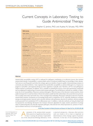
  • 4. GRADIENT DIFFUSION METHODS The M.I.C.Evaluator (Oxoid, Cambridge, UK) and Etest (bioMérieux, Durham, NC), are commercially available gradient diffusion systems for quantitative AST. Both systems use preformed antimicrobial gra- dients applied to 1 face of a plastic strip to generate diffusion of drug into an agar-based medium. The assays are performed in a manner similar to that for disk diffusion using a suspension of test organism equivalent in turbidity to that of a 0.5 McFarland standard to inoculate the surface of an agar plate. Following recommendations for incubation temper- atures, times, and atmospheric conditions, the MIC is read directly from a preprinted scale on the top of the strip at the point at which the ellipse of organism growth inhibition intercepts the strip. An example of an isolate tested for antimicrobial susceptibility by both Etest and disk diffusion is shown in the Figure. Several strips containing different antimi- crobial agents may be applied in a radial arrange- ment to the surface of large round plates, or they may be placed in opposite directions on large rect- angular plates. Minimum inhibitory concentrations generated by these methods generally agree well with those obtained using standard agar and broth dilution methods.10-12 Such gradient methods are similar in flexibility and simplicity to the disk diffu- sion approach, but they enable one to determine quantitative MICs. Significant advantages of the agar gradient diffusion systems include the ability to gen- erate quantitative MIC results for infrequently tested antimicrobial agents and the option to test fastidious and anaerobic organisms, for which reliable disk diffusion methods and/or commercial systems are not available, through the use of specific enriched media. Gradient diffusion strips are, however, con- siderably more expensive than the paper disks used for diffusion testing. METHODS FOR FASTIDIOUS BACTERIA Many fastidious bacterial species do not grow satis- factorily using standard in vitro susceptibility test- ing approaches with unsupplemented media. For several of the more frequently encountered patho- gens (eg, Streptococcus pneumoniae and Streptococcus spp other than S pneumoniae, N gonorrhoeae, and Neisseria meningitidis, and H influenzae and Hae- mophilus parainfluenzae), modifications have been made to the standard CLSI MIC and disk diffusion methods to allow laboratories to perform reliable AST. Such modifications typically involve the use of test media with supplemental nutrients, prolonged incubation times, and/or incubation in an atmo- sphere with an increased concentration of carbon dioxide. Specific MIC and zone diameter break- points have been established by the CLSI for such organisms, as have recommended acceptable ranges for the testing of applicable quality control strains. The CLSI has also published guidelines for AST of the fastidious and/or infrequently recovered bacteria listed in the Table.13 Methods for the standardized testing of potential agents of bioterrorism (eg, Bacil- lus anthracis, Francisella tularensis, Brucella spp, Yer- sinia pestis, and Burkholderia pseudomallei) have also been developed, and specific conditions for their testing are defined in Table 2 of the CLSI M45-A2 document.13 Currently, specific recommendations for several other fastidious bacteria, including Legion- ella and Bordetella spp, do not exist, partially because infections caused by these species typically respond well to the recommended drugs of choice, they are relatively uncommon isolates in clinical laborato- ries, and they require special complex media for re- covery in vitro, presenting unique problems in the development of AST assays. For the most part, breakpoints for these bacteria were predicated on interpretive criteria established for other organisms as published in CLSI standards and adapted based on published literature and the experience of the authors of the document. This is in sharp contrast to the extensive body of clinical mi- crobiologic, pharmacokinetic, and pharmacody- namic information typically used for the establish- ment of breakpoints as published in other CLSI standards. Because of the limited testing and nature of the potential agents of bioterrorism and Helico- bacter pylori, the interpretive recommendations and testing approaches for these organisms were re- cently transferred from the standard CLSI M100 documents to the M45 guideline.13 As mentioned, in addition to standard disk and MIC methods, many species of fastidious bacteria FIGURE. Manual susceptibility testing of Pseu- domonas aeruginosa by Etest and disk diffusion. Various patterns of susceptibility and resis- tance are seen. LABORATORY TESTING TO GUIDE ANTIMICROBIAL THERAPY Mayo Clin Proc. Ⅲ March 2012;87(3):290-308 Ⅲ doi:10.1016/j.mayocp.2012.01.007 293 www.mayoclinicproceedings.org
  • 5. may be tested by a gradient agar technique. The Etest method permits placement of strips on media optimal for the growth of the organism being tested and allows the use of various incubation conditions. A major limitation, however, to such an approach is lack of approval for such testing by the FDA. When FDA clearance has not been awarded, the results of such testing should be interpreted with caution, and an applicable qualifying comment should be an integral component of any resultant patient report. SUSCEPTIBILITY TESTING OF ANAEROBIC BACTERIA The importance of anaerobic bacteria as participants in and causes of significant infections and the need for specific antibiotic therapy for bacteremia and surgical prophylaxis against anaerobes are well documented.14-19 As a rule, AST is considered a necessity for effective guidance of antibiotic ther- apy, but how and when susceptibility testing of anaerobic bacteria should be performed have been topics of debate, in part owing to a number of misconceptions and confounding factors.20-24 Specimens collected from infections in which anaer- obes are involved are typically polymicrobic, ren- dering isolation and identification of individual or- ganisms slow and the results of AST too delayed to have a consistent positive effect on individual pa- tient outcomes. For clinicians, the combination of surgical intervention and broad-spectrum antibiot- ics has limited the correlation of potential antibac- terial resistance with outcome, directing many clin- ical microbiology laboratories away from the routine performance of anaerobic susceptibility testing. There is considerable evidence, though, that antibi- otic resistance is common among many anaerobic species and that patient treatment with inactive agents results in poor clinical responses and in- creased rates of mortality.14,16,19,25,26 Results of AST have also indicated that substantial differences exist in resistance patterns among hospitals on a lo- cal, regional, and national basis, suggesting that one medical center’s patterns may not be applicable to those of other facilities.27-30 Therefore, the need for anaerobic AST is of far more importance today than in the past. If practical, individual hospitals should estab- lish antibiograms for the more frequently recovered anaerobes on a periodic basis and test individual patient isolates as needed to assist in patient care. For purposes of presenting cumulative antimicro- bial susceptibility data, an attempt should be made to include the results from 80 to 100 anaerobic iso- lates with recognized important resistance mecha- nisms (eg, clindamycin resistance among members of the Bacteroides fragilis group). Ideally, following CLSI guidelines for preparation of antibiograms, 30 isolates for each genus or species should be in- cluded.31 When this is not possible, an effort should be made to present data for 30 isolates from the B fragilis group and at least 10 strains for other genera. Antibiotics in the report should reflect the hospital formulary. A recent CLSI document included an an- tibiogram for members of the B fragilis group gener- ated from the results of testing of isolates collected at many health care facilities across the United States by 3 reference laboratories.32 Clinicians TABLE. Clinical and Standards Laboratory Institute Published Guidelines for Antimicrobial Susceptibility Testing of Fastidious and/or Infrequently Recovered Bacteria Abiotrophia and Granulicatella spp (once referred to as thiol-dependent, pyridoxal-dependent, or nutritionally variant streptococci) Aeromonas spp Bacillus spp other than Bacillus anthracis Campylobacter coli and Campylobacter jejuni Corynebacterium spp Erysipelothrix rhusiopathiae The group of bacteria previously referred to as the HACEK organisms (Aggregatibacter actinomycetemcomitans, Aggregatibacter aphrophilus, Cardiobacterium hominis, Eikenella corrodens, and Kingella kingae) Facultatively anaerobic Lactobacillus spp Leuconostoc spp Listeria monocytogenes Moraxella catarrhalis Pasteurella spp Pediococcus spp Plesiomonas shigelloides MAYO CLINIC PROCEEDINGS 294 Mayo Clin Proc. Ⅲ March 2012;87(3):290-308 Ⅲ doi:10.1016/j.mayocp.2012.01.007 www.mayoclinicproceedings.org
  • 6. may refer to this document when prescribing em- pirical therapy for suspected or proven B fragilis group infections in settings in which anaerobic AST is not available. To assist in management, anaerobic susceptibil- ity testing should be performed when (1) the selec- tion of an antibiotic to which the isolate is suscepti- ble is critical for treatment of the patient, (2) long- term therapy is under consideration, (3) anaerobes are recovered from specific, usually sterile, body sites (eg, bone, blood, joint, or brain), or (4) treat- ment with an antimicrobial agent typically active against the organism has failed. The agar dilution susceptibility testing ap- proach, which uses Brucella blood agar as the me- dium, has been designated the reference method by the CLSI anaerobe working group.33 Because of the time-consuming, labor-intensive nature of this method, it is not generally considered practical for routine use in most clinical microbiology laborato- ries but serves as the reference method to which other more practical testing approaches can be com- pared. Alternative testing methods currently used include BMD (only standardized for members of the B fragilis group), limited agar dilution, and gradient strip diffusion assays, such as Etest. Disk diffusion and broth disk elution testing should not be used because the results generated from such methods do not correlate with the CLSI reference agar di- lution method.33 Although of limited use, ␤-lac- tamase testing may be of value for some organisms if therapy with ampicillin or penicillin is being considered. METHODS FOR SUSCEPTIBILITY TESTING OF NOCARDIA SPP AND OTHER AEROBIC ACTINOMYCETES Susceptibility testing of Nocardia spp and other aer- obic actinomycetes (Rhodococcus spp, Streptomyces spp, Gordonia spp, and Tsukamurella spp) should be performed on clinically significant isolates. Suscep- tibility testing results serve to guide initial therapeu- tic choices and may document emergence of drug resistance. No commercially available broth sys- tems have yet been cleared by the FDA for Nocar- dia spp or other aerobic actinomycetes. The CLSI BMD is the reference method for testing.34 Rec- ommended drugs for primary testing are amika- cin, amoxicillin-clavulanate, ceftriaxone, cipro- floxacin, clarithromycin, imipenem, linezolid, minocycline, moxifloxacin, trimethoprim-sulfa- methoxazole, and tobramycin. Second-line drugs for testing include cefepime, cefotaxime, and doxy- cycline. Vancomycin and rifampin results should also be reported on isolates of Rhodococcus equi be- cause these drugs are particularly useful therapeuti- cally. Microbiological breakpoints were established in 2003 and are available for Nocardia spp only.34 When aerobic actinomycetes other than Nocardia spp are tested, the susceptibility categories should be listed as tentative. The breakpoints for S aureus should be adapted as provided in the current CLSI M100 document (M100-S21) for R equi, but they should also be reported as tentative. Susceptibility testing results for most aerobic actinomycetes are available within 3 to 5 days, whereas results for R equi and some isolates of Tsukamurella spp may be read at 24 or 48 hours. Challenges in the identification and AST of No- cardia spp have become apparent within the past 10 years. Before the year 2000, conventional pheno- typic methods were largely used to identify Nocardia spp; however, molecular identification is now the preferred means of reliable identification to the spe- cies level.35 Both 16s ribosomal RNA sequencing and matrix-assisted laser desorption/ionization-time of flight are among the techniques currently used, but limitations exist.36 Because routine molecular test- ing is difficult to implement in many clinical micro- biology laboratories, isolates typically must be sent to a reference laboratory for identification. To ad- dress this issue, Wallace et al37 proposed a series of various susceptibility patterns that could predict placement of Nocardia spp within particular groups, but, as new strains are identified, these groupings have not proved to be valid, particularly for the No- cardia nova complex. The method for susceptibility testing of Nocar- dia spp also presents a challenge to many clinical microbiology laboratories because BMD is some- what impractical owing to cost, availability of sup- plies, and expertise needed to perform and interpret the results. Moreover, false resistance with BMD has been noted for ceftriaxone when testing Nocardia brasiliensis and imipenem when testing Nocardia far- cinica.34 In addition, sulfonamide results are incon- sistent when using BMD; for this compound, disk diffusion testing with sulfisoxazole may be per- formed concurrently. Investigators have evaluated use of the Etest method compared with BMD, with varying results.38 However, both lack of agreement with BMD and the absence of standardized methods have restricted routine use of the Etest. The CLSI has proposed that the agar proportion method be used to confirm questionable results from commercial broth systems and to test additional antibiotics or concentrations of drugs. The major taxonomic revisions within the last 10 years have further complicated testing because clinical and epidemiological differences exist among species. Furthermore, resistance to trim- ethoprim-sulfamethoxazole and other antimicro- bials is increasing among various species; thus, LABORATORY TESTING TO GUIDE ANTIMICROBIAL THERAPY Mayo Clin Proc. Ⅲ March 2012;87(3):290-308 Ⅲ doi:10.1016/j.mayocp.2012.01.007 295 www.mayoclinicproceedings.org
  • 7. identification of isolates to the species level re- mains imperative.39 METHODS FOR SUSCEPTIBILITY TESTING OF MYCOBACTERIA According to the most recent Centers for Disease Control and Prevention mycobacterial suscepti- bility testing guidelines, initial isolates from pa- tients with tuberculosis should be tested for sus- ceptibility to isoniazid, rifampin, ethambutol, and pyrazinamide.40 This guidance overrides the prior practice of performing susceptibility testing for only 3 drugs (isoniazid, rifampin, and ethambutol) and then only when a pulmonary or infectious disease clinician requested it. Current guidelines also state that susceptibility testing should be repeated after 3 months if the patient remains culture-positive de- spite appropriate therapy. However, susceptibility testing may be performed earlier if the patient ap- pears to be failing to respond to therapy or if in- tolerance to the drug regimen is evident. First-line susceptibility test results should be available for isolates of the Mycobacterium tuberculosis complex within 15 to 30 days of original receipt of the specimen in the laboratory.41 However, ideally, susceptibility results should be available within 7 to 14 days of specimen receipt.34 If resistance to any of the 4 initially tested agents is discovered, testing of secondary drugs should be performed as soon as possible. If the isolate is resistant only to pyrazinamide, Mycobacterium bovis should be ruled out because most M tuberculosis isolates are susceptible to pyrazinamide. Further specific guidelines regarding secondary drug testing and follow-up are outlined in CLSI M24-A2.34 The agar proportion approach has traditionally been considered the standard method for antimyco- bacterial susceptibility testing, but as an agar-based system, the time to result reporting is lengthy (ap- proximately 3 weeks).42 Both the agar proportion method and the radiometric method define resis- tance as growth of more than 1% of the inoculum of bacterial cells in the presence of an antitubercular drug. The antitubercular drugs are inoculated at specific in vitro concentrations, the values of which correlate to clinical responsiveness. If more than 1% of the bacterial population grows in the presence of a drug, that particular drug will not be of therapeutic utility.42 The agar proportion method is used primarily to confirm results from commercial liquid broth systems and to test addi- tional drugs that may not be available for testing using other systems. The FDA-cleared broth sys- tems for M tuberculosis testing have shorter incu- bation times than the agar proportion method; however, these commercial testing systems are only cleared for certain drugs.43 There are several molecular assays that have been developed for the detection of mutations asso- ciated with drug resistance in M tuberculosis, includ- ing both real-time polymerase chain reaction (PCR) methods and line probe assays.44 Molecular-based methods for detection of M tuberculosis drug resis- tance are more rapid than traditional methods of susceptibility testing. Mutations associated with re- sistance to isoniazid or rifampin are typically de- tected by such methods. These tests may be run using growth from positive cultures but may also be performed directly on acid-fast smear-positive spec- imens if the patient is highly suspected of having drug-resistant tuberculosis. It is currently suggested that molecular testing of drug susceptibility be backed up by culture, with performance of pheno- typic culture-based drug susceptibility testing when the isolate is retrieved from culture.34 Susceptibility testing of nontuberculous myco- bacteria (NTM) should be performed on isolates considered clinically significant. The American Tho- racic Society criteria for clinical significance of NTM are positive cultures from at least 2 sputum speci- mens or 1 bronchial wash or bronchial lavage spec- imen. Alternatively, a transbronchial or lung biopsy with histopathologic findings consistent with myco- bacteria and positive on culture for NTM is suffi- cient to be interpreted as clinically significant. In addition, NTM isolates from usually sterile body sites, such as cerebrospinal fluid, are considered clinically significant. However, routine susceptibil- ity testing need not be performed on Mycobacterium marinum because acquired resistance is uncommon with this organism.34 Initial susceptibility testing of Mycobacterium kansasii to isoniazid, rifampin, and ethambutol need not be performed but can be of- fered if treatment has failed. The standard susceptibility testing method for NTM is BMD. The American Thoracic Society and CLSI guidelines exist for susceptibility testing of members of the Mycobacterium avium complex, M kansasii, M marinum, and the rapidly growing my- cobacteria.34 However, accurate susceptibility predic- tions for other slowly growing mycobacteria cannot be made. The macrolides are the only antimicrobial agents that should be tested against M avium complex be- cause they are the only agents for which correlations have been demonstrated between in vitro suscepti- bility tests and clinical response.45 Because the mu- tation leading to resistance is the same for clarithro- mycin and azithromycin, only 1 drug need be tested. Generally, clarithromycin is tested because azithromycin demonstrates poor solubility. If the isolate is macrolide resistant, testing for susceptibil- ity to the secondary agents moxifloxacin and lin- ezolid may be considered. Susceptibility testing of M avium complex may also be performed if the patient MAYO CLINIC PROCEEDINGS 296 Mayo Clin Proc. Ⅲ March 2012;87(3):290-308 Ⅲ doi:10.1016/j.mayocp.2012.01.007 www.mayoclinicproceedings.org
  • 8. relapsed while undergoing macrolide therapy. In addition, susceptibility testing should be repeated after 3 months of therapy for patients with dissem- inated M avium complex disease and after 6 months of therapy for patients with chronic pul- monary disease caused by M avium complex. Commercially available broth systems have not yet been cleared by the FDA for slowly growing NTM. PHENOTYPIC AND GENOTYPIC METHODS FOR DETECTION OF ANTIMICROBIAL RESISTANCE A number of phenotypic tests are available to the clinical microbiology laboratory to characterize a pathogen’s susceptibility to an antibiotic by screen- ing for a specific resistance mechanism or pheno- type. Although such screening tests do not result in determination of an MIC, some have sufficient spec- ificity and sensitivity that confirmatory testing is not required and the screening test result can be re- ported without further testing. Other assays require additional or confirmatory testing. For example, tests for inducible clindamycin resistance among staphylo- cocci and screening tests for high-level gentamicin and streptomycin resistance in enterococci are gen- erally considered to be comparable to standard methods for the detection of clinically significant resistance, and they do not require confirmatory testing. By comparison, laboratories that use ertap- enem resistance as a surrogate marker for carbapen- emase production among certain species of Entero- bacteriaceae must confirm resistance to meropenem, imipenem, or doripenem by another more standard- ized approach because ertapenem-resistant strains are not always resistant to these other agents. In addition, methods for the direct detection of antibiotic-resistant bacteria in clinical samples have progressed rapidly in recent years, largely because of the continued evolution and spread of multidrug- resistant pathogens, such as methicillin-resistant S aureus (MRSA). The development of commercial as- says that facilitate rapid detection of such pathogens directly from clinical specimens, often generating results in a few hours or less, has positively en- hanced surveillance efforts and patient manage- ment. Other genotypic assays for detection of spe- cific antimicrobial resistance genes in gram-negative bacteria (eg, blaKPC-containing Klebsiella pneu- moniae) have the potential to improve therapeutic patient decisions and assist in epidemiological in- vestigations of resistance gene dissemination in the hospital and community setting. ␤-LACTAMASE TESTS A positive ␤-lactamase test result indicates that the organism is resistant to applicable ␤-lactam agents, but a negative reaction is inconclusive because other mechanisms of resistance to the ␤-lactams may ex- ist. For example, a positive ␤-lactamase test result for a strain of N gonorrhoeae means that the isolate is resistant to penicillin, ampicillin, and amoxicillin and that these drugs would not be appropriate ther- apeutic choices. However, a ␤-lactamase test only detects one form of penicillin resistance in N gonor- rhoeae. Strains with chromosomally mediated resis- tance with penicillin-binding protein modifications can only be detected by the disk diffusion or the agar dilution MIC method.46,47 Three direct ␤-lactamase tests—the acidomet- ric, iodometric, and chromogenic methods—have been widely used. All 3 methods involve the testing of isolates grown on nonselective media, and results are typically available within 1 to 60 minutes. Al- though some bacteria (eg, N gonorrhoeae and H in- fluenzae) produce ␤-lactamase constitutively, oth- ers (eg, staphylococci) may produce detectable levels of enzyme only after exposure to an induc- ing agent, typically a ␤-lactam.48 Even after induc- tion, though, direct ␤-lactamase tests may not be sufficiently sensitive to detect ␤-lactamase produc- tion in all staphylococci.49 Therefore, for serious in- fections that require penicillin therapy, clinical mi- crobiology laboratories should perform both MIC and induced ␤-lactamase tests on all subsequent iso- lates from the same patient. In addition, PCR testing of the isolate for the presence of the blaZ ␤-lacta- mase gene may also be an option. DETECTION OF METHICILLIN RESISTANCE IN STAPHYLOCOCCUS SPP The most common currently used method for the detection of MRSA is culture.50 Traditional MRSA detection methods consist of culture from a selective liquid or solid medium. Recently, chromogenic agars have shown improved sensitivity and specific- ity over nonchromogenic media for detection of MRSA.51 Chromogenic agars contain selective anti- biotic(s) and various chromogenic substrates, which provide easy visual identification of colonies. An ad- ditional advantage of chromogenic agar is the faster time to detection of the organism. MRSA is often detected within 20 to 48 hours on chromogenic me- dia, with a high percentage of cases identified within 24 hours.52 Several chromogenic media are avail- able from different manufacturers. The use of an overnight preenrichment step with selective broth medium before inoculation of the agar has been shown to increase sensitivity of the testing by 15% to 30%.53 However, the delay in detection of an additional 18 to 24 hours and the cost of the selective broth medium represent disadvantages. LABORATORY TESTING TO GUIDE ANTIMICROBIAL THERAPY Mayo Clin Proc. Ⅲ March 2012;87(3):290-308 Ⅲ doi:10.1016/j.mayocp.2012.01.007 297 www.mayoclinicproceedings.org
  • 9. Early diagnosis of MRSA in the laboratory is cru- cial in guiding appropriate antimicrobial therapy. Rapid molecular methods of MRSA detection are increasingly used and are available for testing from a variety of sources.54 Traditional detection of MRSA from automated blood culture instruments is time- consuming because 24 to 72 hours are required for subculture, biochemical identification, and AST of the isolate once the result turns positive. Several techniques are available for identification of MRSA directly from blood culture bottles that have flagged as positive for growth when gram-positive cocci in clusters are seen on Gram stain.55 Several commer- cial molecular methods are available, including nu- cleic acid amplification and hybridization assays.51 A commercial penicillin-binding protein 2a latex ag- glutination kit, which facilitates the detection of the protein product expressed by the mecA gene, is available for detection of MRSA directly from blood culture.56 Other systems are currently un- der investigation.50 HIGH-LEVEL AMINOGLYCOSIDE RESISTANCE IN ENTEROCOCCI Successful treatment of enterococcal endocarditis and other serious enterococcal infections requires the use of an aminoglycoside with a cell wall–active agent, such as ampicillin, penicillin, or vancomycin. All enterococci demonstrate innate low-level resis- tance to aminoglycosides because of their facultative anaerobic metabolism, which reduces transmem- brane potential, thereby limiting drug uptake. How- ever, the bactericidal combination of an aminogly- coside and a cell wall–active antimicrobial, which allows for markedly enhanced uptake of the amino- glycoside, leads to enhanced killing of the organism in the absence of high-level aminoglycoside resis- tance (HLAR). The CLSI recommends HLAR screening of en- terococci with both gentamicin and streptomycin from blood cultures or other specimens submitted for the evaluation of endocarditis, such as heart valve tissue. The BMD may be performed by assess- ing growth of the organism in the presence of 1000 ␮g/mL of streptomycin or 500 ␮g/mL of gentamicin in brain heart infusion broth.5 The recommended screening concentrations for streptomycin and gentamicin using other methods are also cov- ered.57 Performance data of commercially avail- able media and systems for HLAR screening have been reviewed.58 In enterococci, HLAR is mediated by aminogly- coside-modifying enzymes (AMEs), which modify the aminoglycoside by acetylation, adenylation, or phosphorylation. The most prevalent AME gene among enterococci with HLAR to gentamicin is aac(6’)-Ie-aph(2”)-Ia, which has both acetyltrans- ferase and phosphotransferase activity.59 This com- mon AME confers resistance to all available amino- glycosides, except streptomycin. Commonly, HLAR to streptomycin is mediated by either ant(6’)-Ia or ant(3’’)-Ia; in such cases, streptomycin should not be used in combination with a ␤-lactam agent. Detec- tion of HLAR to both gentamicin and streptomycin precludes the use of aminoglycosides for synergism in any clinical situation. Enterococcus faecium possesses a naturally oc- curring AME, resulting in moderate resistance to to- bramycin (MICs, 64-1000 ␮g/mL). The presence of the aac(6’)-Ii gene precludes synergistic treatment with tobramycin, kanamycin, netilmicin, or sisomi- cin. In addition, many enterococci possess the aph(3’)-IIIa gene, which confers high-level resis- tance to kanamycin and abolishes any synergistic effect with amikacin. Thus, gentamicin and strepto- mycin are the only 2 aminoglycosides to test and consider for synergistic therapy. Novel genes carry- ing AMEs mediating resistance to gentamicin, such as aph(2’’)-Ib, aph(2’’)-Ic, aph(2’’)-Id, and aph(2’’)-Ie, have been discovered and may further complicate HLAR testing because the susceptibilities to various aminoglycosides differ.60 Because of the large num- bers of AME-encoding genes, molecular HLAR screening in enterococci remains investigational and has not yet been widely available in clinical micro- biology laboratories.61 Issues that continue to challenge HLAR screen- ing in enterococci include the isolation of multiple AMEs within single enterococcal isolates, isolates that harbor infrequent AMEs but do not demon- strate the HLAR phenotype, and the increasing prev- alence of various HLAR enzymes in enterococci. DETECTION OF VANCOMYCIN-RESISTANT ENTEROCOCCI The vancomycin-resistant enterococci (VRE) are im- portant opportunistic pathogens in many health care facilities and common colonizers of the gastro- intestinal tract. The VRE are among the most com- mon causes of hospital-acquired infections in the United States, and patients colonized with VRE in the gastrointestinal tract may serve as reservoirs for nosocomial transmission.62,63 Two common patterns of enterococcal resis- tance exist, both of which result in elevated vanco- mycin MICs. The first, and most clinically impor- tant, is vancomycin resistance due to acquisition of genetic information, usually on a plasmid or other transmissible genetic element.64 This acquired trait is most commonly observed in strains of E faecium and Enterococcus faecalis harboring the vanA or vanB genes that encode for high-level vancomycin resis- tance. Expression of the vanA gene results in ele- vated vancomycin MICs (Ͼ128 ␮g/mL) and is the MAYO CLINIC PROCEEDINGS 298 Mayo Clin Proc. Ⅲ March 2012;87(3):290-308 Ⅲ doi:10.1016/j.mayocp.2012.01.007 www.mayoclinicproceedings.org
  • 10. dominant resistance factor in enterococci. By com- parison, expression of the vanB gene results in lower vancomycin MICs, typically in the range of 16 to 64 ␮g/mL. The second pattern of resistance, intrinsic (inherent) in nature, is characteristically seen in En- terococcus gallinarum and Enterococcus casseliflavus. Most frequently encoded by vanC, this pattern of resistance may also be due to the expression of genes other than vanC (eg, vanE, vanG, and vanL) and re- sults in either low-level resistant or intermediate MICs, typically in the 2- to 16-␮g/mL range.65 Con- tact precautions are generally only required for pa- tients harboring enterococci with acquired resis- tance, such as E faecium or E faecalis. Guidelines for susceptibility testing of enterococcal pathogens when grown in culture from blood or other sites have remained fairly standard over time. How- ever, there have been several recent advances in both culture-based and molecular-based screen- ing of stool for VRE, facilitating identification of patients who are potential reservoirs for infection and transmission. The CLSI guidelines for vancomycin suscepti- bility testing of enterococci isolated from various sites suggest the use of standard BMD or disk diffu- sion testing.5 If disk diffusion or Etest is performed, the susceptibility plates must be held for a total of 24 hours to obtain accurate readings. Organisms for which vancomycin MICs are in the range of 8 to 16 ␮g/mL should be further identified by biochemical testing because infection control precautions will differ based on identification of the organism as an Enterococcus sp other than E faecalis or E faecium. Accordingly, isolates with intermediate zones on disk testing should be tested by an MIC method and/or further identified by biochemical and other identification tests to guide appropriate infection control practices. A variety of culture-based and molecular methods have been studied to support active sur- veillance efforts to identify VRE from the gastroin- testinal tract.66,67 Although culture-based methods are not as rapid as molecular-based screening methods, isolates obtained from culture can be stored for fur- ther study or identification. Molecular-based VRE screening methods decrease the time to identifica- tion but are costly. Culture remains the screening method of choice for VRE stool screening, but mo- lecular methods are becoming increasingly recog- nized and used. Some investigators advocate the use of a broth enrichment step before inoculation of a culture plate or a molecular assay to increase sensitivity.66 Traditional screening agars for VRE from stool specimens include Campylobacter medium with 5 antibiotics, including 10 ␮g/mL of vancomycin, and various types of bile esculin azide agar with vanco- mycin.68,69 The traditional VRE screening agars re- quire 24 to 48 hours to identify colonies preliminar- ily, with additional time required for confirmatory identification and susceptibility testing and up to 5 days to final identification. Various chromogenic VRE media demonstrate adequate sensitivity and specificity, with reduced turnaround time to results through early visual colony identification. There are many selective and differential chromogenic VRE agars that appear promising for use in VRE stool screening. However, performance data vary accord- ing to whether prior overnight broth enrichment of the specimen in liquid media was performed.67 Real-time PCR is a sensitive and rapid approach to the identification of VRE from gastrointestinal tract specimens.66 Recently, the BD GeneOhm VanR (BD Diagnostics, Spark, MD) and Xpert vanA/vanB (Cepheid, Sunnydale, CA) assays, which detect iso- lates carrying vanA and vanB genes within 2 to 4 hours, have been introduced.70 Most FDA-cleared assays have been marketed for direct detection of VRE from rectal or perianal swabs, although detec- tion from stool specimens has shown comparable results. Some studies have demonstrated improved performance of molecular assays after overnight aer- obic or anaerobic preenrichment of stool in broth media; however, the preenrichment step increases the turnaround time to results.70 Occasionally, lower specificity has been shown with the use of perianal sampling due to the presence of anaerobes that carry the vanB gene.69 In summary, there are a variety of VRE screen- ing methods available, the most promising of which appear to be chromogenic media and molecular as- says, due to rapid result reporting. However, some assays require an overnight preenrichment step to maximize sensitivity. CLINDAMYCIN RESISTANCE IN STREPTOCOCCI AND STAPHYLOCOCCI Clindamycin and erythromycin resistance in strep- tococci may either be due to erm genes, which lead to the production of macrolide ribosomal methyl- ases, or to expression of the mef gene, which encodes an efflux pump targeting only the macrolides. By comparison, erm enzymes methylate the 23S ribo- somal RNA component of the 50S bacterial ribo- somal subunit, which causes decreased binding of macrolides, lincosamides (clindamycin), and strep- togramin B antibiotics (designated the MLSB pheno- type). In staphylococci, inducible clindamycin resis- tance is also due to the MLSB phenotype, but its efflux pump is encoded by the msrA gene. The MLS resistance phenotype can be either in- duced or constitutively expressed. Inducible clinda- mycin resistance cannot be detected by routine MIC or disk testing of clindamycin.71 Therefore, the 14- LABORATORY TESTING TO GUIDE ANTIMICROBIAL THERAPY Mayo Clin Proc. Ⅲ March 2012;87(3):290-308 Ⅲ doi:10.1016/j.mayocp.2012.01.007 299 www.mayoclinicproceedings.org
  • 11. and 15-member macrolides, which are better induc- ers of clindamycin resistance than is clindamycin itself, must be used for induction of clindamycin in the clinical laboratory.72 Testing of streptococci and staphylococci for inducible clindamycin resistance is important because clindamycin is frequently used to treat staphylococcal, group B streptococcal, and group A streptococcal infections. In addition, resis- tance to clindamycin is increasing in prevalence, with a recent estimate of 12.8% inducible clindamy- cin resistance among S aureus in the United States.73 Both inducible and constitutive clindamycin resis- tance has likewise become increasingly common among ␤-hemolytic streptococci, with less resis- tance reported for group A streptococcal than for group B streptococcal infections.74 The CLSI has recommended 2 different meth- ods for detection of inducible clindamycin resis- tance in staphylococci and ␤-hemolytic strepto- cocci.5 One method is the disk approximation test, also known as the D-zone test. With this approach, separate erythromycin and clindamycin disks are placed specific distances apart on an agar plate, de- pending on whether staphylococci or streptococci are being tested. If there is flattening of the zone of inhibition between the 2 disks and the zone resem- bles the letter “D,” the test result is interpreted as positive for induction of clindamycin resistance. The second method suggested by the CLSI is the use of a single-well microdilution test containing both erythromycin and clindamycin. Whenever induc- ible MLSB resistance is detected, clindamycin treat- ment should be avoided, if possible. However, the CLSI states in their guidelines that clindamycin may still be clinically effective in some patients, despite a positive induction test result. Automated methods for identification of induc- ible clindamycin resistance have also recently been introduced.75 Molecular assays using primers for various erm genes have been used in various re- search settings for the purpose of following resis- tance trends or validating BMD assays.71 However, PCR assays for inducible clindamycin resistance are not currently considered the standard of care in the clinical microbiology laboratory. Constitutive or inducible resistance to clinda- mycin may also be seen in S pneumoniae due to ex- pression of a ribosomal methylase encoded by the ermB gene. The methylase alters the binding site on the ribosomes for the macrolides and clindamycin, similar to that seen for ␤-hemolytic streptococci and staphylococci.76 Clindamycin is recommended as a second- or third-line antibiotic choice for pediatric patients receiving long-term antibiotic therapy for some conditions, such as pneumococcal osteomy- elitis and/or joint infections.77 Because pneumo- cocci are capable of expressing inducible clindamy- cin resistance, some authors suggest that isolates of S pneumoniae be tested for erm-mediated resis- tance in certain clinical situations involving pedi- atric patients. Jorgensen et al77 recently assessed performance of the CLSI-suggested disk and BMD methods for detection of inducible clindamycin resistance in pneumococci. DETECTION OF EXTENDED-SPECTRUM ␤-LACTAMASE PRODUCTION AMONG ENTEROBACTERIACEAE Members of Enterobacteriaceae (and other organ- isms, including P aeruginosa) can produce ␤-lacta- mases, referred to as extended-spectrum ␤-lactam- ases (ESBLs), capable of hydrolyzing penicillins, the monobactam aztreonam, and cephalosporins (in- cluding expanded-spectrum cephalosporins, such as cefotaxime, ceftriaxone, ceftizoxime, and ceftazi- dime).78 The CLSI guidelines specify screening cri- teria and confirmatory testing approaches for detec- tion of ESBL production by Escherichia coli, K pneumoniae, and Proteus mirabilis.5 For organisms such as Enterobacter spp and Serratia spp, which produce AmpC-type enzymes, ESBL screening should not be performed because false-negative re- sults can occur. These screening and confirmatory tests were necessary because standard disk diffusion and MIC tests did not uniformly identify isolates producing ESBLs. Because ESBLs are usually inhib- ited by clavulanic acid, the CLSI made use of this property in developing the tests recommended to clinical laboratories for their detection and recom- mending that isolates producing ESBLs be reported as resistant to all penicillins, cephalosporins, and aztreonam. On establishment of new, lower inter- pretive criteria for many of these compounds, largely based on pharmacokinetic and pharmacody- namic principles and limited clinical data, the CLSI revised their recommendations for reporting.5 When the new breakpoints are adopted by clinical laboratories, the CLSI recommends that results for specific cephalosporins and aztreonam be reported and interpreted as they are tested and that the ESBL screening and confirmatory tests need only be per- formed for epidemiological and infection control purposes. By comparison, although the European Committee on Antimicrobial Susceptibility Testing (EUCAST) breakpoints for the cephalosporins are similar to those now recommended by the CLSI, EUCAST recommends that laboratories continue to screen and confirm ESBL production due to the lim- ited supporting clinical data and that cephalosporin reports be changed from susceptible to intermediate or intermediate to resistant if an isolate tests positive for ESBL production.79 MAYO CLINIC PROCEEDINGS 300 Mayo Clin Proc. Ⅲ March 2012;87(3):290-308 Ⅲ doi:10.1016/j.mayocp.2012.01.007 www.mayoclinicproceedings.org
  • 12. DETECTION OF CARBAPENEMASE ACTIVITY AMONG ENTEROBACTERIACEAE Carbapenemases, enzymes that hydrolyze carba- penem class antibiotics (ertapenem, imipenem, meropenem, and doripenem), usually hydrolyze all other currently available ␤-lactams, with the exception of aztreonam for some metallo-␤- lactamases. The genome encoding for the produc- tion of these enzymes may be located on plasmids (eg, K pneumoniae carbapenemases), a feature that makes them of particular concern from an infec- tion control perspective. There are 3 classes of carbapenemases: serine class A (including KPC, SME, IMI, GES, and NMC), class B enzymes known as the metallo-␤-lactamases (such as VIM, IMP, and NDM), and the class D OXA enzymes.80 Carbapenemases have been identified in a wide range of gram-negative genera. The KPC enzyme, the most frequently identified class A carbapen- emase in the United States, is most often found in the Enterobacteriaceae but has also been detected in P aeruginosa.81 Metallo-␤-lactamases are most frequently seen in Acinetobacter spp and P aerugi- nosa, but recently NDM has become widespread in some regions of the world among species of Enterobacteriaceae, particularly K pneumoniae. OXA carbapenemases are most frequently found in Acinetobacter spp but have also been reported among isolates of Enterobacteriaceae. In 2009 the CLSI Subcommittee on Antimi- crobial Susceptibility Testing (SAST) recom- mended the modified Hodge test for the detection of carbapenemase activity in Enterobactericeae.82 Advantages of this assay include its ease of perfor- mance, the ability to test several isolates on a sin- gle plate, and the detection of different classes of carbapenemases with one test.83,84 The primary disadvantages are subjectivity in reading the re- sults, its inability to differentiate the various car- bapenemases (potentially useful from an epidemi- ological perspective), and the false-positive results that can occur with some organisms pro- ducing AmpC or ESBL enzymes. The modified Hodge test was originally rec- ommended for the detection of carbapenemases in bacteria for which carbapenem MICs were ele- vated but still fell within the susceptible range. When isolates tested positive, the SAST recom- mended that they be designated carbapenemase- producing strains in the patient report with a warning indicating that the therapeutic outcomes of patients infected with such organisms and treated with the relevant carbapenem were un- known, particularly when alternative dosing reg- imens were used (eg, continuous or prolonged infusion). In 2010, though, the SAST lowered the carbapenem breakpoints to capture most carba- penemase-producing strains, which would now test either as resistant or intermediate to these compounds.5 Implementation of the revised break- points eliminates the need for laboratories to rou- tinely perform the modified Hodge test, although such testing may in some cases still be of value from an epidemiological or infection control perspective. A number of phenotypic tests allow detection and differentiation of class A (eg, inhibition by boronic acid) and class C (eg, inhibition by chelating agents such as ethylenediaminetetraacetic acid) carbapen- emases, but these tests fail to detect class D (OXA) and are primarily used for strain characterization rather than for clinical purposes. DETECTION OF PLASMID-MEDIATED AMPC-TYPE ␤-LACTAMASES Chromosomally mediated, inducible, AmpC-type ␤-lactamases are produced by several gram-negative species. Often referred to as the ASPACE or ASPICE organisms, these include, but are not restricted to, Aeromonas spp, Serratia spp, P aeruginosa, Acineto- bacter spp indole-positive Proteae (Providencia spp, Morganella morganii, and Proteus vulgaris), Citrobac- ter spp, and Enterobacter spp.85 Resistant to the cur- rently available ␤-lactamase inhibitors, such as tazobactam, sulbactam, and clavulanic acid, these en- zymes result in resistance to a wide range of ␤-lactam antibiotics. The combination of a porin deletion with an AmpC-type enzyme can result in resistance to car- bapenems. The genome encoding AmpC-type ␤-lacta- mases may also be harbored on transmissible plasmids and has been reported among a number of species that do not naturally produce inducible chromosomally mediated AmpC-type enzymes, including K pneu- moniae,Ecoli, Pmirabilis,and Salmonella spp.86 Similar to the plasmids carrying the genes encoding ESBLs, those with AmpC-type genes often harbor resistance determinants for multiple classes of drugs. Because detection of plasmid-mediated AmpC- producing pathogens may have epidemiological and infection control importance, several assays have been developed in an attempt to accurately detect this resistance type.87-94 None, however, has been sufficiently standardized or tested against an ade- quate number of organisms producing plasmid-me- diated AmpC-type enzymes to be considered supe- rior in performance to the other assays. TESTING TO DETECT BACTERICIDAL ACTIVITY The AST methods used in the clinical microbiology laboratory typically assess only the inhibitory activ- ities of antimicrobial agents. This is generally suffi- cient for patient management for most bacterial in- fections encountered by clinicians. In some, albeit rare, situations it may also be of value to determine LABORATORY TESTING TO GUIDE ANTIMICROBIAL THERAPY Mayo Clin Proc. Ⅲ March 2012;87(3):290-308 Ⅲ doi:10.1016/j.mayocp.2012.01.007 301 www.mayoclinicproceedings.org
  • 13. the bactericidal activity of an antibiotic against a specific patient bacterial pathogen. Serious infec- tions in which a bactericidal effect is generally con- sidered necessary for optimal treatment include bac- teremia in neutropenic patients, patients with chronic osteomyelitis, and patients with bacterial endocarditis.95-97 Minimum Bactericidal Concentration Testing After determination of MICs in a broth system under standard conditions, measured aliquots of growth media may be subcultured quantitatively to solid media to assess bactericidal activity. To calculate the extent of killing at each antibiotic concentration, plates are incubated under appropriate conditions, colony counts are performed, and results are com- pared to that of the growth control tube or well. The accepted definition of the minimum bactericidal concentration (MBC) is the lowest concentration of antibiotic at which a 99.9% (3 log) or greater reduc- tion in growth compared with the initial inoculum is observed.98 As with the MIC, the antibiotic MBC is reported in micrograms per milliliter or in some re- gions of the world in milligrams per liter. Determi- nation of the MBC allows one to detect potential tolerance by the patient’s specific isolate to an anti- biotic usually considered bactericidal, a phenome- non reportedly leading to clinical failure among some patients.99-101 Tolerance occurs when the an- tibiotic MIC for an organism is low (within the sus- ceptible range) but the MBC is elevated, frequently at a concentration beyond that generally considered clinically achievable from a pharmacokinetic per- spective. Tolerance is specifically defined as an MBC 32-fold or more higher than the MIC for the antibi- otic under consideration.98,100 Failure to demon- strate at least a 3-log10 decrease in colony-forming units per milliliter in the time-kill assay is also con- sidered to represent tolerance.98 Time-Kill Kinetic Assays Time-kill assays allow one to assess the rate of bac- tericidal activity at varying antibiotic concentrations over time. Although these assays are time-consum- ing and laborious to perform because they require subculture of media at specific times during a 24- hour period, results of combinations of antimicro- bial agents can also be assessed. In 1999, standard methods for performance of the assay were pub- lished by the CLSI (previously the National Com- mittee for Clinical Laboratory Standards).98 Results of time-kill assays are typically presented graphi- cally, plotting colony counts for each antimicrobial agent and concentration tested at each time point at which subcultures were performed (usually at 0, 4, 8, 12, and 24 hours). As with the MBC, bactericidal activity is defined as a 99.9% or greater killing at a specified time. When antibiotics are tested in com- bination using the time-kill approach, synergy is typically defined as a 2-log decrease or more in the number of colony-forming units achieved with the combination of antibiotics when compared with that achieved by the most active agent tested alone. TESTS FOR ASSESSMENT OF INTERACTIONS AMONG ANTIMICROBIAL AGENTS The value of in vitro testing to assess antibiotic in- teractions (ie, synergy testing) remains highly con- troversial. With a few bacterial species and antibi- otic combinations, synergistic bactericidal activity is actually predictable and need not be routinely de- termined (eg, ampicillin, penicillin, or a glycopep- tide plus gentamicin or streptomycin against sus- ceptible strains of enterococci). When synergy testing is performed, the results should be inter- preted with caution because they do not take into account drug interactions from a pharmacokinetic or a patient safety (adverse effect) perspective. The checkerboard BMD test, wherein 2 antimi- crobial agents are serially diluted in a 2-dimensional fashion to include all combinations during a speci- fied clinically relevant range, is a somewhat less la- bor-intensive approach to assessing antibiotic inter- actions in vitro than the time-kill assay. This same approach to drug interaction testing can be taken using an agar dilution approach, although the method is even more laborious. Using these meth- ods, one is able to recognize synergistic, additive, indifferent, or antagonistic interactions occurring with the agents being tested. By this method, a frac- tional inhibitory concentration (⌺FIC) is calculated by comparing the MIC of each drug alone to the MIC of that drug in combination with the second agent. Synergy is usually defined as a 4-fold decrease in the MIC of the agents in combination when compared with the antibiotics tested alone.102 The FIC is cal- culated and interpreted as follows: ⌺FIC ϭ FIC of agent A ϩ FIC of agent B FIC of agent A ϭ MIC of agent A in combination MIC of agent A alone FIC of agent B ϭ MIC of agent B in combination MIC of agent B alone Synergy is defined as ⌺FIC Յ 0.5 Indifference is defined as 0.5 Ͻ ⌺FIC Յ 4 Antagonism is defined as ⌺FIC Ͼ 4 Some investigators consider compounds additive when Ͼ0.5 ⌺FIC Յ1. MAYO CLINIC PROCEEDINGS 302 Mayo Clin Proc. Ⅲ March 2012;87(3):290-308 Ⅲ doi:10.1016/j.mayocp.2012.01.007 www.mayoclinicproceedings.org
  • 14. The time-kill approach appears to correlate more closely with in vivo studies of combined antibiotic ef- fects than does the checkerboard method.103,104 A simpler approach, albeit less standardized, is the use of Etest strips. Using this method, synergy is again de- fined by the FIC as described earlier.105 SERUM BACTERICIDAL TESTING (SCHLICHTER ASSAYS) Another approach to assessing bactericidal activity and determining the effects of antibiotic combina- tions is serum bactericidal testing. Determinations of serum inhibitory titers and serum bactericidal ti- ters (SBTs) are performed in a manner analogous to that for MIC and MBC testing, except that the patient’s serum, rather than serially diluted concen- trations of antibiotic, is used. A guideline outlining the specifics for performance of SBT testing has been published by the CLSI.106 To perform the testing, 1 or more blood samples are collected from the pa- tient (usually attempting to draw the specimens when antibiotic peak and trough levels are achieved). Serial 2-fold dilutions of the patient se- rum in pooled, pretested human serum are then pre- pared in tubes or wells of microtiter plates. Each tube or well is then inoculated with a standardized suspension of the patient’s infecting pathogen in an applicable growth medium. The serum inhibitory titer is defined as the highest dilution of the patient’s serum preventing visible growth after an appropri- ate incubation period. Similar to MBC testing, quan- titative subcultures are then performed from each dilution of the patient’s serum that prevented visible growth. The SBT is defined as that dilution resulting in a 99.9% or greater decrease in the original inoc- ulum based on standardized rejection value tables. Results are then reported as titers (dilutions) of the patient’s serum. The purpose of this assay is to de- termine whether the dosage regimen chosen for treatment of the infection results in sufficient bacte- ricidal activity in the patient’s blood.96,107 Higher serum titers suggest that adequate patient dosing has been achieved, unexpected antibiotic elimina- tion has not occurred, and the bacterial pathogen is not tolerant. A peak SBT of 1:64 or higher and a trough SBT of 1:32 or higher have been shown to correlate with rapid bacterial eradication from the blood and optimal time to cardiac vegetation steril- ization in cases of endocarditis.96,108 Lower bacteri- cidal titers, however, do not necessarily predict a poor clinical response. Limited data have indicated that in cases of acute and chronic osteomyelitis a peak SBT of 1:16 or higher and a trough SBT of 1:4 or higher are predictive of therapeutic efficacy.96,109 Major disadvantages of this assay include its labor- intensive nature and the requirement to obtain, pre- pare, and pretest pooled human serum for use in the procedure. ANTIFUNGAL SUSCEPTIBILITY TESTING There have been major advances in the standardiza- tion and clinical interpretation of antifungal suscep- tibility testing in recent years. The number of anti- fungal agents is increasing as the incidence of systemic and other infections due to Candida spp, Aspergillus spp, Zygomycetes, and other filamentous fungi is increasing.110 Susceptibility testing of clin- ically significant isolates, especially those from usu- ally sterile sites, is important both epidemiologically and clinically for guiding treatment. Antifungal sus- ceptibility testing standards for yeasts and molds have been developed by both the CLSI and Euro- pean Committee on Antimicrobial Susceptibility Testing (EUCAST) for a variety of antifungal agents.111-115 The standard antifungal reference testing method against which many other susceptibility testing meth- ods are measured is the BMD method.112 The BMD methods proposed by different organizations vary by the media used, supplements and inoculum added, incubation conditions, and end point inter- pretations. The CLSI provides MIC breakpoint and interpretive data for Candida spp for several antifun- gal agents, including fluconazole, itraconazole, vori- conazole, anidulafungin, caspofungin, micafungin, and flucytosine.111 The CLSI breakpoints for flu- conazole, voriconazole, and the echinocandins have recently been revised for Candida species.116-118 The susceptible dose-dependent category for the azoles infers that susceptibility to these antifungals by Candida spp is dependent on achieving the max- imal possible blood level. In addition, the flucona- zole data for the CLSI breakpoints were gathered from studies involving patients with oropharyngeal candidiasis and invasive candidal infections in non- neutropenic patients. Therefore, the clinical rele- vance of fluconazole breakpoints in clinical situa- tions other than those mentioned has not been established. Interpretive data for the echinocandin class of drugs are based primarily on experience with nonneutropenic patients with candidemia, and clinical relevance of these data in other patient pop- ulations is uncertain. Finally, itraconazole data are based on experience with mucosal infections only, and data supporting the CLSI breakpoints for inva- sive infections are not available. Although there are no CLSI breakpoints currently approved for ampho- tericin B when testing Candida spp, isolates for which the MICs are greater than 1.0 ␮g/mL are gen- erally considered resistant.111 Although the CLSI documents only provide guidelines for Candida spp, some investigators have applied the CLSI break- points to Cryptococcus spp, and correlations have LABORATORY TESTING TO GUIDE ANTIMICROBIAL THERAPY Mayo Clin Proc. Ⅲ March 2012;87(3):290-308 Ⅲ doi:10.1016/j.mayocp.2012.01.007 303 www.mayoclinicproceedings.org
  • 15. been demonstrated between higher MICs and treat- ment failures.119 Because the BMD testing method is difficult to perform in daily practice in clinical microbiology laboratories, other testing approaches have been in- vestigated. Disk diffusion antifungal susceptibility testing is a simple and cost-effective method for both yeasts and molds. Fluconazole disk diffusion testing of Candida spp has been available for several years. The CLSI has also established guidelines for disk diffusion testing of filamentous fungi, which is a relatively simple, rapid, and cost-effective alterna- tive to BMD testing.115 Etests are likewise less labor intensive than BMD and are relatively simple and reproducible for the testing of antifungal agents, es- pecially against molds.120 Sensititre YeastOne (Trek Diagnostic Systems) is a colorimetric antifungal sus- ceptibility testing MIC plate that exhibits high agree- ment with the CLSI BMD method.121 A new disk agar diffusion method, the Neo-Sensitabs tablet dif- fusion assay (Rosco, Taastrump, Denmark) has also been developed and tested for antifungal testing of molds and yeasts.122 Finally, commercial auto- mated systems for MIC determination of yeasts are simple alternative methods and are comparable to other established antifungal susceptibility testing methods.123 Antifungal susceptibility testing of Candida albi- cans and other Candida spp is fairly simple to per- form, with determination of fluconazole susceptibil- ity as the most important first step. If the C albicans isolate is fluconazole resistant or if the isolate is non–C albicans, further susceptibility testing will of- ten be required. In contrast, antifungal susceptibility testing of molds is not currently as useful clinically because of both the long turnaround time of such testing and the difficulties in developing accurate breakpoints for molds.124 CONCLUSION The goal of this article was to provide a review of current concepts in laboratory methods and ap- proaches that serve to assist clinicians in making optimal antibiotic decisions for treatment of infec- tions in this era of ever-evolving antimicrobial resis- tance. By its nature, the review could not be all- inclusive. For example, a standard has been developed for the susceptibility testing of Myco- plasma spp and Ureaplasma urealyticum.125 Until re- cently, M pneumoniae, an important cause of com- munity-acquired pneumonia, was thought to be universally susceptible to the macrolide class of an- tibiotics. Not only has macrolide resistance now been reported, but it is in fact widespread in some countries, including China and Israel, rendering availability of standardized methods for testing, heretofore of limited value but now of clinical im- portance.126,127 A topic that warrants consideration is the influence and effect of standards-setting orga- nizations (eg, CLSI, EUCAST, and FDA) on AST and reporting. This is, however, beyond the scope of this article but has been addressed in another recent publication.128 Many of the testing approaches re- viewed in these discussions have been used, essen- tially unchanged, for decades. As molecular tech- niques increasingly become a part of the daily routine in clinical laboratories, the day may come when such highly sensitive and specific methods replace many of the assays currently being used to predict antimicrobial susceptibility and resistance. As bacterial pathogens continue to exhibit increas- ing antibiotic resistance and appropriate empirical antibiotic decisions become more and more diffi- cult, AST will take on an even more important role in managing patient infections. Correspondence: Address to Stephen G. Jenkins, PhD, Department of Pathology, Weill Cornell Medical College, East 68th Street, Starr 737A, New York, NY 10065 (stj2005@med.cornell.edu). Individual reprints of this article and a bound reprint of the entire Symposium on Antimi- crobial Therapy will be available for purchase from our Web site www.mayoclinicproceedings.org. The Symposium on Antimicrobial Therapy will continue in an upcoming issue. REFERENCES 1. Boucher HW, Talbot GH, Bradley JS, et al. Bad bugs, no drugs: no ESKAPE! an update from the Infectious Disease Society of America. Clin Infect Dis. 2009;48(1):1-12. 2. Clinical and Laboratory Standards Institute. Methods for Dilu- tion Antimicrobial Susceptibility Tests for Bacteria That Grow Aerobically. Approved standard, 9th ed. Wayne, PA: Clinical and Laboratory Standards Institute; 2012. CLSI publication M7-A9. 3. International Organization for Standardization. Susceptibility Testing of Infectious Agents and Evaluation of Performance of Antimicrobial Susceptibility Devices, Part 1: Reference Method for Testing the In Vitro Activity of Antimicrobial Agents against Bacteria Involved in Infectious Diseases. Vol. ISO/DIS 20776-1. Geneva, Switzerland: International Organization for Stan- dardization; 2006. 4. Clinical and Laboratory Standards Institute. Performance Stan- dards for Antimicrobial Disk Susceptibility Tests. Approved stan- dard, 11th ed. Wayne, PA: Clinical and Laboratory Standards Institute; 2012. CLSI publication M02-A11. 5. Clinical and Laboratory Standards Institute. Performance Stan- dards for Antimicrobial Susceptibility Testing. 21st informational supplement. Wayne, PA: Clinical and Laboratory Standards Institute; 2012. CLSI publication M100-S22. 6. Barry AL, Badal RE, Hawkinson RW. Influence of inoculum growth phase on microdilution susceptibility tests. J Clin Mi- crobiol. 1983;18(3):645-651. 7. Metzler CM, DeHaan RM. Susceptibility tests of anaerobic bacteria: statistical and clinical considerations. J Infect Dis. 1974;130(6):588-594. 8. Clinical and Laboratory Standards Institute. Development of In Vitro Susceptibility Testing Criteria and Quality Control MAYO CLINIC PROCEEDINGS 304 Mayo Clin Proc. Ⅲ March 2012;87(3):290-308 Ⅲ doi:10.1016/j.mayocp.2012.01.007 www.mayoclinicproceedings.org
  • 16. Parameters. Approved guideline, 3rd ed. Wayne, PA: Clinical and Laboratory Standards Institute; 2008. CLSI publication M23-A3. 9. Bauer AW, Kirby WM, Sherris JC, Turck M. Antibiotic sus- ceptibility testing by a standardized single disk method. Am J Clin Pathol. 1966;45(4):493-496. 10. Baker CN, Stocker SA, Culver DH, Thornsberry C. Compar- ison of the E Test to agar dilution, broth microdilution, and agar diffusion susceptibility testing techniques by using a spe- cial challenge set of bacteria. J Clin Microbiol. 1991;29(3):533- 538. 11. Huang MB, Baker CN, Banerjee S, Tenover FC. Accuracy of the E Test for determining antimicrobial susceptibilities of staphylococci, enterococci, Campylobacter jejuni, and gram- negative bacteria resistant to antimicrobial agents. J Clin Mi- crobiol. 1992;30(12):3243-3248. 12. Pfaller MA, Jones RN. Performance accuracy of antibacterial and antifungal susceptibility test methods: report from the College of American Pathologists Microbiology Surveys Pro- gram (2001-2003). Arch Pathol Lab Med. 2006;130(6): 767-778. 13. Clinical and Laboratory Standards Institute. Methods for An- timicrobial Dilution and Disk Susceptibility Testing of Infrequently Isolated or Fastidious Bacteria. Approved guideline, 2nd ed. Wayne, PA: Clinical and Laboratory Standards Institute; 2010. CLSI publication M45-A2. 14. Goldstein EJ, Citron DM, Merriam CV, Abramson MA. Infec- tion after elective colorectal surgery: bacteriological analysis of failures in a randomized trial of cefotetan vs. ertapenem prophylaxis. Surg Infect. 2009;10(2):111-118. 15. Lassmann B, Gustafson DR, Wood CM, Rosenblatt JE. Re- emergence of anaerobic bacteremia. Clin Infect Dis. 2007; 44(7):895-900. 16. Robert R, Deraignac A, Le MG, Ragot S, Grollier G. Prog- nostic factors and impact of antibiotherapy in 117 cases of anaerobic bacteremia. Eur J Clin Microbiol Infect Dis. 2008; 27(8):674-678. 17. Salonen JH, Eerola E, Meurman O. Clinical significance and outcome of anaerobic bacteremia. Clin Infect Dis. 1998;26(6): 1413-1417. 18. Wilson JR, Limaye AP. Risk factors for mortality in patients with anaerobic bacteremia. Eur J Clin Microbiol Infect Dis. 2004;23(4):310-316. 19. Zahar JR, Farhat H, Chacaty E, Meshaka P, Antoun S, Niten- berg G. Incidence and clinical significance of anaerobic bac- teremia in cancer patients: a 6-year retrospective study. Clin Microbiol Infect. 2005;11(9):724-729. 20. Baron EJ, Citron DM, Wexler HM. Son of anaerobic suscep- tibility testing – revisited. Clin Microbiol Newsletter. 1990; 12(9):69-70. 21. Dougherty SH. Antimicrobial culture and susceptibility test- ing has little value for routine management of secondary bacterial peritonitis. Clin Infect Dis. 1997;25(suppl 2):S258- S261. 22. Finegold SM. Perspective on susceptibility testing of anaero- bic bacteria. Clin Infect Dis. 1997;25(suppl 2):S251-S253. 23. Rosenblatt JE, Brook I. Clinical relevance of susceptibility testing of anaerobic bacteria. Clin Infect Dis. 1993;16(suppl 4):S446-S448. 24. Wilson SE, Huh J. In defense of routine antimicrobial suscep- tibility testing of operative site flora in patients with perito- nitis. Clin Infect Dis. 1997;25(suppl 2):S254-S257. 25. Golan Y, McDermott LA, Jacobus NV, et al. Emergence of fluoroquinolone resistance among Bacteroides species. J An- timicrob Chemother. 2003;52(2):208-213. 26. Nguyen MH, Yu VL, Morris AJ, et al. Antimicrobial resistance and clinical outcome of Bacteroides bacteremia: findings of a multicenter prospective observational trial. Clin Infect Dis. 2000;30(6):870-876. 27. Hecht DW, Osmolski JR, O’Keefe JP. Variation in the sus- ceptibility of Bacteroides fragilis group isolates from six Chi- cago hospitals. Clin Infect Dis. 1993;16(suppl 4):S357-S360. 28. Snydman DR, Jacobus NV, McDermott LA, et al. National survey on the susceptibility of Bacteroides fragilis group: re- port and analysis of tends in the United States from 1997 to 2004. Antimicrob Agents Chemother. 2007;51(5):1649-1655. 29. Snydman DR, Jacobus NV, McDermott LA, et al. National survey on the susceptibility of Bacteroides fragilis group: re- port and analysis of trends for 1997-2000. Clin Infect Dis. 2002;35(suppl 1):S126-S134. 30. Snydman DR, Jacobus NV, McDermott LA, et al. Multicenter study of in vitro susceptibility of the Bacteroides fragilis group, 1995 to 1996, with comparison of resistance trends from 1990 to 1996. Antimicrob Agents Chemother. 1999;43(10): 2417-2422. 31. Clinical and Laboratory Standards Institute. Analysis and Pre- sentation of Cumulative Antimicrobial Susceptibility Test Data. Approved guideline, 3rd ed. Wayne, PA: Clinical and Labo- ratory Standards Institute; 2009. CLSI publication M39-A3. 32. Clinical and Laboratory Standards Institute. Acceptable Anaer- obe Control Strain Ranges for Minimal Inhibitory Concentration (MIC) Determination by Broth Microdilution and Agar Dilution Testing and Cumulative Antimicrobial Susceptibility Report for Bacteroides fragilis Group Bacteria. Approved standard infor- mational supplement. Wayne, PA: Clinical and Laboratory Standards Institute; 2009. CLSI publication M11-S1. 33. Clinical and Laboratory Standards Institute. Methods for An- timicrobial Susceptibility Testing of Anaerobic Bacteria. Ap- proved standard, 7th ed. Wayne, PA: Clinical and Laboratory Standards Institute; 2007. CLSI publication M11-A7. 34. Clinical and Laboratory Standards Institute. Susceptibility Test- ing of Mycobacteria, Nocardia spp., and Other Aerobic Actino- mycetes. Approved standard, 2nd ed. Wayne, PA: Clinical and Laboratory Standards Institute; 2011. CLSI publication M24-A2. 35. Brown-Elliott BA, Brown JM, Conville PS, Wallace RJ Jr. Clinical and laboratory features of the Nocardia spp. based on current molecular taxonomy. Clin Microbiol Rev. 2006;19(2): 259-282. 36. Verroken A, Janssens M, Berhin C, et al. Evaluation of matrix- assisted laser desorption ionization-time of flight mass spec- trometry for identification of Nocardia species. J Clin Microbiol. 2010;48(11):4015-4021. 37. Wallace RJ Jr, Steele LC, Sumter G, Smith JM. Antimicrobial susceptibility patterns of Nocardia asteroides. Antimicrob Agents Chemother. 1988;32(12):1776-1779. 38. Ambaye A, Kohner PC, Wollan PC, Roberts KL, Roberts GD, Cockerill FR III. Comparison of agar dilution, broth microdi- lution, disk diffusion, E-test, and BACTEC radiometric meth- ods for antimicrobial susceptibility testing of clinical isolates of the Nocardia asteroides complex. J Clin Microbiol. 1997;35(4): 857–852. 39. Udhe KB, Pathak S, McCullum I Jr, et al. Antimicrobial-resis- tant Nocardia isolates, United States, 1995-2004. Clin Infect Dis. 2010;51(12):1445-1448. LABORATORY TESTING TO GUIDE ANTIMICROBIAL THERAPY Mayo Clin Proc. Ⅲ March 2012;87(3):290-308 Ⅲ doi:10.1016/j.mayocp.2012.01.007 305 www.mayoclinicproceedings.org
  • 17. 40. Centers for Disease Control and Prevention. Initial therapy for tuberculosis in the era of multidrug resistance: recom- mendations of the Advisory Council for the Elimination of Tuberculosis. MMWR Recomm Rep. 1993;42(RR-7):1-8. 41. Styrt BA, Shinnick TM, Ridderhof JC, Crawford JT, Tenover FC. Turnaround time for mycobacterial cultures. J Clin Micro- biol. 1997;35(4):1041-1042. 42. Kent PT, Kubica GP. Public Health Mycobacteriology: A Guide for the Level III Laboratory. Atlanta, GA: Centers for Disease Control and Prevention; 1985. In: Clinical and Laboratory Standards Institute. Susceptibility Testing of Mycobacteria, No- cardiae, and Other Aerobic Actinomycetes. Approved standard, 2nd ed. Wayne, PA: Clinical and Laboratory Standards Insti- tute; 2011. CLSI publication M24-S2 43. Scarparo C, Ricordi P, Ruggiero G, Piccoli P. Evaluation of the fully automated BACTEC MGIT 960 system for testing sus- ceptibility of Mycobacterium tuberculosis to pyrazinamide, streptomycin, isoniazid, rifampin, and ethambutol and com- parison with the radiometric BACTEC 460TB method. J Clin Microbiol. 2004;42(3):1109-1114. 44. Hoek K, Van Rie A, van Helden PD, Warren RM, Victor TC. Detecting drug-resistant tuberculosis: the importance of rapid testing. Mol Diagn Ther. 2011;15(4):189-194. 45. Griffith DE, Askamit T, Brown-Elliott BA, et al. An official ATS-IDSA statement: diagnosis, treatment, and prevention of nontuberculous mycobacterial diseases. Am J Respir Crit Care Med. 2007;175(4):367-416. 46. Hindler J. Antimicrobial susceptibility tests. In: Isenberg HD, ed. Essential Procedures in Clinical Microbiology. Washington, DC: American Society for Microbiology; 1997:105-154. 47. Leitch C, Boonlayangoor S. ␤-Lactamase tests. In: Isenberg HD, ed. Clinical Microbiology Procedures Handbook. Washing- ton, DC: American Society for Microbiology; 1992:5.3.1- 5.3.8. 48. Dyke JW. ␤-Lactamases of Staphylococcus aureus. In: Hamil- ton-Miller JM, ed. ␤-Lactamases. London, England: Academic Press; 1979:291-310. 49. Kaase M, Lenga S, Friedrich S, Szabados F, Sakinc T, Feibelkorn KR. Comparison of phenotypic methods for pen- icillinase detection in Staphylococcus aureus. Clin Microbiol Infect. 2008;14(6):614-616. 50. Tang YW, Stratton CW. Staphylococcus aureus: an old patho- gen with new weapons. Clin Lab Med. 2010;30(1):179-208. 51. Malhotra-Kumar S, Haccuria K, Michiels M, et al. Current trends in rapid diagnostics for methicillin-resistant Staphylo- coccus aureus and glycopepetide-resistant Enterococcus spe- cies. J Clin Microbiol. 2008;46(5):1577-1587. 52. Flayhart D, Hindler JF, Bruckner DA, et al. Multicenter eval- uation of BBL CHROMagar MRSA medium for direct detec- tion of methicillin-resistant Staphylococcus aureus from sur- veillance cultures of the anterior nares. J Clin Microbiol. 2005; 43(11):5536-5540. 53. Struelens MJ, Hawkey PM, French GL, Witte W, Tacconelli E. Laboratory tools and strategies for methicillin-resistant Staph- ylococcus aureus screening, surveillance and typing: state of the art and unmet needs. Clin Microbiol Infect. 2009;15(2): 112-119. 54. Marlowe EM, Bankowski MJ. Conventional and molecular methods for the detection of methicillin-resistant Staphylo- coccus aureus. J Clin Microbiol. 2011;49(9):S53-S56. 55. Paule SM, Pasquariello AC, Thomson RB Jr, Kaul KL, Peterson LR. Real-time PCR can rapidly detect methicillin-susceptible and methicillin-resistant Staphylococcus aureus directly from positive blood culture bottles. Am J Clin Pathol. 2005;124(3):404-407. 56. Carey BE, Nicol L. The combined oxacillin resistance and coagulase (CORC) test for rapid identification and prediction of oxacillin resistance in Staphylococcus species directly from blood culture. J Clin Pathol. 2008;61(7):866-868. 57. Swenson JM, Ferraro MJ, Sahm DF, Clark NC, Culver DH, Tenover FC; National Committee for Clinical Laboratory Standards Study Group on Enterococci. Multilaboratory eval- uation of screening methods for detection of high-level ami- noglycoside resistance in enterococci. J Clin Microbiol. 1995; 33(11):3008-3018. 58. Chen Y, Marshall SA, Winokur PL, et al. Use of molecular and reference susceptibility testing methods in a multicenter eval- uation of MicroScan Dried Overnight Gram-Positive MIC panels for detection of vancomycin and high-level aminogly- coside resistances in enterococci. J Clin Microbiol. 1998; 36(10):2996-3001. 59. Chow JW. Aminoglycoside resistance in enterococci. Clin Infect Dis. 2000;31(2):586-589. 60. Watanabe S, Kobayashi N, Quinones D, Nagashima S, Ue- hara N, Watanabe N. Genetic diversity of enterococci har- boring the high-level gentamicin resistance gene aac(6’)-Ie- aph(2’’)-Ia or aph(2’’)-Ie in a Japanese hospital. Microb Drug Resist. 2009;15(3):185-194. 61. Yean CY, Yin LS, Lalitha P, Ravichandran M. A nanoplex PCR assay for the rapid detection of vancomycin and bifunctional aminoglycoside resistance genes in Enterococcus species. BMC Microbiol. 2007;7:112. 62. Salgado CD. The risk of developing a vancomycin-resistant Enterococcus bloodstream infection for colonized patients. Am J Infect Control. 2008;36(10):S175-S178. 63. Tacconelli E, Cataldo MA. Vancomycin-resistant enterococci (VRE): transmission and control. Int J Antimicrob Agents. 2008; 31(2):99-106. 64. Cetinkaya Y, Falk P, Mayhall CG. Vancomycin-resistant en- terococci. Clin Microbiol Rev. 2000;13(4):686-707. 65. Courvalin P. Vancomycin resistance in gram positive-cocci. Clin Infect Dis. 2006;42(suppl 1):S25-S34. 66. Gazin M, Lammens C, Goossens H, Malhotra-Kumar S. Eval- uation of GeneOhm VanR and Xpert vanA/vanB molecular assays for the rapid detection of vancomycin-resistant en- terococci [published online ahead of print June 12, 2011]. Eur J Clin Microbiol Infect Dis. doi 10.1007/s10096-011-1306-y. 67. Stamper PD, Shulder S, Bekalo P, et al. Evaluation of BBL CHROMagar VanRE for detection of vancomycin-resistant enterococci in rectal swab specimens. J Clin Microbiol. 2010; 48(11):4294-4297. 68. Shigei J, Tan G, Shiao A, de la Maza LM, Peterson EM. Comparison of two commercially available selective media to screen for vancomycin-resistant enterococci. Am J Clin Pathol. 2002;117(1):152-155. 69. Marner ES, Wolk DM, Carr J, et al. Diagnostic accuracy of the Cepheid GeneXpert vanA/vanB assay ver. 1.0 to detect the vanA and vanB vancomycin resistance genes in Enterococcus from perianal specimens. Diagn Microbiol Infect Dis. 2011; 69(4):382-389. 70. Malhotra-Kumar S, Haccuria K Michiels M, et al. Current trends in rapid diagnostics for methicillin-resistant Staphylo- coccus aureus and glycopeptide-resistant enterococcus spe- cies. J Clin Microbiol. 2008;46(5):1577-1587. 71. Bowling JE, Owens AE, McElmeel ML, et al. Detection of inducible clindamycin resistance in beta-hemolytic strepto- MAYO CLINIC PROCEEDINGS 306 Mayo Clin Proc. Ⅲ March 2012;87(3):290-308 Ⅲ doi:10.1016/j.mayocp.2012.01.007 www.mayoclinicproceedings.org