
Recommended
More Related Content
What's hot
What's hot (20)
Similar to tertalogy of fallot
Similar to tertalogy of fallot (20)
More from Patel Dharmendra
More from Patel Dharmendra (13)
Recently uploaded
Recently uploaded (20)
tertalogy of fallot
- 2. INTRODUCTION Tetralogy of Fallot refers to a type of congenital heart defect. Congenital means present at birth. Tetralogy of Fallot (TOF or "TET") is a complex condition involving several associated congenital heart defects that occur due to abnormal development of the fetal heart.
- 3. ANATOMY & PHYSIOLOGY OF THE CARDIOVASCULAR SYSTEM
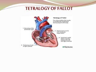
- 4. DEFINITION : Tetralogy is a congenital heart defect. A congenital heart defect is a problem with the heart's structure that’s present at birth. This type of heart defect changes the normal flow of blood through the heart. Tetralogy of Fallot involves four heart defects: A large ventricular septal defect (VSD) Pulmonary stenosis Right ventricular hypertrophy An overriding aorta or dextraposition of aorta
- 7. EPIDEMIOLOGY : Tetralogy of Fallot occurs in approximately 400 per million live births. Tetralogy of Fallot is a rare, complex heart defect that occurs in about 5 out of every 10,000 babies. Occurs equally in girls and boys.
- 8. ventricular septal defect (VSD)
- 9. A hole between the two bottom chambers (ventricles) of the heart. The defect is centered around the most superior aspect of the ventricular septum (the outlet septum), and in the majority of cases is single and large. In some cases thickening of the septum (septal hypertrophy) can narrow the margins of the defect.
- 11. A narrowing of the right ventricular outflow tract and can occur at the pulmonary valve (valvular stenosis) or just below the pulmonary valve (infundibular stenosis). Infundibular pulmonic stenosis is mostly caused by overgrowth of the heart muscle wall (hypertrophy of the septoparietal trabeculae), however the events leading to the formation of the overriding aorta are also believed to be a cause. The pulmonic stenosis is the major cause of the malformations, with the other associated malformations acting as compensatory mechanisms to the pulmonic stenosis. The degree of stenosis varies between individuals with TOF, and is the primary determinant of symptoms and severity. This malformation is infrequently described as sub- pulmonary stenosis or subpulmonary obstruction
- 13. The right ventricle is more muscular than normal, causing a characteristic boot-shaped appearance as seen by chest X-ray. Due to the misarrangement of the external ventricular septum, the right ventricular wall increases in size to deal with the increased obstruction to the right outflow tract. This feature is now generally agreed to be a secondary anomaly, as the level of hypertrophy
- 14. Overriding aorta
- 15. An aortic valve with biventricular connection, that is, it is situated above the ventricular septal defect and connected to both the right and the left ventricle. The degree to which the aorta is attached to the right ventricle is referred to as its degree of "override." The aortic root can be displaced toward the front (anteriorly) or directly above the septal defect, but it is always abnormally located to the right of the root of the pulmonary artery. The degree of override is quite variable, with 5-95% of the valve being connected to the right ventricle.
- 16. CAUSES : Certain conditions or factors that occur during pregnancy may raise your risk for having a child with tetralogy of Fallot. These conditions and factors include: Poor nutrition Overuse of alcohol Age (being older than 40) Diabetes Genetic factors : chromosome 22 deletions and DiGeorge syndrome, Down syndrome Heredity
- 17. SIGNS AND SYMPTOMS : CYANOSIS TET SPELLS HEART MURMUR FAILURE TO GAIN WEIGHT CLUBBING
- 18. DIAGNOSTIC TESTS Doctors diagnose tetralogy of Fallot based on a baby’s signs and symptoms, a physical exam, and the results from tests and procedures. Signs and symptoms : The heart defect usually occur during the first weeks of life. Your infant's doctor may notice signs or symptoms during a routine check-up. Some parents also notice cyanosis (a bluish tint to the skin, lips, and fingernails) or poor feeding and bring the baby to the doctor.
- 19. Physical Exam During a physical exam, the doctor may: A physical examination with a stethoscope almost always reveals a heart murmur. Look for signs and symptoms, such as a bluish tint to the skin, lips, or fingernails and rapid breathing. Look at your baby’s general appearance. Some children who have tetralogy of Fallot have characteristic facial traits because they have DiGeorge syndrome
- 20. Diagnostic Procedures Echocardiography EKG (Electrocardiogram) Pulse Oximetry
- 21. Chest X Ray Cardiac Catheterization
- 22. SURGICAL TREATMENT A) COMPLETE INTRACARDIAC REPAIR : The first total repair of Tetralogy of Fallot was done by a team led by C. Walton Lillehei at the University of Minnesota in 1954 on a 11-year-old boy. Total repair on infants has had success from 1981, with research indicating that it has a comparatively low mortality rate. Widen the narrowed pulmonary blood vessels : The pulmonary valve is widened or replaced, and the passage from the right ventricle to the pulmonary artery is enlarged. These procedures improve blood flow to the lungs. This allows the blood to get enough oxygen to meet the body's needs.
- 23. Close the ventricular septal defect (VSD) : A patch is used to cover the hole in the septum. This patch stops oxygen-rich and oxygen-poor blood from mixing between the ventricles.
- 24. MEDICAL MANAGEMENT Beta blocker : Propranolol Analgesic : Morphine Vasopressor : epinephrine, phenylephrine, norepinephrine
- 25. NURSING MANAGEMENT Feeding and Nutrition : Small, frequent meals may be easier for your baby to handle. Child also may need extra nutrition. A supplement or an extra feeding can give the baby more calories, vitamins, or iron. Your child's doctors will work with you to determine what extra nutrition your baby needs.
- 26. Activity Restrictions Child needs to restrict activity or exercise Child can play in organized sports, especially contact sports club. You need a note for child’s school or coaches about limiting your child's exercise
- 27. Nursing diagnosis Impaired gas exchange r/t disturbed pulmonary blood flow Decreased cardiac output r/t reduce myocardial function Activity intolerance r/t hypoxia Imbalance nutrition less than body requirement r/t excessive energy demand required by increased cardiac workload Parental anxiety/fear r/t life threatening illness Ineffective family coping r/t crisis situation and fear of losing child Knowledge deficit r/t long term problem & prevention of complications Risk for infection r/t chronic illness
- 28. COMPLICATIONS Leaking Heart Valves Arrhythmias Pulmonary Artery Branch Stenoses Right Ventricular Aneurysms Residual Ventricular Septal Defects Coronary Heart Disease