Anatomy and Physiology on the musculoskeletal system
•
114 likes•60,395 views

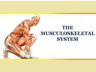
The musculoskeletal system is made up of bones, cartilage, ligaments, tendons and muscles, which form a framework for the body. Tendons, ligaments and fibrous tissue bind the structures together to create stability, with ligaments connecting bone to bone, and tendons connecting muscle to bone.

Recommended
More Related Content
What's hot
What's hot (20)
Similar to Anatomy and Physiology on the musculoskeletal system
Similar to Anatomy and Physiology on the musculoskeletal system (20)
More from DR .PALLAVI PATHANIA
More from DR .PALLAVI PATHANIA (20)
Recently uploaded
Recently uploaded (20)
Anatomy and Physiology on the musculoskeletal system
- 2. PRESENTATION ON THE MUSCULOSKELETAL SYSTEM SUBMITTED TO:- SUBMITTED BY:- DR. PALLAVI PATHANIA KAJAL CHANDEL ASSOCIATE PROFESOR MSC(N) 1ST YEAR SHIMLA NURSING COLLEGE ROLL-NO:- 2
- 3. INDEX Introduction The Skeletal System Bones- Structure, Bone cells, Classification Bone development and growth Healing of bone Bone remodeling and repair Blood and Nerve supply of bone The Axial Skeleton The Appendicular Skeleton The joints The tendons The ligaments The Muscular system The Muscles-types The Musculoskeleton disorders Bone grafting
- 4. INTRODUCTION The Musculoskeletal System consists of two different systems- The Muscular System The Skeletal System. The System includes bones of the skeleton, their joints and the skeleton Muscles that move the body.
- 6. CONTINUE…. The Human Skeletal system is the body system composed of bones, cartilage, tendons, and ligaments and other tissues that perform essential function for the human body. Altogether, the skeleton makes up about 20% of person’s body weight. The human skeleton system is composed of around 270 bones at birth, this total decrease to around 206 bones by adulthood after some bones get fused.
- 7. CONTINUE…. The human skeleton can be divided into the Axial skeleton and the Appendicular skeleton. The axial skeleton is formed by the vertebral column, the rib cage, the skull and others associated bones. The Appendicular skeleton, which is attached to the Axial skeleton, is formed by the shoulder girdle, the, the pelvic girdle and the bones of the upper and lower limb. There are about 650 muscles in the human body and responsible for the movement . Joints are regions of the human skeleton where two or more bones meet and There are about 360 joints in the human body.
- 8. The human body has approximately 900 ligaments. Ligaments are the band of tough elastic tissue around joints. They connect bone to bone, give support, and limit their movement There are approximately 4,000 Tendons in the human body. It is a fibrous connective tissue which attach muscle to bone. Cartilage is a soft, gel like padding between bones that protects joints and facilitates movement.
- 9. BONES
- 10. INTRODUCTION The Basic unit of human skeleton is Bone. Human body contains 206 bones. Bone is living tissue that provides shape and support for the body, as well as protection of some organs. Although bones are often thought to be static or permanent, they are highly vascular living structure that are continuously being remodeled.
- 11. DEFINITION A bone is a rigid tissue that constitutes part of the skeleton in most vertebrate animals. Bones protect the various organ of the body, produce red and white blood cells, store minerals, Provide structure and support for the body, and enable mobility. Bones come in variety of shapes and sizes and have a complex internal and external structure.
- 12. Bone is living tissue, which makes up the body skeleton and is one of the hardest structure of the human body. Bone is a substance that form the skeleton of the body. It is composed chiefly of calcium phosphate and calcium carbonate. It also serves as a storage area for calcium, playing a large role in calcium balance in the blood. The 206 bones in the body serve several other purposes. They support and protect internal organs , Muscles pull against bones to make the body move. Bone marrow, the soft, spongy tissue in the center of many bones, make and stores blood cells.
- 13. MICROSCOPIC STRUCTURE OF BONE
- 14. CONTINUE…. The basic microscopic unit of bone is an Osteon. Osteon are roughly cylindrical structures that can measure several millimeters long and around 0.2 mm in diameter. Each Osteon consists of a lamellae of compact bone tissue that surround a central canal (Haversian canal). Bone is a strong and durable type of connective tissue. Its major constituent (65%) is a mixture of calcium salts and calcium phosphate. This inorganic matrix gives bone great hardness. The remaining third is organic material, called osteoid, which is composed mainly of collagen. Collagen is very strong and gives bone slight flexibility.
- 15. Haversian canals are microscopic tubes or tunnel in cortical bone which generally contains capillaries and nerve fibers. Volkmann’s canals are channels that assist with blood and nerve supply from the periosteum to the haversian canals.
- 16. The periosteum is a membranous tissue that covers the surfaces of your bones. The only areas it doesn't cover are those surrounded by cartilage and where tendons and ligaments attach to bone. Osteon are formations characteristic of mature bone and take shape during the process of bone remodeling, or renewal.
- 17. Canaliculus is a small canal or duct or passageway in the body. It is a small channel in ossified bone, particularly between the lacunae of ossified bone.
- 18. BONE CELLS There are three types of bone cell: Osteoblast (Bone formation) Osteocyte ( Maintain mineral concentration of Matrix) Osteoclast ( Bone resorption)
- 19. A) OSTEOBLASTS OSTEOBLASTS are the large cells responsible for the synthesis and mineralization of bone during both initial bone formation and later bone remodeling that form new bone. They produce new bone called "osteoid" which is made of bone collagen and other protein.
- 20. B) OSTEOCYTES OSTEOCYTES are the longest living bone cell, making up 90–95% of cells in bone tissue in contrast to osteoclasts and osteoblasts making up ~5% (40). Osteocytes form when osteoblasts become buried in the mineral matrix of bone and develop distinct features.
- 21. C) OSTEOCLAST OSTEOCLAST are specialized multinucleated giant cells that reabsorb bone, allowing for the development of new bone and maintenance of bone strength.
- 22. There are two types of bone tissue: 1) Compact bone 2) Spongy bone CONTINUE….
- 23. 1.COMPACT BONE Compact bone (cortical bone) forms the hard external layer of all bones and surrounds the medullary cavity, or bone marrow. It provides protection and strength to bones. This tissue gives bones their smooth, white, and solid appearance, and accounts for 80% of the total bone mass of an adult skeleton. E.g. long bones of the arm and leg.
- 24. 2.SPONGY BONE Spongy bone also known as cancellous bone or trabecular bone, is a very porous type of bone found in animals. It is highly vascularized and contains red bone marrow. Spongy bone is usually located at the ends of the long bones (the epiphyses), with the harder compact bone surrounding it. E.g. It is found at the ends of long bones and in the vertebrae.
- 25. BONE MARKINGS Bone markings are projections and depressions found on bones, which help us to identify the location of other body structures, such as muscles. Their importance comes when we try to describe the shape of the bone or to understand how the muscles, ligaments and other structures affect this bone and vice versa.
- 28. CONTINUE…. The Bones of the body came in a variety of sizes and shapes. The four principal types of bones are long , short , flat and irregular.
- 29. 1.LONG BONES Bones that are longer than they are wide are called long bones. They consist of a long shaft with two bulky ends or extremities. They are primarily compact bone. Example:- long bones include bones of thigh, leg, arms, and forearm. The longest bone in the human body is called the femur, or thigh bone.
- 30. 2.SHORT BONES Short bones are shaped roughly as a cube and contain mostly spongy bone. The outside surface is comprised of a thin layer of compact bone. Short bones are located in the hands and feet. Example:-Short bones include the bones of wrist and ankle. The stapes is the smallest bone in the human body.
- 32. 3.FLAT BONES Flat bones are thin, flattened, and usually curved. Flat bones are bones whose principal function is either extensive protection or the provision of broad surfaces for muscular attachment. Example:- The occipital, parietal, frontal, nasal, lacrimal, vomer, hip bone, sternum, ribs, and scapulae are flat bones.
- 33. 4.IRREGULAR BONES These are bones in the body which do not fall into any other category, due to their non-uniform shape. They primarily consist of cancellous bone, with a thin outer layer of compact bone. Example:- Good examples of these are the Vertebrae, Sacrum, and Mandible (lower jaw).
- 34. THE BONE DEVELOPMENT AND GROWTH The terms Osteogenesis and ossification are often used synonymously to indicate the process of bone formation. Parts of the skeleton form during the first few weeks after conception. By the end of the eighth week after conception, the skeletal pattern is formed in cartilage and connective tissue membranes and ossification begins.
- 35. Bone development continues throughout adulthood. Even after adult stature is attained, bone development continues for repair of fractures and for remodeling to meet changing lifestyles. Osteoblasts, osteocytes and osteoclasts are the three cell types involved in the development, growth and remodeling of bones.
- 36. There are two types of ossification:- 1. Intramembranous ossification 2. Endochondral. ossification 1. Intramembranous ossification:- Intramembranous ossification involves the replacement of sheet-like connective tissue membranes with bony tissue. Bones formed in this manner are called intramembranous bones. They include certain flat bones of the skull and some of the irregular bones. The future bones are first formed as connective tissue membranes. Osteoblasts migrate to the membranes and deposit bony matrix around themselves. When the osteoblasts are surrounded by matrix they are called osteocytes.
- 38. The osteoblasts form a collar of compact bone around the diaphysis. At the same time, the cartilage in the center of the diaphysis begins to disintegrate. Osteoblasts penetrate the disintegrating cartilage and replace it with spongy bone. This forms a primary ossification center. Ossification continues from this center toward the ends of the bones. After spongy bone is formed in the diaphysis, osteoclasts break down the newly formed bone to open up the medullary cavity.
- 39. 2. Endochondral Ossification:- involves the replacement of hyaline cartilage with bony tissue. Most of the bones of the skeleton are formed in this manner. These bones are called endochondral bones. In this process, the future bones are first formed as hyaline cartilage models. During the third month after conception, the perichondrium that surrounds the hyaline cartilage "models" becomes infiltrated with blood vessels and osteoblasts and changes into a periosteum.
- 40. BONE GROWTH Bones grow in length at the epiphyseal plate by a process that is similar to endochondral ossification. The cartilage in the region of the epiphyseal plate next to the epiphysis continues to grow by mitosis.Osteoblasts move in and ossify the matrix to form bone.
- 41. This process continues throughout childhood and the adolescent years until the cartilage growth slows and finally stops. When cartilage growth ceases, usually in the early twenties, the epiphyseal plate completely ossifies so that only a thin epiphyseal line remains and the bones can no longer grow in length. Bone growth is under the influence of growth hormone from the anterior pituitary gland and sex hormones from the ovaries and testes.
- 42. Even though bones stop growing length around early adulthood, they can continue to increase their thickness or diameter throughout life with regards to stress from increased muscle activity or to weight. The increased diameter is called appositional growth. Osteoblasts inside the periosteum form compact bone around the external bone surface. At the same time, osteoclasts within the endosteum break down bone on the internal bone surface, around the medullary cavity. These two processes together increase the diameter of the bone and, at the same time, keep the bone from becoming excessively heavy and bulky.
- 43. HEALING OF BONE
- 44. INRODUCTION All broken bones go through the same healing process. This is true whether a bone has been cut as part of a surgical procedure or fractured through an injury.
- 45. HEALING PROCESS The bone healing process has three overlapping stages: inflammation, bone production and bone remodeling. Inflammation starts immediately after the bone is fractured and lasts for several days. When the bone is fractured, there is bleeding into the area, leading to inflammation and clotting of blood at the fracture site. This provides the initial structural stability and framework for producing new bone.
- 46. Bone production begins when the clotted blood formed by inflammation is replaced with fibrous tissue and cartilage (known as soft callus). As healing progresses, the soft callus is replaced with hard bone (known as hard callus), which is visible on x-rays several weeks after the fracture.
- 47. Bone remodeling, the final phase of bone healing, goes on for several months. In remodeling, bone continues to form and becomes compact, returning to its original shape. In addition, blood circulation in the area improves. Once adequate bone healing has occurred, weight bearing (such as standing or walking) encourages bone remodeling.
- 48. BONE REMOLDING AND REPAIR Bone renewal continues after birth into adulthood. Bone remodeling is the replacement of old bone tissue by new bone tissue. It involves the processes of bone deposition or bone production done by osteoblasts and bone resorption done by osteoclasts, which break down old bone. Normal bone growth requires vitamins D, C, and A, plus minerals such as calcium, phosphorous, and magnesium. Hormones such as parathyroid hormone, growth hormone, and calcitonin are also required for proper bone growth and maintenance.
- 49. STAGES OF BONE REPAIR A fractured or broken bone undergoes repair through four stages: 1.Hematoma formation: Blood vessels in the broken bone tear and hemorrhage, resulting in the formation of clotted blood, or a hematoma, at the site of the break. The severed blood vessels at the broken ends of the bone are sealed by the clotting process. Bone cells deprived of nutrients begin to die.
- 50. 2. Bone generation: (fibrocartilaginous callus formation)Within days of the fracture, capillaries grow into the hematoma, while phagocytic cells begin to clear away the dead cells. Though fragments of the blood clot may remain, fibroblasts and osteoblasts enter the area and begin to reform bone. Fibroblasts produce collagen fibers that connect the broken bone ends, while osteoblasts start to form spongy bone. The repair tissue between the broken bone ends, the fibrocartilaginous callus, is composed of both hyaline and fibrocartilage. Some bone spicules may also appear at this point.
- 51. 3.Bony callous formation: The fibrocartilaginous callus is converted into a bony callus of spongy bone. It takes about two months for the broken bone ends to be firmly joined together after the fracture. This is similar to the endochondral formation of bone when cartilage becomes ossified; osteoblasts, osteoclasts, and bone matrix are present.
- 52. 4.Bone remodeling:- The bony callus is then remodelled by osteoclasts and osteoblasts, with excess material on the exterior of the bone and within the medullary cavity being removed. Compact bone is added to create bone tissue that is similar to the original, unbroken bone. This remodeling can take many months; the bone may remain uneven for years.
- 53. BLOOD SUPPLY OF BONE
- 54. CONTINUE…. Nutrient Artery - It enters the shaft through the nutrient foramen, runs through the cortex, and divides into ascending and descending branches in the medullary cavity. Each branch divides into a number of small parallel channels which terminate in the adult metaphysis by anastomosing with the epiphysial, metaphysial and periosteal arteries. The nutrient artery supplies medullary cavity, inner 2/3 of cortex and metaphysis.
- 55. CONTINUE…. Periosteal Arteries -The periosteal artery system is a low-pressure system that supplies the outer 1/3 of bone and is connected through Haversian and Volkmann canals. These canals are part of the osteon structure of the cortex. Epiphysial Arteries - These are derived from periarticular vascular arcades found on the nonarticular bony surface. Metaphysial Arteries - These are derived from the neighbouring systemic vessels. They pass directly into the metaphysis and reinforce the metaphysial branches from the primary nutrient artery.
- 56. NERVE SUPPLY OF BONE Nerves accompany the blood vessels. Most of them are sympathetic and vasomotor in function. A few of them are sensory which are distributed to the articular ends and periosteum of the long bones, to the vertebra, and to large flat bones.
- 57. FUNCTIONS OF BONE Shape and support to the body, and resist any forms of stress. Provide surface for the attachment of muscles, tendons, ligaments, etc. Levers for muscular actions. Protective in function. Bone marrow manufactures blood cells.
- 58. CONTINUE…. Stores 97% of the body calcium and phosphorus. Bone marrow contains reticuloendothelial cells which are phagocytic in nature and take part in immune responses of the body. Allowing movement of the body as a whole and of parts of the body, by forming joints that are moved by muscle.
- 59. CLASSIFICATION OF SKELETON The bones of the skeleton are divided into two groups: 1. The Axial Skeleton 2. The Appendicular Skeleton
- 60. 1. THE AXIAL SKELETON The Axial skeleton is the part of the skeleton that consists of the bones of the head and trunk of a vertebrate. The human skeleton consists of 80 bones and is composed of six parts; the skull, also the ossicles of the middle ear, the hyoid bone, the rib cage, sternum and the vertebral column. The axial skeleton together with the appendicular skeleton form the complete skeleton.
- 61. A. The Skull Bones
- 62. Continue…. The skull is a bony structure that supports the face and forms a protective cavity for the brain. It is comprised of many bones, which are formed by intramambranous ossification, and joined by sutures(fibrous joints). Skull is the bony framework, that gives the head, its characteristic shape. The function of the skull is to protect the soft and the vital tissues of the head, particularly the brain. The Skull consists of the Cranium (the bony box housing the brain) and the face. The skull is composed of 22 bones, 8 in the cranium and 14 in the face.
- 63. 1.CRANIUM The cranium is formed by a number of flat and irregular bones that provides a bony protection for the brain. It has a base upon which the brain rest and a vault that surround and covers.
- 64. The Bone Of Cranium The bone of the cranium are: 1 Frontal Bone 2 Parietal Bone 2 Temporal Bone 1 Occipital Bone 1 Sphenoid Bone 1 Ethmoid Bone
- 65. a) Frontal bone The frontal bone is a bone of the skull found in the forehead region. It is one of eight bones that form the cranium, or brain case. The frontal bone plays a vital role in supporting and protecting the delicate nervous tissue of the brain. It gives shape to the skull and supports several muscles of the head. The frontal bone is a bowl-shaped bone in the frontal (forehead) region of the skull. It is located superior to the nasal bones and maxillae and anterior to the parietal bones.
- 66. b)Parietal bone The Parietal bones forms a large part of the cranial vault and extend from the frontal bone to the occipital bone. The two bones join at the midline on the top of the cranium and form the saggital suture. From this suture, these bones extend down and out to about the level of the top of the external ear where they meet the temporal bones.
- 67. c)Temporal bone Temporal bones complete the sides and part of the base of the cranium. These bones contain organs of hearing and equilibrium. The external acoustic meatus in the side of each bone forms a passage from the external ear to the middle ear which lies within each bone.
- 68. d) Occipital bone The Occipital bone forms the posterior (back) part of the floor and vault of the cranium. It is the bone which supports the head upon the spinal column. The spinal cord leaves the cranium through an opening in the occipital bone called the foramen magnum.
- 69. e) Sphenoid bone The sphenoid bone is the central part of the base of the cranium. It forms part of the orbits, transmits the optic nerve and supports the posterior part of the maxilla. The sphenoid air sinuses lie in this bone. The pituitary gland lies in a bony socket called the sella turcica, located on the superior aspect of the sphenoid bone.
- 70. f) Ethmoid bone The Ethmoid bone lies between the eyes and extends from the frontal bone to the sphenoid bone. It forms the anterior part of the skull, the medial wall of each orbit, part of the nasal septum and the roof of the nose. It transmits the olfactory nerve .
- 71. 2. THE FACE There are 14 bones in the face. (i) Mandible (1) (ii) Maxillae (2) (iii) Zygomatic Bones (2) (iv) Lacrimal Bones (2) (v) Nasal Bones (2) (vi) Inferior conchae (2) (vii) Palatine Bones (2) (viii) Vomer (1)
- 72. a) Zygomatic bone The right and left zygomatic bones form the lower and outer edges of each orbit and that part of each zygomatic arch nearest the eye. The Zygomatic bone and Zygomatic process of the temporal bone form the Zygomatic arch. The anterior edge of the zygomatic bone joins the maxilla. That part of the maxilla which joins the zygomatic bone is called the zygomatic process.
- 73. b) Maxilla The right and left maxillary bones forms the upper jaw and palate of the mouth. The two halves are fused at the intermaxillary suture to form the upper jaw.
- 74. c) Nasal bone The Nasal bones (right and left) are long, thin pieces of bone that form the upper part of the bridge of the nose. The anterior lower part of the nasal septum is composed of cartilage.
- 75. d) Lacrimal bone The paired (right and left) Lacrimal bones form small parts of the medial walls of the orbits. The lacrimal bones transmits the naso-lacrimal duct from the eye to the nose or nasal fossa.
- 76. e) Vomer The vomer forms the inferior part of the nasal septum, the vertical partition separating the right and left nasal cavities.
- 77. f) Palatine bone The palatine bones (right and left) join in the midline to form the posterior part of the hard palate . Palatine bones also form part of the floor and lateral walls of the nasal cavity and part of the floor of the orbits.
- 78. g) Inferior conchae The inferior nasal conchae (right and left) are scroll like bones lying horizontally along the lateral walls of the nasal cavity. The bony elements of the middle and superior conchae are extensions of the lateral parts of the ethmoid bones.
- 79. h) Mandible The horse shoe- shaped bone forming the lower jaw, articulating with the skull at the temporomandibular joint. Mandible is the largest, strongest and lowest bone in the face.
- 80. B. The Vertebrae
- 81. Vertebrae are the 33 individual, interlocking bones that form the spinal column. The spinal column consists of seven cervical, twelve thoracic, and five lumbar vertebrae in addition to five fused vertebrae of the sacral region and four fused vertebrae forming the coccyx.
- 82. Continue…. The Spine is divided into several sections. The Cervical Vertebrae make up the neck. The Thoracic vertebrae comprise the chest section and have ribs attached.
- 83. The Lumber Vertebrae are the remaining vertebrae below the last thoracic bone and the top of the sacrum. The Sacral Vertebra are caged within the bones of the pelvis, and the coccyx represents the terminal vertebrae or vestigial tail.
- 84. C. The Ribs
- 85. CONTINUE…. The Ribs are a set of twelve paired bones which form the protective ‘cage’ of the thorax. They articulate with the vertebral column posteriorly, and terminate anteriorly as cartilage(known as costal cartilage). As part of the bony thorax, the ribs protect the internal thoracic organs.
- 86. D. The Sternum The Sternum is a long flat bone located in the central part of the chest. It connects to the Ribs Via cartilage and forms the front of the ribcage, thus helping to protect the heart, lungs, and major blood vessels from injury.
- 88. CONTINUE…. The Appendicular skeleton is composed of the bones of the upper limbs (which function to grasp and manipulate objects) and the lower limbs (which permit locomotion). It also includes the pectoral girdle, or shoulder girdle, that attaches the upper limbs to the body, and the pelvic girdle that attaches the lower limbs to the body.
- 89. A. The Upper/Lower Limbs Bones THE UPPER LIMB:-The upper limbs consists of the arm(the upper arm), the forearm(the lower arm),and the hand. The arm consists of a single bone, the humerus.The forearm consists of two bones, the ulna and radius. And the hand consists of 27 bones, which are grouped into the phalanges, metacarpals, and carpals.
- 91. Continue…. THE LOWER LIMB:- The lower limb consists of the thigh(the upper leg), the leg(the lower leg), and the foot. The leg consists of two long bones, the tibia and fibula, and the sesamoid bone, the patella, that serves as the knee cap.
- 93. Continue…. The Shoulder Girdle or Pectoral Girdle is the set of bones in the Appendicular skeleton which connects to the arm on each side. In humans it consists of the clavicle and scapula.
- 94. C. Pelvic Girdle
- 95. Continue…. The Pelvic Girdle is composed of the Appendicular hip Bones( ileum, ischium and pubis) oriented in a ring, and connects the pelvic region of the spine to the lower limbs. The pelvic spine consists of the sacrum and coccyx.
- 96. THE JOINTS A joints is the point where two or more bones meet. There are three main types:
- 97. A joint or articulation (or articular surface) is the connection made between bones in the body which link the skeletal system into a functional whole.They are constructed to allow for different degrees and types of movement. Some joints, such as the knee, elbow, and shoulder, are self- lubricating, almost frictionless, and are able to with stand compression and maintain heavy loads while still executing smooth and precise movements. Other joints such as sutures between the bones of the skull permit very little movement (only during birth) in order to protect the brain and the sense organs. The connection between a tooth and the jaw bone is also called a joint, and is described as a fibrous joint known as a gomphosis. Joints are classified both structurally and functionally.
- 98. CONTINUE…. Fibrous (immovable)-Eg:-skull joint. Cartilaginous (partially moveable)- Eg:-intervertebral disc of spinal column. Synovial(freely moveable) – E.g.:- knee joint.
- 99. 1.FIBROUS JOINT Fibrous joints are connected by dense connective tissue consisting mainly of collagen. These joints are also called fixed or immovable joints because they do not move. Fibrous joints have no joint cavity and are connected via fibrous connective tissue. The skull bones are connected by fibrous joints called sutures.
- 100. 2.CARTILAGENOUS JOINT Cartilaginous joints are connected entirely by cartilage (fibrocartilage or hyaline).Cartilaginous joints allow more movement between bones than a fibrous joint but less than highly mobile synovial joint.
- 101. 3.SYNOVIAL JOINT There are six types of synovial joints: 1.Pivot joint, 2.Ball-and-socket joint, 3.Hinge joint, 4.Condyloid joint, 5.Saddle joint, 6.Gliding joint.
- 102. a) Pivot joints Pivot joints, also known as rotary joints, are a type of synovial joint that permit axial rotation. The moving bone rotates within a ring formed by the concave surface of a second bone and an adjoining ligament. example of a pivot joint is the joint of the first and second vertebrae of the neck that allows the head to move back and forth .
- 103. b) Ball-and-socket joint Ball-and-socket joint, also called spheroidal joint, in vertebrate anatomy, a joint in which the rounded surface of a bone moves within a depression on another bone, allowing greater freedom of movement than any other kind of joint. Examples of ball-and-socket joints are the shoulder and hip joints.
- 104. c) Hinge joint Hinge joint is a type of synovial joint that exists in the body and serves to allow motion primarily in one plane. The hinge joint is made up of two or more bones with articular surfaces that are covered by hyaline cartilage and lubricated by synovial fluid. The best examples of hinge joints are the Interphalangeal joints of the hand and those of the foot and the joint between the humerus and ulna.
- 105. d) Condyloid joints Condyloid joints are a type of synovial joint where the articular surface of one bone has an ovoid convexity sitting within an ellipsoidal cavity of the other bone. Examples of condyloid joints are Radiocarpal joint and Metacarpo-phalangeal joint.
- 106. E) Saddle joints Saddle joints are a type of synovial joint that allow articulation by reciprocal reception. Both bones have concave-convex articular surfaces which interlock like two saddles opposed to one another. The prime example of a saddle joint is the trapeziometacarpal joint at the base of your thumb.
- 107. F) gliding joint Gliding joint, also known as a plane joint or planar joint, is a common type of synovial joint formed between bones that meet at flat or nearly flat articular surfaces. Gliding joints allow the bones to glide past one another in any direction along the plane of the joint — up and down, left and right, and diagonally. Examples are the intermetacarpal joints and the acromioclavicular joint (between the acromion of the scapula and the clavicle).
- 108. CONTINUE….
- 109. THE MAIN MOVEMENT POSSIBLE AT SYNOVIAL JOINTS
- 110. THE TENDONS A tendon is tough but flexible structure made of fibrous tissue that joins a bone to a muscle. When a muscle contracts it pulls on a bone to cause movement. The tendon transmits the force from the muscle to the bones. The tendonitis is the inflammation of a tendon.
- 111. THE LIGAMENTS Ligaments are bands of connective tissues that link two or more bones to make joints stable are prevent from excessive movements. Ligaments are bands of tough elastic tissue around the joints. They connect bone to bone, give joints support, and limit their movement. ligaments present around the knees, ankles, elbows, shoulders, and other joints. Stretching or tearing them can make joints unstable.
- 112. THE MUSCULAR SYSTEM
- 113. CONTINUE…. The Muscular System is an organ system consisting of skeletal, smooth and cardiac muscles. It permits movement of the body, maintains posture and circulates blood throughout the body. The muscular systems in vertebrates are controlled through the nervous system although some muscles (such as the cardiac muscle) can be completely autonomous. Together with the skeletal system in the human, it forms the musculoskeletal system, which is responsible for movement of the body.
- 114. THE MUSCULAR TISSUE Muscular cells are called muscle fibers. Every fibers contain thousand of myofibrils.
- 115. CONTINUE…. Inside each myofibril there are many myofilaments that are made of two proteins: the actin and the myosin .The myofibrils are divided in subunits called sarcomeres.
- 116. TYPES OF MUSCLES There are three types of muscles: Skeletal muscle Cardiac muscle Smooth muscle
- 117. 1.SKELETAL MUSCLES The skeletal muscles are also known as striated or voluntary. They are attached to bones by tendons providing movement. Their contraction is quick and variable from powerful to precise. It is controlled by the CNS.
- 118. 2.CARDIAC MUSCLE Cardiac muscle is only found in heart. Cardiac muscle tissue is one of the three types of muscle tissue in our body. Cardiac muscle tissue is only found in your heart, where it performs coordinated contractions that allow heart to pump blood through circulatory system.
- 119. 3.SMOOTH MUSCLE It is a type of muscles that contracts without any voluntary control, and it is made of a thin form of layers which is made up of spindle-shaped, unstriated cells with only one nucleus and present in inner organs walls like bladder, intestine, stomach, blood vessels, etc. excluding the heart.
- 120. CONTINUE…. It covers the hollow walls of many organs such as the oesophagus, the broncchi, the uterus or stomach. It contracts slowly.
- 121. The calf is the back portion of lower leg in human anatomy. The muscles within the calf correspond to the posterior compartment of leg. The two largest muscles within this compartment are known together as the calf muscle and attach to the heel via the Achilles tendon. CALF MUSCLE
- 122. The Second Heart ( Calf Muscle) The second heart calf muscle is a system of muscles, veins, and valves in the foot that work together to push deoxygenated blood back up to the heart and lungs. Vein valves act as trapdoors that open and close with each muscle contraction to prevent the backflow of blood. When valves become defective or weak the second heart can be leads to varicose veins, spider veins, and Chronic Venous Insufficiency (CVI) because of blood pool in veins.
- 123. The calf muscle, on the back of the lower leg, is actually made up of two muscles: The gastrocnemius is the larger calf muscle, forming the bulge visible beneath the skin. The gastrocnemius has two parts or "heads," which together create its diamond shape. The soleus is a smaller, flat muscle that lies underneath the gastrocnemius muscle.
- 124. Functions of calf muscles Plantar flexion:- The superficial muscles planter flex the ankle, which is when move foot and toes downward while lift heel up. This movement is important for walking, running and biking because we need to push the ball of foot of the ground for each step. Curling toes:-Two of the deep calf muscles have tendons that start up in the calf and run down the leg and the bottom of foot to attach the toes. The flexor hallucis longus flexes big toe while the flexor digitorum longus has tendon that attach to other four toes. Flexing the toes cause them to curl under, which helps the feet push of the ground when walk and run and is also important for gymnastic and dance moves, which require a pointed toe.
- 125. Continue…. Bending knee:- The hamstring muscles in the back part of the thigh. Or upper leg, do most of work when we bend our knee, but two of the calf muscles help. The gastrocnemius of the superficial group and the planters of the deep group work as knee flexors to help bend the knee, which is necessary for movement such as walking, running and squatting. Returning blood to the heart:- Vein are the blood vessels that return blood to the heart. The veins that start down in the feet divide into superficial and deep veins that run up through the legs and thighs before meeting at the major vein that connect to the heart. Blood flow from the feet and legs often has to work against gravity, so contractions of the calf muscles help build pressure that moves the blood through the vein. This is called the calf muscle pump.
- 127. INTRODUCTION Musculoskeletal Disorders are injuries and disorders that affect the human body’s movement or musculoskeletal system (i.e. muscles, tendons, ligaments, nerves, discs, blood vessels, etc.).
- 128. DEFINITION:-This is an inflammation of a tendon – the fibrous tissues that connect a muscle to a bone. It can especially affect your shoulder, elbow, ankle, or wrist. Cause:-injury, aging, certain antibiotics,Athletes who participate in certain sports, certain diseases, such as diabetes or rheumatoid arthritis Sign and symptoms:- Pain, tenderness, mild swelling Diagnostic Evaluation:- Health history, Physical Examination, X- rays, MRI scan. Management:- Resting or elevating the tendon as advised by doctor Applying heat or ice Taking medications, such as the pain reliever acetaminophen (tylenol) and the anti-inflammatory drugs aspirin , ibuprofen and naproxen 1.TENDONITIS
- 129. 2.OSTEOARTHRITIS DEFINITION:- Usually referred to as just “arthritis” this is a condition in which cartilage – the rubbery protective tissue at the end of bones – gradually wears down. This can result in joint pain in the hands, neck, lower back, knees, or hips. Cause:- Injury. Obesity. Heredity, Joint overuse, Other diseases. Sign and symptoms:- Stiffness after periods of rest, Bony enlargements in the middle and end joints of the fingers (which may or may not be painful), Joint swelling Pain after overuse or after long periods of inactivity. Diagnostic evaluation:- Health examination, physical examination, X- rays Treatment:- Osteoarthritis usually is treated by a combination of treatments, including exercise, weight loss if needed, medications, physical therapy with muscle strengthening exercises, hot and cold compresses to the painful joint.
- 130. 3.RHEUMATOID ARTHRITIS DEFINITION:-With rheumatoid arthritis (RA), body’s immune system attacks its own cells. If left untreated, RA can erode the bones and cause deformity in the joints, such as the fingers. RA affects joint cartilage first, but the inflammation can spread to other organs throughout the body. Cause:- Infection, Obesity, Smoking, Family history Sign and symptoms:-Tender, warm, swollen joints, Joint stiffness that is usually worse in the mornings and after inactivity, Fatigue, fever and loss of appetite Diagnostic Evaluation:- Health history, Physical examination, Blood test, Imaging test Management:-here is no cure for rheumatoid arthritis. But clinical studies indicate that remission of symptoms is more likely when treatment begins early with medications known as disease-modifying antirheumatic drugs (DMARDs). Physical therapy Surgical management:- Total joint replacement, Joints fusion, Tendon Repair
- 131. 4)BONE FRACTURES DEFINITION:- Trauma, overuse, and disease can weaken bones – sometimes to the point of causing a complete or partial break. Not only is a bone fracture painful, but it can also result in a temporary loss of functionality in an arm, leg, foot, or hand. Types:-Open Fracture, Close fracture, Greenstick Fracture, Impact fracture , Oblique Fracture, Comminuted Fracture Cause:- Bad fall, People age, Osteoporosis, Infection, tumor Sign and symptoms:- pain, swelling, bruising, discolored skin around the affected area, the patient cannot move the affected area Diagnostic evaluation:- Health history, Physical examination, X-rays, MRI, CT- SCAN. Management:- Plaster cast wrap the break with hard protection, Immobilization, physical therapy, surgical treatment
- 132. 5.CARPAL TUNNEL SYNDROME DEFINITION:- Carpal tunnel syndrome is a common condition that causes pain, numbness, tingling, and weakness in the hand and wrist. It happens when there is increased pressure within the wrist on a nerve called the median nerve. This nerve provides sensation to the thumb, index, and middle fingers, and to half of the ring finger. The small finger (the “pinky”) is typically not affected. Risk -Factors :-High-force (hammering), Long-term use, Extreme wrist motions, Vibration. Sign and symptoms:-Tingling in the fingers, Decreased feeling in the fingertips, Difficulty using the hand for small tasks Diagnostic evaluation:- Physical examination, Wrist flexion test, X-rays Management-Wearing a wrist splint at night, Taking nonsteroidal anti- inflammatory drugs, such as ibuprofen.
- 133. 6.MUSCULAR DYSTROPHY DEFINITION:-Muscular dystrophy is a group of inherited muscle diseases. These conditions all cause muscle loss and weakness. Some appear in infancy or childhood, and others may not appear until middle age or even later. Cause:- mutations (alterations) in the genes responsible for healthy muscle structure and function. Symptoms :- pain and stiffness in the muscles, difficulty with running and jumping, walking on toes, difficulty sitting up or standing, learning disabilities, such as developing speech later than usual, frequent falls. Diagnostic evaluation:- Genetic testing, Heart monitoring, Lung monitoring, Treatments:-include medications, physical therapy, speech therapy, orthopedic devices, and surgery.
- 134. BONE GRAFTING Bone grafting is a surgical procedure that uses transplanted bone to repair and rebuild diseased or damaged bones. A bone graft is a choice for repairing bones almost anywhere in body. Surgeon might take bone from hips, legs, or ribs to perform the graft. Sometimes, surgeons also use bone tissue donated from cadavers to perform bone grafting
- 135. Most of skeleton consists of bone matrix. This is the hard material that helps give the bones their strength. Inside the matrix are living bone cells. These make and maintain this matrix. The cells in this matrix can help repair and heal bone when necessary. When bone is break, the healing process begins. As long as the break bone is not too large, bone cells can repair it. Sometimes, though, a fracture results in a large loss of bone, like when a large chunk of the bone crumbles away. In these cases, your bone might not fully heal without a bone graft.
- 136. TYPES OF BONE GRAFT The two most common types of bone grafts are: Allograft, which uses bone from a deceased donor or a cadaver that has been cleaned and stored in a tissue bank. Autograft, which comes from a bone inside your body, such as your ribs, hips, pelvis, or wrist
- 137. Allografts are commonly used in hip, knee, or long bone reconstruction. Long bones include arms and legs. The advantage is there’s no additional surgery needed to acquire the bone. It also lowers risk of infection since additional incisions or surgery aren’t required. Allograft bone transplant involves bone that has no living cells so that the risk of rejection is minimal as opposed to organ transplants, in which living cells are present. Since the transplanted bone doesn’t contain living marrow, there is no need to match blood types between the donor and the recipient.
- 138. The risks of a bone graft All surgical procedures involve risks of bleeding, infection, and reactions to anesthesia. Bone grafts carry these risks and others, including: Pain Swelling Nerve injury Rejection of the bone graft Inflammation Reabsorption of the graft
- 139. WHY BONE GRAFTING IS PERFORMED? Bone grafting is done for numerous reasons, including injury and disease. There are four main reasons bone grafts are used: A bone graft may be used in the case of multiple or complex fractures or those that don’t heal well after initial treatment. Fusion helps two bones heal together across a diseased joint. Fusion is most often done on the spine. Regeneration is used for bone lost to disease, infection, or injury. This can involve using small amounts of bone in bone cavities or large sections of bones. A graft can be used to help bone heal around surgically implanted devices, like joint replacements, plates, or screws.
- 140. HOW A BONE GRAFT IS PERFORMED Doctor will decide which type of bone graft to use before surgery. Given general anesthesia, which will put patient into a deep sleep. An anesthesiologist will monitor the anesthesia and recovery. Surgeon will make an incision in the skin above where the graft is needed. They’ll then shape the donated bone to fit the area. The graft will be held in place using any of the following: Pins Plates Screws Wires Cables
- 141. Once the graft is securely in place, surgeon will close the incision or wound with stitches and bandage the wound. A cast or splint may be used to support the bone while it heals. Many times, no casting or splint is necessary. Bone fixation and repair devices traditionally are fabricated with metals and used clinically. Stainless steel, titanium and its alloys have been employed for the majority of fracture fixation treatments. Alloplastic grafts may be made from hydroxyapatite, a naturally occurring mineral (main mineral component of bone), made from bioactive glass. Hydroxyapatite is a synthetic bone graft, which is the most used now due to its osteoconduction, hardness, and acceptability by bone.
- 142. An alloplastic graft is composed of material that is not taken from an animal or human source. Alloplastic grafts can be derived from natural sources (such as an elements or minerals), synthetic (man-made) substances, or a combination of the two. One reason many dentists prefer alloplastic grafts is that they do not require tissue to be harvested from another source.
- 143. Alloplastic grafts can be made of hydroxyapatite (HA), calcium carbonate, and tricalcium phosphate. Hydroxyapatite is the most frequently used due to its strength, durability, and ability to integrate well with bone. In fact, a large percentage of human bone is composed of a form of hydroxyapatite. Calcium carbonate is becoming less popular because it tends to resorb more quickly and make the bone susceptible to breakage.
- 144. ASSIGNMENT Draw the structue of skeletal. Draw the Microscopic structure of bone. Draw the structure of compact and spongy Bone. Make a table of bone marking. Make the paper project on classification of skeleton system. Make a diagram of Skull Draw the structure of synovial joints. Draw the structure of muscles.
- 145. SUMMARIZATION
- 146. CONCLUSION Bones are an important part of the musculoskeletal system and serve many core functions, as well as supporting the body’s structure and facilitating movement.Togethers our bones, muscles , and joints along with other tendons, ligaments, and cartilage from our musculoskeletal system and enable us to do everyday physical examination.
- 147. RECAPTULIZATION Question. Define bone Answer. The Basic unit of human skeleton is bone.Human body contains 206 bones. Bone is living tissue that provides shape and support for the body, as well as protection of some organs. Question. Enlist the type of bone cell Answer. Osteoblast, Osteoclast, Osteocyte Question. Bones of the skeleton are divided into how many groups.Enlist them. Answer. Divided into two groups: - The axial skeleton , The Appendicular system
- 148. CONTINUE… Question. How many bones are present in face .Enlist them. Answer. There are 14 bones in the face. (i) Mandible (1) (ii) Maxillae (2) (iii) Zygomatic Bones (2) (iv) Lacrimal Bones (2) (v) Nasal Bones (2) (vi) Inferior conchae (2) (vii) Palatine Bones (2) (viii) Vomer (1)
- 149. CONTINUE… Question. Enlist the types of synovial joints Answer. There are six types of synovial joints: 1.Pivot joint, 2.Ball-and-socket joint, 3.Hinge joint, 4.Condyloid joint, 5.Saddle joint, 6.Gliding joint. Question. Enlist the types of Muscle Answer. There are three types of muscles: Skeletal muscle Cardiac muscle Smooth muscle
- 150. Question. Define bone grafting. Answer. Bone grafting is a surgical procedure that uses transplanted bone to repair and rebuild diseased or damaged bones. A bone graft is a choice for repairing bones almost anywhere in body. Surgeon might take bone from hips, legs, or ribs to perform the graft. Sometimes, surgeons also use bone tissue donated from cadavers to perform bone grafting
- 151. BIBLIOGRAPHY BOOKS REFERENCES:- Wilson and Ross. Anatomy and Physiology in Health and Illness 12th edition (International Edition) Anne Waugh Allison Grant. ELSEVIER INTERNET REFERENCES:- https://www.slideshare.net https://www.birdvilleschools.net https://www.soinc.org