Primary Maternal Care: Assessment of fetal growth and condition during pregnancy
•
3 gefällt mir•1,042 views

Primary Maternal Care addresses the needs of healthcare workers in level 1 district hospitals and clinics who provide antenatal and postnatal care, but do not conduct deliveries. It is adapted from theory chapters and skills workshops from Maternal Care. This book complements the national protocol of antenatal care in South Africa. It covers: booking for antenatal care, assesing fetal growth and wellbeing, hypertensive disorders of pregnancy, antepartum haemorrhage, preterm labour, important medical conditions

Empfohlen
Empfohlen
Weitere ähnliche Inhalte
Was ist angesagt?
Was ist angesagt? (19)
Ähnlich wie Primary Maternal Care: Assessment of fetal growth and condition during pregnancy
Ähnlich wie Primary Maternal Care: Assessment of fetal growth and condition during pregnancy (20)
Mehr von Saide OER Africa
Mehr von Saide OER Africa (20)
Kürzlich hochgeladen
Kürzlich hochgeladen (20)
Primary Maternal Care: Assessment of fetal growth and condition during pregnancy
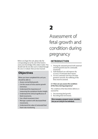
- 1. 2 Assessment of fetal growth and condition during pregnancy Before you begin this unit, please take the INTRODUCTION corresponding test at the end of the book to assess your knowledge of the subject matter. You 1. During the antenatal period, both maternal should redo the test after you’ve worked through and fetal growth must be continually the unit, to evaluate what you have learned. monitored. 2. Individualised care will improve the Objectives accuracy of antenatal observations. 3. At every antenatal visit from 28 weeks When you have completed this unit you gestation onwards, the wellbeing of the should be able to: fetus must be assessed. • Assess normal fetal growth. • List the causes of intra-uterine growth 2-1 How can you assess the condition of the fetus during pregnancy? restriction. • Understand the importance of The condition of the fetus before delivery is measuring the symphysis-fundus height. assessed by: • Understand the clinical significance of 1. Documenting fetal growth. fetal movements. 2. Recording fetal movements. • Use a fetal movement chart. When managing a pregnant woman, remember • Manage a patient with decreased fetal that you are caring for two individuals. movements. • Understand the value of antenatal fetal heart-rate monitoring.
- 2. 60 PRIMAR Y MATERNAL CARE FETAL GROWTH Poor maternal weight gain is of very little value in diagnosing intra-uterine growth restriction. 2-2 What is normal fetal growth? 2. Fetal factors: • Multiple pregnancy. If the assessed fetal weight is within the • Chromosomal abnormalities, expected range for the duration of pregnancy, e.g. trisomy 21. then the fetal growth is regarded as normal. • Severe congenital malformations. • Chronic intra-uterine infection, To determine fetal growth you must have an e.g. congenital syphilis. assessment of both the duration of pregnancy 3. Placental factors: and the weight of the fetus. • Poor placental function (placental insufficiency) is usually due to a 2-3 When may fetal growth maternal problem such as pre- appear to be abnormal? eclampsia. • Smoking. Fetal growth will appear to be abnormal when Poor placental function is uncommon in a the assessed fetal weight is greater or less than healthy woman who does not smoke. that expected for the duration of pregnancy. Remember that incorrect menstrual dates If severe intra-uterine growth restriction is are the commonest cause of an incorrect present, it is essential to look for a maternal or assessment of fetal growth. fetal cause. Usually a cause can be found. 2-4 When is intra-uterine growth 2-6 How can you estimate fetal weight? restriction suspected? The following methods can be used: When the weight of the fetus is assessed as 1. Measure the size of the uterus on being less than the normal range for the abdominal examination. duration of pregnancy, then intra-uterine 2. Palpate the fetal head and body on growth restriction (fetal growth restriction) is abdominal examination. suspected. 3. Measure the size of the fetus using antenatal ultrasonography (ultrasound). 2-5 What maternal and fetal factors are associated with intra- 2-7 How should you measure uterine growth restriction? the size of the uterus? Intra-uterine growth restriction may be 1. This is done by determining the associated with either maternal, fetal and symphysis-fundus height (S-F height), placental factors: which is measured in centimetres from the 1. Maternal factors: upper edge of the symphysis pubis to the • Low maternal weight, especially a top of the fundus of the uterus. low body mass index resulting from 2. The S-F height in centimetres should be undernutrition. plotted against the gestational age on the • Tobacco smoking. S-F growth curve. • Alcohol intake. 3. From 36 weeks onwards, the presenting • Strenuous physical work. part may descend into the pelvis and • Poor socio-economic conditions. measurement of the S-F height will not • Pre-eclampsia and chronic accurately reflect the size of the fetus. A hypertension. reduction in the S-F height may even be observed.
- 3. ASSESSMENT OF FETAL GROWTH AND CONDITION DURING PREGNANC Y 61 2-8 What is the symphysis- 1. Slow increase in uterine size until one fundus growth curve? measurement falls under the 10th centile. 2. Three successive measurements ‘plateau’ The symphysis-fundus growth curve compares (i.e. remain the same) without necessarily the S-F height to the duration of pregnancy. crossing below the 10th centile. The growth curve should preferably form 3. A measurement which is less than that part of the antenatal card. The solid line of recorded 2 visits previously without the growth curve represents the 50th centile, necessarily crossing below the 10th centile. and the upper and lower dotted lines, the 90th and 10th centiles, respectively. If intra-uterine Note that a measurement that was originally growth is normal, the S-F height will fall normal, but on subsequent examinations between the 10th and 90th centiles. The ability has fallen to below the 10th centile, indicates to detect abnormalities from the growth curve intra-uterine growth restriction and not is much increased if the same person sees the incorrect dates. patient at every antenatal visit. Between 18 and 36 weeks of pregnancy, the 2-10 How can you identify severe S-F height normally increases by about 1 cm intra-uterine growth restriction? a week. With severe intra-uterine growth restriction, the difference between the actual duration of 2-9 When will the symphysis-fundus height pregnancy and that suggested by plotting S-F suggest intra-uterine growth restriction? height is 4 weeks or more. If any of the following are found: SIGNATURE: DATE: GESTATION 12 13 14 15 16 17 18 19 20 21 22 23 24 25 26 27 28 29 30 31 32 33 34 35 36 37 38 39 40 41 42 43 45 GESTATION EST. BY: 45 Dates Sonar 40 40 Both SF-measurement 35 LW. 0. = Weight 35 x = measurement 30 30 25 25 20 20 15 15 10 10 Start SF measurement Repeat examination of breasts at 34 weeks 5 Uterine size using PRESENTING PART 5 anatomical landmarks HEAD ABOVE PELVIS (fifths) GESTATION 12 13 14 15 16 17 18 19 20 21 22 23 24 25 26 27 28 29 30 31 32 33 34 35 36 37 38 39 40 41 42 43 Figure 2-1: The symphysis-fundus growth chart.
- 4. 62 PRIMAR Y MATERNAL CARE GESTATION 12 13 14 15 16 17 18 19 20 21 22 23 24 25 26 27 28 29 30 31 32 33 34 35 36 37 38 39 40 41 42 43 45 GESTATION EST. BY: 45 Dates Sonar 40 40 Both SF-measurement 35 LW. 0. = Weight 35 x = measurement 30 30 25 25 20 20 15 15 10 10 Start SF measurement Repeat examination of breasts at 34 weeks 5 Uterine size using PRESENTING PART 5 anatomical landmarks HEAD ABOVE PELVIS (fifths) GESTATION 12 13 14 15 16 17 18 19 20 21 22 23 24 25 26 27 28 29 30 31 32 33 34 35 36 37 38 39 40 41 42 43 Figure 2-2: One measurement below the 10th centile GESTATION 12 13 14 15 16 17 18 19 20 21 22 23 24 25 26 27 28 29 30 31 32 33 34 35 36 37 38 39 40 41 42 43 45 GESTATION EST. BY: 45 Dates Sonar 40 40 Both SF-measurement 35 LW. 0. = Weight 35 x = measurement 30 30 25 25 20 20 15 15 10 10 Start SF measurement Repeat examination of breasts at 34 weeks 5 Uterine size using PRESENTING PART 5 anatomical Vx landmarks HEAD ABOVE PELVIS (fifths) 5 GESTATION 12 13 14 15 16 17 18 19 20 21 22 23 24 25 26 27 28 29 30 31 32 33 34 35 36 37 38 39 40 41 42 43 Figure 2-3: Three successive measurements that remain the same
- 5. ASSESSMENT OF FETAL GROWTH AND CONDITION DURING PREGNANC Y 63 GESTATION 12 13 14 15 16 17 18 19 20 21 22 23 24 25 26 27 28 29 30 31 32 33 34 35 36 37 38 39 40 41 42 43 45 GESTATION EST. BY: 45 Dates Sonar 40 40 Both SF-measurement 35 LW. 0. = Weight 35 x = measurement 30 30 25 25 20 20 15 15 10 10 Start SF measurement Repeat examination of breasts at 34 weeks 5 Uterine size using PRESENTING PART 5 anatomical VxVxVx landmarks HEAD ABOVE PELVIS (fifths) 5 5 5 GESTATION 12 13 14 15 16 17 18 19 20 21 22 23 24 25 26 27 28 29 30 31 32 33 34 35 36 37 38 39 40 41 42 43 BLOOD- Syst. PRESSURE Diast. P P Urine S S OEDEMA RRT 2/01 Fetal movements Antenatal Haemoglobim (g/dl) card B ENG Figure 2-4: A measurement less than that recorded 2 visits before 2-11 Does descent of the presenting diet. If possible, patients must be given part of the fetus affect your food supplements (food parcels). interpretation of the growth curve? 3. Exclude pre-eclampsia as a cause. 4. If the gestational age is 28 weeks or more, Yes. Descent of the presenting part may occur careful attention must be paid to counting in the last 4 weeks of pregnancy. Therefore, the fetal movements. after 36 weeks the above criteria are no 5. The patient should be followed up weekly longer valid, if at subsequent antenatal visits at a level 1 hospital. progressively less of the fetal head is palpable above the pelvic inlet. 2-13 Which special investigation is of great value in the further 2-12 What action would you take if the management of this patient? symphysis-fundus height measurement suggests intra-uterine growth restriction? The patient must be referred to a fetal evaluation clinic or level 2 hospital for a 1. The patient should stop smoking and rest Doppler measurement of blood flow in the more, while attention must be given to her umbilical arteries: diet. It may be necessary to arrange sick leave and social support for the patient. 1. Good flow (low resistance) indicates good 2. A poor diet which is low in energy placental function. As a result the woman (kilojoules) may cause intra-uterine growth can receive further routine management restriction, especially in a patient with a as a low-risk patient. Spontaneous onset of low body mass index. Therefore, ensure that patients with suspected intra-uterine growth restriction receive a high-energy
- 6. 64 PRIMAR Y MATERNAL CARE labour can be allowed. Induction of labour patient to patient. Therefore, it is only useful at 38 weeks is not needed. as an approximate guide to the duration of 2. Poor flow (high resistance) indicates poor pregnancy. placental function. Antenatal electronic fetal heart rate monitoring must be done. 2-17 What is the value of The further management will depend on assessing fetal movements? the result of the monitoring. Fetal movements indicate that the fetus is well. If Doppler measurement is not available, the By counting the movements, a patient can, patient must be managed as given in 2-14. therefore, monitor the condition of her fetus. 2-14 What possibilities must be 2-18 From what stage of pregnancy considered if, after taking the above will you advise a patient to become steps, there is still no improvement aware of fetal movements in order in the symphysis-fundus growth? to monitor the fetal condition? 1. Intra-uterine death must be excluded From 28 weeks, because the fetus can now by the presence of a fetal heart beat on be regarded as potentially viable (i.e. there is auscultation. a good chance that the infant will survive if 2. With moderate intra-uterine growth delivered). All patients should be encouraged restriction and good fetal movements, the to become aware of the importance of an patient must be followed up weekly and adequate number of fetal movements. delivery at 38 weeks should be considered. 3. If the above patient also has poor social Asking the patient if the fetus is moving normally circumstances, an admission to hospital on the day of the visit is an important way of will need to be considered. This should monitoring the fetal wellbeing. ensure that the patient gets adequate rest, a good diet and stops smoking. 4. If there are decreased or few fetal move- 2-19 What is a fetal movement chart? ments, the patient should be managed as A fetal movement chart records the frequency described in sections 2-25 and 2-26. of fetal movements and, thereby, assesses the 5. When there is severe intra-uterine growth condition of the fetus. The name “kick chart” restriction, the patient must be referred is not correct, as all movements must be to a level 2 or 3 hospital for further counted, e.g. rolling and turning movements, management. as well as kicking. 2-20 Which patients should use FETAL MOVEMENTS a fetal movement chart? A fetal movement chart need not be used 2-15 When are fetal movements first felt? routinely by all antenatal patients, but only when: 1. At about 20 weeks in a primigravida. 2. At about 16 weeks in a multigravida. 1. There is concern about the fetal condition. 2. A patient reports decreased fetal 2-16 Can fetal movements be movements. used to determine the duration of pregnancy accurately? No, because the gestational age when fetal movements are first felt differs a lot from
- 7. ASSESSMENT OF FETAL GROWTH AND CONDITION DURING PREGNANC Y 65 2-21 How should you advise a patient 2-24 What would you advise if the to use the fetal movement chart? fetal movements suggest that the fetal condition is not good? Fetal movements should be counted and recorded on the chart over a period of an hour 1. The mother should lie down on her side for per day after breakfast. The patient should another hour and repeat the count. preferably rest on her side for this period. 2. If the number of fetal movements improves, there is no cause for concern. 2-22 How accurate is a fetal 3. If the number of fetal movements does movement count? not improve, she should report this to her clinic or hospital as soon as possible. A good fetal movement count always indicates a fetus in good condition. A distressed fetus A patient who lives far away from her nearest will never have a good fetal movement count. hospital or clinic should continue with bed rest, However, a low count or a decrease in fetal but if the movements are 3 or fewer over a 6 movements may also be the result of periods hour period, then arrangements must be made of rest or sleep in a healthy fetus. The rest and for her to be moved to the nearest hospital. sleep periods can last several hours. 2-25 What should you do if a patient Tests with electronic equipment have shown arrives at the clinic or hospital without that mothers can detect fetal movements a cardiotocograph (CTG machine) accurately. With sufficient motivation, the fetal with reduced fetal movements? movement chart can be an accurate record of fetal movements. It is, therefore, not necessary 1. Listen to the fetal heart with a fetal to listen to the fetal heart at antenatal clinics stethoscope or a doptone to exclude intra- if the patient reports an adequate number of uterine death. fetal movements, or an adequate number of 2. The patient should be allowed to rest and fetal movements has been recorded for the day. count fetal movements over a 6 hour period. With 4 or more movements during the next A uterus which increases in size normally, and 6 hours, repeat the fetal movement count an actively moving fetus, indicate that the fetus the next day, after breakfast. If there are 3 or is well. fewer movements over the next 6 hours, the patient should see the responsible doctor. 2-23 What is the least number The patient should be given a drink of movements per hour which containing sugar (e.g. tea) to drink to exclude indicates a good fetal condition? hypoglycaemia as the cause of the decreased fetal movements. 1. The number of movements during an observation period is less important than a decrease in movements when compared to previous observation periods. If the CASE STUDY 1 number of movements is reduced by half, it suggests that the fetus may be at increased A patient is seen at the antenatal clinic at 37 risk of fetal distress. weeks gestation. She is clinically well and 2. If a fetus normally does not move much, reports normal fetal movements. The S-F and the count falls to 3 or fewer per hour, height was 35 cm the previous week and is the fetus may be in danger. now 34 cm. The previous week the fetal head was ballotable above the brim of the pelvis and it is now 3/5 above the brim. The fetal heart rate is 144 beats per minute. The patient is reassured that she and her fetus are healthy,
- 8. 66 PRIMAR Y MATERNAL CARE and she is asked to attend the antenatal clinic measurements have remained the same even again in a week’s time. though the S-F height measurement has not fallen below the 10th centile. 1. Are you worried about the decrease in the S-F height since the last antenatal visit? 2. What are the probable causes of the poor fundal growth? No, as the fetal head is descending into the pelvis. The head was 5/5 above the brim of the Hard physical labour and smoking. Both pelvis and is now 3/5 above the brim. these factors can cause intra-uterine growth restriction. 2. What is your assessment of the fetal condition? 3. What is the possibility of fetal distress or death in the next few days? The fetus is healthy as the S-F height is normal for 37 weeks and the fetus is moving normally. Both these possibilities are most unlikely as the patient has reported normal fetal movements. 3. What is the value of a normal fetal heart rate during the antenatal period? 4. What can be done to improve fetal growth? The fetal heart rate is not a useful measure of the fetal condition before the onset of labour. Arrangements should be made, if possible, for If the fetus moves well during the antenatal the patient to stop working. She must also stop period, there is no need to listen to the fetal smoking, get enough rest and have a good diet. heart. 5. How should this patient be managed? 4. What is the value of fetal movements She must be given a fetal movement chart and during the antenatal period? you must explain clearly to her how to use Active fetal movements, noted that day, the chart. She must be placed in the high-risk indicate that the fetus is healthy. The patient category and, therefore, seen at the clinic every can, therefore, monitor the condition of her week. If the fundal growth does not improve, fetus by taking note of fetal movements. the patient must be hospitalised and labour should be induced at 38 weeks. If a Doppler blood flow measurement of the CASE STUDY 2 umbilical arteries indicates normal placental function, routine management of a low-risk You examine a 28 year old gravida 4 para 3 patient can be given. Induction at 38 weeks is patient who is 34 weeks pregnant. She has no not needed. particular problems and mentions that her fetus has moved a lot, as usual, that day. The S- F height has not increased over the past three CASE STUDY 3 antenatal visits but only the last S-F height measurement has fallen to the 10th centile. The A patient, who is 36 weeks pregnant with patient is a farm labourer and she smokes. suspected intra-uterine growth restriction, is asked to record her fetal movements on 1. What do the S-F height a fetal movement chart. She reports to the measurements indicate? clinic that her fetus, which usually moves 20 They indicate that the fetus may have intra- times per hour, only moved 5 times during an uterine growth restriction, as the last three hour that morning.
- 9. ASSESSMENT OF FETAL GROWTH AND CONDITION DURING PREGNANC Y 67 1. What should the patient have done? 4. What should you do if the fetus moves fewer than 10 times during the hour? Rather than come to the clinic, she should have counted the number of fetal movements If the fetal movement count remains less than for a further hour. half the previous count, the patient should be transferred to a hospital where antenatal 2. What is the correct management electronic fetal heart monitoring can be done. of this patient? Further management will depend on the result of the monitoring. She must not go home unless you are sure that her fetus is healthy. She should lie on her side 5. What is the correct management and count the number of fetal movements if electronic fetal heart during one hour. If she has not had breakfast, monitoring is not available? give her a cold drink or a cup of sweetened tea to make sure that she is not hypoglycaemic. Fetal movements should be counted for a full 6 hours. If the fetus moves fewer than 4 3. What should you do if the fetus moves times, there is a high chance that the fetus is more than 10 times during the hour? distressed. A doctor must now examine the patient and decide whether the fetus should If the number of fetal movements returns to be delivered and what would be the safest more than half the previous count (i.e. more method of delivery. than 10 times per hour), she can go home and return to the clinic in a week. In addition, she must count the fetal movements daily.
- 10. 68 PRIMAR Y MATERNAL CARE Gestation 28 Gestation 28 weeks or more weeks or more with normal with normal fetal growth fetal growth No Yes Concern about Concern about fetal wellbeing fetal wellbeing 1. Inform patient about Good fetal Use fetal movements importance of fetal movements, or 4 or chart for 1 hour each movements more movements morning 2. Routine low-risk care per hour Decrease of 50% or more, or 3 or fewer movements per hour Repeat movements count Good fetal Repeat count for a the next day movements further hour Send patient to nearest clinic or Fetal movements hospital still poor Flow diagram 2-I: The management of a patient with decreased fetal movements