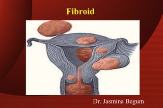
2. Fibroid
• Synonyms : Myoma, Leiomyoma,
Fibromyoma
• Most common benign neoplasm in the
female.
• Incidence : 20 to 40% of reproductive age
women.
3. Fibroid
Etiology : It arises from smooth muscle cell
of myometrium.
* Exact etiology not known.
* Monoclonal origin ( arising from single cell)
* Genetic basis definite.
* Various growth factors like TGFβ , EGF, IGF-1, IGF-
2, BFGF
4. Fibroid - Etiology
Epidemiological risk factors :-
• Increased risk age 35 to 45 years , nulliparous or
low parity , Black women, strong family history,
obesity, early Menarche, Diabetes, hypertension.
• Decreased risk ↑↑ parity, exercise, ↑↑intake of
green vegetables, Prog.only contraceptives, cigarette
smoking
5. Fibroid - Etiology
Genetic basis: Responsible for 40 % cases of
fibroids
• Translocation between Chromo. 12 & 14,
• Trisomy 12,
• Rearrangement of short arm of Chromo 6
• Rearrangement of long arm of Ch. 10,
• Deletion of Ch.3 or Ch.7
6. Fibroid - Etiology
Estrogen although not proved for causing
myoma definitely implicated in its growth.
• Not detected before puberty & regresses after
menopause.
• May increase during pregnancy
• Estrogen receptors are in higher concent.ns
• Common fifth decade due to anovulatory cycles with
high or unopposed estrogen.
9. Fibroid
Submucous fibroids are
classified by European society
for gynec endoscopy ( ESGE ):
Type 0 – No intramural extension
Type I – Intramural extension < 50 %
Type II – Intramural extension > 50 %
19. Fibroid Signs
G/E – Pallor
P/A – If > 12 weeks size , firm, nodular, arising from
pelvis, lower limit can’t be reached, relatively well
defined, mobile from side to side, nontender, dull
on percussion, no free fluid in abdomen
P/S – Cervix pulled higher up
P/V – Uterus enlarged, nodular.
D/D from ovarian tumour Uterus not
separately
felt , transmitted movement present, notch not felt.
20. Fibroid Diagnosis
• Clinical : From symptoms & signs
• USG : Well defined hypoechoic
lesions. Peripheral calcification
with distal shadowing in old fibroids
21. TAS&TVS
size, site and number of fibroids
differentiates the tumour from other
swellings as ovarian tumour
25. (4) Intra venous pyelogram (IVP)
In cervical and broad ligament fibroid
- Course of ureter.
- Hydroureter & hydroneprosis
- Kidney function.
26. Fibroid Diagnosis
5. MRI : Most accurate imaging modality for diagnosis of
fibroid. It does precise fibroid mapping &
characterization Detects all fibroids accurately
D/D from adenomyosis
D/D from adnexal pathology
Ovaries are easily seen
Detects small myomas(0.5 cm)
6. H S G : Not done for diagnosis , Done for infertility
evaluation filling defects may be seen.
29. Fibroid D/D
• Pregnancy
• Adenomyosis
• Ovarian tumour
• Ectopic pregnancy
• Endometriosis
• T O mass
30. Fibroid Management
Expectant :
asymptomatic ,
Size < 12 weeks,
near menopause .
• Regular follow up every 6 months
• Recent guidelines suggest upto 16 wks size
however difficult to practice
31.
32. Medical Management
• Not a definitive Rx
• For symptomatic relief
• Preoperatively to decrease the size
• Progestogens, antiprogestogens
( Miefpristone ) androgens ( Danazol,
Gestrinone ) & GnRH analogues are used
33. GnRH analogues
• Agonists are commonly used drugs :-
• Triptorelin ( Decapeptyl) 3.75 mg or leuprolide depot
3.75 mg I/M or Goseraline ( Zoladex) 3.6 mg SC for 3
months
• Advantages : Decrease in size of myoma by 20 to 50 %
Decrease in bleeding increases Hb level
Decreases blood loss during surgery
Converts hysterectomy into myomectomy
Converts Abd. hyst into
vag.hysterectomy
34. GnRH analogues
• Disadvantages : High cost
Hypoestrogenic side effects
Effect is reversible
Rarely ↑↑ bleeding due to degeneration
Occasionally difficulty in enucleation
• Antagonist
Cetrorelix is used
60 mg I/M repeated after 3-4 months if necessary
Initial flare up does not occur
36. Surgical Management
Vaginal hysterectomy is favoured in following
if
• Uterus < 16 wks, preferably < 14 wks
• No associated pathology like endometriosis , PID,
adhesions
• Uterus mobile & adequate
lateral space in pelvis
• Experienced vaginal surgeon
37. Surgical Management
Myomectomy is done in following :-
• Infertility,
• Recurrent pregnancy loss
& no other cause
• Young patients
• Patients who wish to preserve
their uterus
38. Hysteroscopic myomectomy
• For submucous myoma causing infertility, Recurrent
pregnancy loss, AUB or pain
• Criteria :- < 5 cm in size
< 50 % intramural component
< 12 cm2
uterine size
39. Laparoscopic myomectomy
• In 3 phases excision of myoma, repair of myometrium
& extraction
• Suitable for subserous & intramural fibroids upto 10 cm
size
• Complications are those of operative laparoscopy +
myomectomy
40. Abdominal myomectomy
• Other factors for infertility should be ruled out
• Consent for hysterectomy
• Blood cross matched & ready
• Pap’s smear & endometrial sampling to rule out
malignancy
• Medical or mechanical means to control blood loss
Bonney’s Myomectomy clamp, rubber tourniquet,
manual ( finger compression) pressure at isthmic
region or use of vasopressin 10 – 20 units diluted in
100ml saline infiltrated before putting the incision .
41. Abdominal myomectomy
• Minimum incisions are kept – preferably single
midline vertical, lower, anterior wall .
• Removal of as many fibroids as possible through one
incision & secondary tunnelling incisions.
• Meticulous closure of all dead space.
• Proper haemostasis
• Multiple small fibroids can be removed enbloc by
wedge resection.
• Measures for adhesion prevention should be taken.
43. Vaginal myomectomy
• Submucous pedunculated or small sessile
cervical fibroids are removed vaginally.
• Ligation of pedicle if accessible
• Twisting off the fibroids if pedicle not accessible
in case of small & medium size fibroids
• To gain access to pedicle of higher & big fibroid
incision on the cervix can be made.
44. Surgical Management
Laparoscopic myolysis :
• By ND-YAG laser or long bipolar needle
electrode through laparoscope, blood supply of
myoma is coagulated.
• Without blood supply, myoma atrophies.
• Applicable to 3 -10 cm size & myomas < 4 in
number
* Cryomyolysis is under investigation
45. Uterine artery embolization
• By interventional radiologist
• Catheter is passed retrograde thro. Right femoral
artery to bifurcation of aorta & then negotiated down
to opposite uterine artery first.
• Polyvinyl alcohol ( PVA ) particles ( 500-700 um) or
gelfoam are used for embolization.
• 60 – 65 % reduction in size of fibroid
• 80 – 90 % have improvements in menorrhagia &
pressure symptoms
47. Uterine artery embolization
• High vascularity & solitary fibroid are associated with
greater chance of long term success.
• Pregnancy, active infection & suspicion of malignancy
are absolute C I .
• Desire for fertility is also a contraindication
• The risk of ovarian failure must be counselled
• Post embolization syndrome ( fever, vomiting,
pain) can occur
51. MCQs in Fibroid
• Most common type of uterine leiomyoma .
a) Intramural.
b)Subserosal.
c) Submucosal.
d)Broad ligament.
• Which of the following is for symptomatic treatment of fibroid.
a) OCPs
b)Testosterone.
c) GnRH agonist.
d)GnRH antagonist.
52. MCQs
• Most common symptom of fibroid
a) Abnormal uterine bleeding.
b) Pelvic pain.
c) Mass in abdomen.
d) Abdominal discomfort
• Most common pelvic tumor of reproductive age group is
a) Uterine fibroid
b) Dermoid cyst .
c) Ovarian cysts.
d) Ovarian tumor.