Bitemporal hemianopsia
•
8 likes•25,835 views

Bitemporal hemianopsia is a type of partial blindness where vision is missing in the outer half of both the right and left visual fields, caused by lesions or compression affecting the center of the optic chiasm. It results in inability to view the peripheral vision. The most common cause is tumors located at the mid-optic chiasm, such as pituitary adenomas or craniopharyngiomas. Other potential causes include meningiomas or aneurysms of the anterior communicating artery. The detailed anatomy and neurocircuitry of the visual pathway empower understanding of how specific lesions can lead to different visual field deficits.

Recommended
More Related Content
What's hot
What's hot (20)
Similar to Bitemporal hemianopsia
Similar to Bitemporal hemianopsia (20)
Recently uploaded
Recently uploaded (20)
Bitemporal hemianopsia
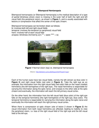
- 1. 1 Bitemporal Hemianopsia Bitemporal hemianopsia (or Bitemporal hemianopia) is the medical description of a type of partial blindness where vision is missing in the outer half of both the right and left visual field (the peripheral vision), as shown in Figure 1, and is usually associated with lesions or compression that affect the center of the optic chiasm. Bitemporal hemianopsia can be broken down as follows: bi: involves both left and right visual fields temporal: involves the temporal (or peripheral) visual field hemi: involves half of each visual field anopsia: blindness (formed by a(n) no + opsis vision + ia) Figure 1 Normal vision (top) vs. bitemporal hemianopsia (Source: http://pituitary.ucla.edu/images/site/Visual3.3.jpg) Each of the human eyes have two visual fields, namely the left (shown as blue color in Figure 2) and right visual fields (red color in Figure 2). Take the right eye as an example, the information from the right visual field (red color) falls on the nasal (medial) hemiretina (the left hemisphere of the right eye). The nasal hemiretina is responsible for carrying the information along the optic nerve, and crosses to the other side at the optic chiasm and eventually, the information will reach the left primary visual cortex. On the other hand, the information from the left visual field (blue color) of the right eye falls on the temporal hemiretina (the right hemisphere of the right eye, blue color). The temporal hemiretina is responsible for carrying the information along the optic nerve and eventually the information will reach the right primary visual cortex. When there is compression at optic chiasm (site of lesion 2 shown in Figure 2) the visual information from both nasal hemiretina are affected, leading to inability to view the peripheral vision, i.e. loss of the right visual field of the right eye and the left visual field of the left eye.
- 3. Bitemporal hemianopsia most commonly occurs as a result of tumors located at the mid-optic chiasm. Since the adjacent structure is the tumors causing compression are pituitary adenomas craniopharyngiomas. Also another relatively common neoplastic etiology is meningiomas. An etiology of vascular origin is an aneurysm of the anterior communicating artery which arise superior to the chiasm, enlarge, and compress it from above. (Source: http://nicandjacelyn.files.wordpress.com/201 Personal Reflection This course has empowered me t hemianopsia by equipping me with the neurocircuitry of visual signal flow through the optic tract. This course also allows me to develop a neuro-system. Take the example types of disorders can happen causing deficits in different parts of this assignment, bitemporal hemianopsia chiasm while a lesion at the right optic tract will cause contralateral (left) homonymous hemianopsia where the left visual Figure 4 Paris as seen with left homonymous hemianopsi (Source: http://upload.wikimedia.org/wikipedia/commons/thumb/8/81/Lhvf.png/230px 3 Bitemporal hemianopsia most commonly occurs as a result of tumors located at the optic chiasm. Since the adjacent structure is the pituitary gland, some common tumors causing compression are pituitary adenomas (as shown in craniopharyngiomas. Also another relatively common neoplastic etiology is meningiomas. An etiology of vascular origin is an aneurysm of the anterior ch arise superior to the chiasm, enlarge, and compress it from Figure 3 Pituitary adenoma http://nicandjacelyn.files.wordpress.com/2012/03/bitemporal-hemianopia.png has empowered me to analyze and better understand by equipping me with the knowledge in the visual fields and l flow through the optic tract. ws me to develop a sense of awe towards the sophistication example of visual system, as shown in Figure 2, happen when lesion occurs at different sites of the neuro ifferent parts of the visual fields. Like what has been discus itemporal hemianopsia is caused by a lesion at the center of optic e a lesion at the right optic tract will cause contralateral (left) homonymous ianopsia where the left visual field of both eyes will be lost, as shown in Paris as seen with left homonymous hemianopsia http://upload.wikimedia.org/wikipedia/commons/thumb/8/81/Lhvf.png/230px Bitemporal hemianopsia most commonly occurs as a result of tumors located at the pituitary gland, some common (as shown in Figure 3) and craniopharyngiomas. Also another relatively common neoplastic etiology is meningiomas. An etiology of vascular origin is an aneurysm of the anterior ch arise superior to the chiasm, enlarge, and compress it from hemianopia.png) understand bitemporal the visual fields and the sophistication of our , many different of the neuro-system Like what has been discussed in is caused by a lesion at the center of optic e a lesion at the right optic tract will cause contralateral (left) homonymous field of both eyes will be lost, as shown in Figure 4. a http://upload.wikimedia.org/wikipedia/commons/thumb/8/81/Lhvf.png/230px-Lhvf.png)
- 4. 4 I would like to thank Professor Mason and her team for making this course so engaging for me and I have learned a lot from this course despite my limited background in biology. Professor Mason’s explanation is very clear and easy to follow. The exercises in this course are thought provoking and allowed me to appreciate the course materials better. References 1. http://en.wikipedia.org/wiki/Bitemporal_hemianopsia 2. https://umem.org/educational_pearls/1055/ 3. http://www.gpnotebook.co.uk/simplepage.cfm?ID=1872363522