Accidental Femoral fractures
•Download as PPTX, PDF•
1 like•1,680 views

the slide describes femoral fracture with case presentations as well as rediological diagnosis ,when opened and closed .the management from emergency period and through to stabilization

Recommended
More Related Content
What's hot
What's hot (20)
Viewers also liked
Similar to Accidental Femoral fractures
Similar to Accidental Femoral fractures (20)
Accidental Femoral fractures
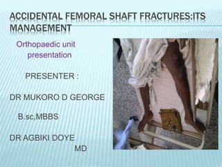
- 1. ACCIDENTAL FEMORAL SHAFT FRACTURES:ITS MANAGEMENT Orthopaedic unit presentation PRESENTER : DR MUKORO D GEORGE B.sc,MBBS DR AGBIKI DOYE MD
- 2. CASE PRESENTATIONS Femoral fracture has been a common presentation in this facility , shaft fractures is the commonest part involved in recent months ,common implicated causes are RTA and fall from heights. They usually associate and present with other injuries, morbidity, and mortality.
- 3. CASE PRESENTATION 1 Miss I.I. ,24 yr old waiteress, Admitted via A/E 27/1/2012, with history of inability to move Left Lower Limb 19 hrs duration following an RTA on a motorcycle). Sustained wound to left knee, thigh swelling . On general exam- , conscious ,not pale, afebrile. PR 126 b/min, Bp 110/70mmhg RR 24c/min. Sutured laceration Lt knee, medial side of Lt leg, swelling of the knee, Marked abduction of the leg at rest X-RAY:Displaced Spiral fracture of distal shaft of the Rt femur with medial condylar and patella fractures. ASS:Rt femoral fracture with intra- articular involvement following a RTA
- 4. MANAGEMENT . HAD resuscitative measures at A/E Along With anti tetanus prophylaxis ,IV fluids ORIF with condylar plate for spiral fracture ,cancellous screw for condylar fragment fixation and fixation of the avulsed posterior cruciate ligament , on 26th day after presentation Analgesics, blood transfusion ,antibiotics, hematinics, antithrombotic Currently on the ward ,immobilized with Above knee synthetic cast
- 5. OPERATION SECTION Distal bone fragment with spiral edge with good exposure . Stay close to the bone as much as possible
- 6. CASE PRESENTATION :TWO Mr O.J ,43 year old Architect Admitted via A/E (22/12/11)with history of multiple injuries following a RTA (motorcycle) ,4 hrs to presentation. Sustained facial swelling ,open injury to left thigh .loss of consciousness which improved within 4 hrs Generally , Conscious but drowsy. GCS 13/15 ,not pale, febrile 37.2 C, receiving oxygen via intranasal prongs ,PR 100 b/min,BP 120/80mmhg, RR 32c/min Hemifacial swelling (left side),enclosing mandibular region ,left thigh swelling and deformity,wound 6cm in dimension X-RAY result:communited segmental fracture of left femur with associated fracture of the mandible Mild head injury with left femoral fracture 2º to RTA
- 7. MANAGEMENT HAD resuscitative measures at A/E Along with cervical collar, anti tetanus prophylaxis ,IV fluids ORIF with condylar plate on 14th day after presentation Blood transfusion, Analgesics, ,antibiotics,,hemtinics Discharged 15th DAY post -op With clutches Follow up –VIA clinic with POST-OP X-ray film.
- 8. IN SUMMARY In the last 2 months we had several cases of femoral fractures , with a few bilateral. Some opt-for surgical intervention . Surgical option should be seen as the best option for management of femoral fracture following RTA ,to allow for early mobilization ,knowing well that: life is movement and movement is life . THANK YOU
- 9. PRESENTATION CONTINUE Introduction • Anatomy of the femur • Epidemiology of femoral fractures • Aetiology • mechanism • Classification of shaft fractures • Clinical features • Investigations • Treatment • complications
- 10. INTRODUCTION A fracture by definition, is a break in the continuity of a bone. It occurs when an external force overcomes the modules of elasticity of the bone. Strongest and largest bone. Femoral shaft fractures ,may be associated with multisystem trauma.
- 11. ANATOMY OF THE FEMUR
- 12. BLOOD SUPPLY
- 14. EPIDEMIOLOGY Common injury : major violent trauma 1 femur fracture/ 10,000 people More common : < 25 y or >65 y RTA , waterway motorcycle, fall from height and gunshot wound accidents are most frequent causes.
- 15. AETIOLOGY . Trauma. RTA (motorcycle races, auto/pedestran accident, auto crash, plane crash, vehicle,). Sports(skiing, football, hockey). Falls(mountain, pole). Gunshot. Pathologic Stress
- 16. MECHANISM High Energy Often high-speed impact or rapid deceleration But may take surprisingly little energy in children Direct blow Proximal - distal compression Twisting/torsion Injury Shear Compression with angulation Fall from height High speed collisions Often seen in combination with other significant injuries
- 19. Winquist and Hansen 66A, 1984 CLASSIFICATION Type 0 - No comminution Type 1 - Insignificant butterfly fragment with transverse or short oblique fracture Type 2 - Large butterfly of less than 50% of the bony width, > 50% of cortex intact Type 3 - Larger butterfly leaving less than 50% of the cortex in contact Type 4 - Segmental comminution
- 20. ACCORDING TO THE PRESENCE/ABSENCE OF WOUND. 1. OPEN FRACTURES 2. CLOSE FRACTURES
- 21. SYNTOMS Age/sex/occupation Duration Severe pain Swelling Inability to move the limb Deformity shortening
- 22. SIGNS tenderness visible deformity shortening crepitus Swollen thigh Signs of vascular compromise should be looked out for to rule out vascular injury. - absent or diminished pulses - expanding haematoma - tachycardia - hypotension
- 23. INVESTIGATIONS Done after the initial resuscitation of the patient. PCV/Hb Radiograph of the affected femur, adjacent joints and hip.(rule of 2s) Wound swab for m/c/s in open fractures. E/U/Cr Depends on the patient’s presentation.
- 24. FIELD MANAGEMENT Control bleeding, treat shock Dress wounds Distal CMS :FACT Manual stabilization Traction splint for mid-shaft fracture Backboard without traction for hip injury Re-check CMS Address other injuries as needed Early coordination with EMS agencies ALS transport criteria per local protocol Frequent vital sign checks and documentation Expedited transport to definitive care
- 25. TREATMENT Initial resuscitation. Definitive treatment. - non operative / conservative - operative Physiotherapy.
- 26. INITIAL RESUSCITATION ABCD of resuscitation. IV Fluid IV antibiotics Oxygen Anti-tetanus prophylaxis Blood transfusion Analgesics Wound care (wound debridement ). Splinting
- 27. DEFINITIVE TREATMENT Non operative /Conservative mgt split traction casting (for children < 8 years)
- 28. HARE TRACTION
- 29. GALLOW SKIN TRACTION THOMAS SPLIT
- 30. OPERATIVE METHODS Operative treatment. 1) ORIF 2) External fixation 3) Minimally invasive method.
- 31. INDICATIONS FOR FEMORAL SHAFT ORIF Inability to secure and maintain reduction by manipulation. Old and frail px. Px with multiple injuries. Pathological fractures. Fractures suitable for nailing. Early ambulation is needed.
- 32. ORIF : 1. Intramedullary nails are used e.g. Kuntcher interlocking nail{Grosse’s and Kempf } This could be done either by antegrade or retrograde ;reamed and non reamed method. 2. Plate and screws.
- 33. ANTEGRADE IM NAILING RETROGRADE IM NAILING
- 34. External fixation is usually used for open fractures of the femoral shaft with severe soft tissue injury.
- 35. Minimally invasive method involves closed method of IM nailing under image intensification .eg :ESIN
- 36. REHABILITATION /PHYSIOTHERAPY This should be started early as soon as the pain begins to settle. Exercises for quadriceps, leg and foot are necessary to preserve muscle tone and prevent deformity. For post surgical patients, it can be started two weeks after surgery but the patient should not bear weight. Physiotherapy continues after discharge from the hospital.
- 37. COMPLICATIONS EARLY Infection Hypovolaemic shock. Fat embolism (1st 72 hrs ). DVT. Pulmonary embolism. LATE Delayed union Malunion Non – union Atrophy of the thigh and gluteal muscles Limb shortening
- 38. A femoral shaft fracture is a serious injury that takes a long time ( 3 to 6 months ) Average of 12 weeks to heal, hence most femoral shaft fractures are treated surgically. The goal of treatment is reliable anatomic stabilization, allowing mobilization as early as possible.