SlideShare ist ein Scribd-Unternehmen logo
1 von 24
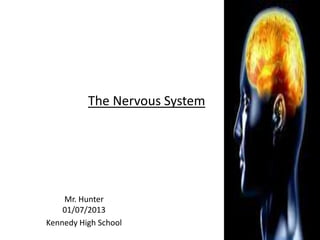
The Nervous System


    Mr. Hunter
    01/07/2013
Kennedy High School
Mr. Hunter
                     Anatomy and Physiology
                           01/15/13

•   Objective(s)
•   SWBAT
•   List the major divisions of the Nervous System
•   Describe the action of a nerve impulse / Action Potential
•   Describe the structure and function of synapses.
•   Review Concepts via Practice Problems

• Bell Ringer: List three possible stimuli that could generate
  a nerve impulse.
• There are two principal divisions of the
                         nervous system.
Organs and Divisions
                       • The Central Nervous System (CNS)
                       • The Peripheral Nervous System (PNS)
                       • The CNS consists of the brain and the
                         spinal cord.
                       • The PNS consists of nerves which
                         extend from the spinal cord to the
                         peripheral parts of the body.
                       • A subdivision of the PNS is the
                         Autonomic Nervous System (ANS).
                       • ANS consists of structures that regulate
                         the body’s involuntary or automatic
                         functions ex. heart rate, smooth muscle
                         contractions, secretion of
                         hormones, enzymes, etc.
• The two types of cells found in the
Organs and Divisions     Nervous System (NS) are neurons
                         and glia cells.
                       • Neurons conduct nerve impulses.
                       • Glia cells support neurons.

                       •   Neuron consists of three main parts:
                       •   Cell body
                       •   Dendrites
                       •   Axon
Cells of the Nervous   • The main part of the neuron is the
System                   cell body.
                       • Branching projections are called
                         dendrites. Dendrites transmit
                         information to the neuronal cell
                         body.
                       • Axons are the long processes that
                         transmit information away from the
                         cell body.
                       • There are three types of neurons
                         named based on the direction in
                         which they transmit information.
                       • Sensory (afferent)
                       • Motor (efferent)
                       • Interneuron
Cells of the Nervous   • Sensory neurons (afferent) transmit
System                   information to the CNS from all
                         regions of the body.
                       • Motor neurons (efferent) transmit
                         information in the opposite
                         direction- away from the CNS.
                       • They conduct impulses to muscle
                         and glandular epithelial tissue.
                       • Interneurons conduct impulses from
                         sensory neurons to motor neurons.
                       • They connect to form central
                         networks of nerve fibers and are
                         called central or connecting neurons.
Cells of the Nervous
                       • The axon is covered by myelin.
System                 • This is a white, fatty substance formed
                         by specialized cells called schwann cells
                         that wrap around axons outside the
                         CNS. The outer membrane of this type
                         of cell is called the neurilemma-plays
                         regenerative part in damaged axons.
                       • Myelin allows for saltatory conduction
                         of electrical transmissions.
                       • Nodes of Ranvier are interrupted
                         segments between schwann cells.

                       • Glia (glue) cells – one function is to hold
                         functioning neurons together and
                         protect them. They also coordinate the
                         functions of the NS.
                       • Glioma- common type of brain tumor.
• Glia vary in size and shape.
Types of glial cells   • Astrocytes: resemble stars. Their
                         threadlike branches attach to neurons
                         and to small blood vessels holding both
                         structures together.
                       • Astrocyte branches form a two-layer
                         structure called the blood brain barrier
                         (BBB).
                       • BBB separates blood tissue and nervous
                         tissue. Protects brain from harmful
                         chemicals that might be found in the
                         blood.
                       • Microglia: smaller than astrocytes. They
                         remain stationary until brain tissue
                         becomes injured. They act as microbe
                         scavengers in the brain via phagocytosis.
• Oligodendrocytes hold nerve fibers
Types of glial cells     together and they also produce
                         myelin sheaths around multiple
                         nerve cell axons in the CNS.
                       • Schwann cells are glia cells that
                         produce myelin sheaths on single
                         axons within the PNS.
                       • Assignment 01/07/13
                       • Pg. 195 # 1-4 Quick Check.
• A nerve can be defined as a group of
                      peripheral nerve fibers (axons) bundled
Nerves and Tracts
                      together.
                    • The fibers have a myelin sheath provided by
                      schwann cells.
                    • Because myelin is white, peripheral nerve
                      fibers often look white.

                    • Bundles of axons in the CNS are called
                      tracts. They are also myelinated and form
                      the white matter of the brain and spinal
                      cord.
                    • Brain and spinal cord tissue composed of
                      unmyelinated axons dendrites and cell
                      bodies is called gray matter due to its
                      appearance.
• Each axon in a nerve is surrounded
Nerves and Tracts     by a thin wrapping of fibrous
                      connective tissue called
                      endoneurium.
                    • Groups of these wrapped axons are
                      called fascicles.
                    • Each fascicle is surrounded by a
                      thin, fibrous perineurium.
                    • A tough, fibrous sheath called the
                      epineurium covers the entire nerve.
• Only neurons can provide the rapid
Reflex Arcs     communication between cells that is
                necessary for sustaining life.
              • Hormonal messages travel much
                more slowly than neuronal
                transmissions within the body. They
                have to circulate via the blood
                stream.

              • Nerve impulses are also called action
                potentials. The routes traveled by
                nerve impulses are called neuron
                pathways.
              • A basic type of neuron pathway is
                called a reflex arc.
• A simple kind of reflex arc consists of
Reflex Arcs     two types of neurons: sensory
                neurons and motor neurons.
              • Three neuron arcs consists of:
                sensory neurons, interneurons and
                motor neurons.
              • Reflex arcs allow impulse conduction
                in only one direction.
              • Impulse conduction starts mainly at
                receptors.
              • Receptors are the beginnings of
                dendrites of sensory neurons. They
                are located some distance away
                from the spinal cord. (tendons,skin
                mucous membranes etc.)
• The knee-jerk reflex is the simplest example
                 of a two neuron reflex arc.
Reflex Arcs   • As a result of a tap on the patellar ligament
                 causes the quadriceps muscle to contract
                 via a neuronal pathway.
              1. A nerve impulse is generated by stretch
                  receptors within the quadriceps.
              2. It travels along the sensory neuron’s
                  dendrites to the cell body located in the
                  dorsal root ganglion (group of nerve cell
                  bodies located in the PNS)
              3. The axon from the sensory neuron travels
                  from the cell body of the dorsal root
                  ganglion and ends near the dendrites of
                  another neuron located in the gray matter
                  of the spinal cord.
              4. The nerve impulse stops at the synapse
                  (space that separates axon and dendrites)
              5. Chemical signals are sent across the
                  synapse and connect with other dendrites
•  and cell bodies located in the CNS gray
                 matter and axons outside the gray matter.
Reflex Arcs   6. The motor neuron synapses with an
                  effector which are muscles or glands – they
                  put the nerve signals” into effect.”
              • The response to the impulse conduction
                  over a reflex arc is called a reflex.

              •   Complex reflexes may involve the actions
                  of three neurons.
              •   The end of the sensory neuron will synapse
                  with the dendrites of an interneuron
              •   located within the gray matter of the spinal
                  cord. The axon of that interneuron will
                  then synapse with the dendrites and cell
                  body of a motor neuron within the spinal
                  cord. The axons of the motor neuron lie
                  outside the spinal cord and will exit via the
                  anterior root of the spinal nerve and
                  terminate in the muscle(effector)
Withdrawl Reflex

• Interneurons are located
within the gray matter of
the brain or spinal cord.
•Three neuron reflex arcs
have two synapses.
•A Two-neuron refex arc
only has a sensory
neuron, a motor neuron
and one synapse between
them.
Nerve Impulses and the   • Nerve impulse: self-propagating
Synapse                    wave of electrical disturbance that
                           travels along the surface of a
                           neuron’s plasma membrane.
                         • It is similar to a spark traveling along
                           a fuse.
                         • Nerve impulses have to be initiated
                           by a stimulus – a change in the
                           neuron’s environment. A stimulus
                           could possibly be on of the
                           following:
                         Environmental change(s)
                         • Pressure
                         • Temperature
                         • Chemical changes
Nerve Impulses and the   • Due to the distribution of Na +
Synapse                    (sodium) and K+ (potassium) ions,
                           the resting neuron has a slight
                           positive charge on the outside of the
                           membrane and a slight negative
                           charge on the inside.
                         • This separation of electrical charges
                           is called polarization
                         • There is normally an excess of
                           sodium on the outside of the cell
                           than inside.
                         • When a portion of the membrane is
                           stimulated, Na+ ions will enter the
                           cell causing that region of the cell to
                           become temporarily positive –
                           outside becomes negative.
• This process is called depolarization.
Nerve Impulses and the
                         • This section of the membrane immediately
Synapse
                           recovers during a process of repolarization.
                         • However, depolarization has already
                           stimulated Na+ channels to open in the next
                           portion of the membrane. The Action
                           Potential continues along the axon in one
                           direction jumping from node to node until it
                           reaches the synaptic terminal of the axon.
Nerve Impulses and the
Synapse
• Transmission of nerve
signals from one neuron to
the next occurs across the
synapse.
• Impulses travel from the
presynaptic neuron to the
postsynaptic neuron.
•Impulses travel across the
synaptic cleft.
•Three structures that
make up a synapse:
•Synaptic knob
•Synaptic cleft
•Plasma membrane of
postsynaptic neuron
Nerve Impulses and the
Synapse
• synaptic knob – terminal
branch at the end of the
post synaptic neuron.
•Small vesicles contain
neurotransmitters.
•When a nerve impulse
arrives at the synaptic
knob, neurotransmitters
are released into the
synaptic cleft.
•The plasma membrane of
the postsynaptic neuron
contains receptors for the
neurotransmitter.
Nerve Impulses and the
Synapse
• The binding of the
neurotransmitter with the
receptors initiates an
impulse in the postsynaptic
neuron opening Na+
channels in the cell.
Therefore, the nerve signal
is transmitted to the
postsynaptic neuron.
•The neurotransmitter can
be :
•Taken back up by the
synaptic knobs
•Metabolized into inactive
chemicals
Neurotransmitters
•At least 30 different substances
have been identified as
neurotransmitters.
•Specific neurotransmitters are
localized in discrete groups of
neurons.
•Acetylcholine is released at some
synapses in the spinal cords and
at neuromuscular junctions.
•Norepinephrine, Dopamine and
Serotonin are grouped into
compounds called catecholamines
– involved in sleep motor
function, mood and pleasure
recognition.
Neurotransmitters
• Endorphins and Enkephalins are
released at spinal cord and
various brain synapses in the pain
conduction pathway.
•They inhibit the conduction of
pain impulses.
•Nitrous Oxide – neurotransmitter
that diffuses directly across the
plasma membrane instead of
being transported by vesicles.

Weitere ähnliche Inhalte

Was ist angesagt?

Was ist angesagt? (20)

Nervous system ( anatomy and physiology)
Nervous system ( anatomy and physiology)Nervous system ( anatomy and physiology)
Nervous system ( anatomy and physiology)
 
Neurons
NeuronsNeurons
Neurons
 
special sense organs (anatomy and physiology) - a brief discussion
special sense organs (anatomy and physiology) - a brief discussion special sense organs (anatomy and physiology) - a brief discussion
special sense organs (anatomy and physiology) - a brief discussion
 
Nervous system notes
Nervous system notesNervous system notes
Nervous system notes
 
Skeletal system
Skeletal systemSkeletal system
Skeletal system
 
Neuroglial cells _neurons
Neuroglial cells _neuronsNeuroglial cells _neurons
Neuroglial cells _neurons
 
The urinary system
The urinary systemThe urinary system
The urinary system
 
Nerves
NervesNerves
Nerves
 
Nervous tissues and types of Nervous Tissue
Nervous tissues and types of Nervous TissueNervous tissues and types of Nervous Tissue
Nervous tissues and types of Nervous Tissue
 
Anatomy of meninges, ventricles, cerebrospinal fluid
Anatomy of meninges, ventricles, cerebrospinal fluidAnatomy of meninges, ventricles, cerebrospinal fluid
Anatomy of meninges, ventricles, cerebrospinal fluid
 
PPT on Nervous System
PPT on Nervous SystemPPT on Nervous System
PPT on Nervous System
 
Skeletal System Anatomy and Physiology
Skeletal System Anatomy and PhysiologySkeletal System Anatomy and Physiology
Skeletal System Anatomy and Physiology
 
Urinary System Notes
Urinary System NotesUrinary System Notes
Urinary System Notes
 
Anatomy nervous tissue
Anatomy nervous tissue   Anatomy nervous tissue
Anatomy nervous tissue
 
Nervous tissue
Nervous tissueNervous tissue
Nervous tissue
 
SENSE ORGANS
SENSE ORGANSSENSE ORGANS
SENSE ORGANS
 
Anatomical terminology
Anatomical terminologyAnatomical terminology
Anatomical terminology
 
Nervous tissue
Nervous tissueNervous tissue
Nervous tissue
 
Anatomy and physiology of nervous system
Anatomy and physiology of nervous systemAnatomy and physiology of nervous system
Anatomy and physiology of nervous system
 
Nervous System
Nervous SystemNervous System
Nervous System
 

Ähnlich wie The Nervous System: Anatomy and Physiology

Ähnlich wie The Nervous System: Anatomy and Physiology (20)

central nervous system
central nervous systemcentral nervous system
central nervous system
 
The nervous system
The nervous systemThe nervous system
The nervous system
 
CNS.pdf
CNS.pdfCNS.pdf
CNS.pdf
 
Nervous system PPT for grade 10 (basic concepts regarding human nervous system)
Nervous system PPT for grade 10 (basic concepts regarding human nervous system)Nervous system PPT for grade 10 (basic concepts regarding human nervous system)
Nervous system PPT for grade 10 (basic concepts regarding human nervous system)
 
CNS.pptx
CNS.pptxCNS.pptx
CNS.pptx
 
Nervous System-ppt.pptx
Nervous System-ppt.pptxNervous System-ppt.pptx
Nervous System-ppt.pptx
 
Lecture 3.. Dr. Hameed
Lecture 3.. Dr. HameedLecture 3.. Dr. Hameed
Lecture 3.. Dr. Hameed
 
central nervous system
central nervous systemcentral nervous system
central nervous system
 
Central nervous system 1
Central nervous system 1Central nervous system 1
Central nervous system 1
 
neuron.pdf
neuron.pdfneuron.pdf
neuron.pdf
 
Introduction to nervous system
Introduction to nervous systemIntroduction to nervous system
Introduction to nervous system
 
Nervous tissue
Nervous tissueNervous tissue
Nervous tissue
 
nervous system
 nervous system nervous system
nervous system
 
Lect 1 introduction to nerve
Lect 1 introduction to nerveLect 1 introduction to nerve
Lect 1 introduction to nerve
 
Nervous system
Nervous systemNervous system
Nervous system
 
Anatomy and physiology of brain ppt, its organization
Anatomy and physiology of brain ppt,  its organizationAnatomy and physiology of brain ppt,  its organization
Anatomy and physiology of brain ppt, its organization
 
Nervous System 1 (63).pptx
Nervous System 1 (63).pptxNervous System 1 (63).pptx
Nervous System 1 (63).pptx
 
muscular - neurvous tissue
muscular - neurvous tissuemuscular - neurvous tissue
muscular - neurvous tissue
 
Nervous system
Nervous system Nervous system
Nervous system
 
NERVOUS SYSTEM
NERVOUS SYSTEMNERVOUS SYSTEM
NERVOUS SYSTEM
 

Mehr von mrhunterspage

Chapter 14 study guide Biology
Chapter 14 study guide BiologyChapter 14 study guide Biology
Chapter 14 study guide Biologymrhunterspage
 
Chap14 studyguide Biology
Chap14 studyguide BiologyChap14 studyguide Biology
Chap14 studyguide Biologymrhunterspage
 
Sense organs Anatomy and Physiology
Sense organs Anatomy and PhysiologySense organs Anatomy and Physiology
Sense organs Anatomy and Physiologymrhunterspage
 
Biology Genetics II 03/05/2013
 Biology Genetics II            03/05/2013 Biology Genetics II            03/05/2013
Biology Genetics II 03/05/2013mrhunterspage
 
Biology RNA and Proteins 02 / 13 / 13
 Biology RNA and Proteins 02 / 13 / 13 Biology RNA and Proteins 02 / 13 / 13
Biology RNA and Proteins 02 / 13 / 13mrhunterspage
 
Biology DNA Chapter 10 02/04/13
 Biology DNA Chapter 10   02/04/13 Biology DNA Chapter 10   02/04/13
Biology DNA Chapter 10 02/04/13mrhunterspage
 
Anatomy and Physiology The Central Nervous System 02 / 04 / 2013
 Anatomy and Physiology The Central Nervous System 02 / 04 / 2013 Anatomy and Physiology The Central Nervous System 02 / 04 / 2013
Anatomy and Physiology The Central Nervous System 02 / 04 / 2013mrhunterspage
 
Anatomy and Physiology Cell Transport and The Cell Cycle
Anatomy and Physiology Cell Transport and The Cell CycleAnatomy and Physiology Cell Transport and The Cell Cycle
Anatomy and Physiology Cell Transport and The Cell Cyclemrhunterspage
 
Anatomy and Physiology RNA and Proteins
Anatomy and Physiology RNA and ProteinsAnatomy and Physiology RNA and Proteins
Anatomy and Physiology RNA and Proteinsmrhunterspage
 
Anatomy and PhysiologySkeletal system II
 Anatomy and PhysiologySkeletal system II Anatomy and PhysiologySkeletal system II
Anatomy and PhysiologySkeletal system IImrhunterspage
 
Anatomy and Physiology Cell organelles
 Anatomy and Physiology Cell organelles Anatomy and Physiology Cell organelles
Anatomy and Physiology Cell organellesmrhunterspage
 
Anatomy and Physiology Muscular System
Anatomy and Physiology Muscular SystemAnatomy and Physiology Muscular System
Anatomy and Physiology Muscular Systemmrhunterspage
 
Anatomy and Physiology Connective Tissue Notes
Anatomy and Physiology Connective Tissue NotesAnatomy and Physiology Connective Tissue Notes
Anatomy and Physiology Connective Tissue Notesmrhunterspage
 
Anatomy and Physiology Connective tissue
Anatomy and Physiology Connective tissueAnatomy and Physiology Connective tissue
Anatomy and Physiology Connective tissuemrhunterspage
 
Anatomy and physiology Tissue Chapter
Anatomy and physiology Tissue ChapterAnatomy and physiology Tissue Chapter
Anatomy and physiology Tissue Chaptermrhunterspage
 
Anatomy and physiology Introduction Chapter 1 Notes
Anatomy and physiology Introduction Chapter 1 NotesAnatomy and physiology Introduction Chapter 1 Notes
Anatomy and physiology Introduction Chapter 1 Notesmrhunterspage
 
Anatomy and Physio NS Notes 01/08/13 Tues
Anatomy and Physio NS Notes 01/08/13 TuesAnatomy and Physio NS Notes 01/08/13 Tues
Anatomy and Physio NS Notes 01/08/13 Tuesmrhunterspage
 
Anatomy and Physio NS notes 01/ 07/13 Mon.
Anatomy and Physio NS notes 01/ 07/13 Mon.Anatomy and Physio NS notes 01/ 07/13 Mon.
Anatomy and Physio NS notes 01/ 07/13 Mon.mrhunterspage
 
Biology General Genetics 01/07/2013 Mon.
Biology General Genetics 01/07/2013 Mon.Biology General Genetics 01/07/2013 Mon.
Biology General Genetics 01/07/2013 Mon.mrhunterspage
 
Anatomy and physiology article nerve signals around body speeds 12 / 20 /12 ...
Anatomy and physiology article nerve signals around body speeds 12 / 20 /12  ...Anatomy and physiology article nerve signals around body speeds 12 / 20 /12  ...
Anatomy and physiology article nerve signals around body speeds 12 / 20 /12 ...mrhunterspage
 

Mehr von mrhunterspage (20)

Chapter 14 study guide Biology
Chapter 14 study guide BiologyChapter 14 study guide Biology
Chapter 14 study guide Biology
 
Chap14 studyguide Biology
Chap14 studyguide BiologyChap14 studyguide Biology
Chap14 studyguide Biology
 
Sense organs Anatomy and Physiology
Sense organs Anatomy and PhysiologySense organs Anatomy and Physiology
Sense organs Anatomy and Physiology
 
Biology Genetics II 03/05/2013
 Biology Genetics II            03/05/2013 Biology Genetics II            03/05/2013
Biology Genetics II 03/05/2013
 
Biology RNA and Proteins 02 / 13 / 13
 Biology RNA and Proteins 02 / 13 / 13 Biology RNA and Proteins 02 / 13 / 13
Biology RNA and Proteins 02 / 13 / 13
 
Biology DNA Chapter 10 02/04/13
 Biology DNA Chapter 10   02/04/13 Biology DNA Chapter 10   02/04/13
Biology DNA Chapter 10 02/04/13
 
Anatomy and Physiology The Central Nervous System 02 / 04 / 2013
 Anatomy and Physiology The Central Nervous System 02 / 04 / 2013 Anatomy and Physiology The Central Nervous System 02 / 04 / 2013
Anatomy and Physiology The Central Nervous System 02 / 04 / 2013
 
Anatomy and Physiology Cell Transport and The Cell Cycle
Anatomy and Physiology Cell Transport and The Cell CycleAnatomy and Physiology Cell Transport and The Cell Cycle
Anatomy and Physiology Cell Transport and The Cell Cycle
 
Anatomy and Physiology RNA and Proteins
Anatomy and Physiology RNA and ProteinsAnatomy and Physiology RNA and Proteins
Anatomy and Physiology RNA and Proteins
 
Anatomy and PhysiologySkeletal system II
 Anatomy and PhysiologySkeletal system II Anatomy and PhysiologySkeletal system II
Anatomy and PhysiologySkeletal system II
 
Anatomy and Physiology Cell organelles
 Anatomy and Physiology Cell organelles Anatomy and Physiology Cell organelles
Anatomy and Physiology Cell organelles
 
Anatomy and Physiology Muscular System
Anatomy and Physiology Muscular SystemAnatomy and Physiology Muscular System
Anatomy and Physiology Muscular System
 
Anatomy and Physiology Connective Tissue Notes
Anatomy and Physiology Connective Tissue NotesAnatomy and Physiology Connective Tissue Notes
Anatomy and Physiology Connective Tissue Notes
 
Anatomy and Physiology Connective tissue
Anatomy and Physiology Connective tissueAnatomy and Physiology Connective tissue
Anatomy and Physiology Connective tissue
 
Anatomy and physiology Tissue Chapter
Anatomy and physiology Tissue ChapterAnatomy and physiology Tissue Chapter
Anatomy and physiology Tissue Chapter
 
Anatomy and physiology Introduction Chapter 1 Notes
Anatomy and physiology Introduction Chapter 1 NotesAnatomy and physiology Introduction Chapter 1 Notes
Anatomy and physiology Introduction Chapter 1 Notes
 
Anatomy and Physio NS Notes 01/08/13 Tues
Anatomy and Physio NS Notes 01/08/13 TuesAnatomy and Physio NS Notes 01/08/13 Tues
Anatomy and Physio NS Notes 01/08/13 Tues
 
Anatomy and Physio NS notes 01/ 07/13 Mon.
Anatomy and Physio NS notes 01/ 07/13 Mon.Anatomy and Physio NS notes 01/ 07/13 Mon.
Anatomy and Physio NS notes 01/ 07/13 Mon.
 
Biology General Genetics 01/07/2013 Mon.
Biology General Genetics 01/07/2013 Mon.Biology General Genetics 01/07/2013 Mon.
Biology General Genetics 01/07/2013 Mon.
 
Anatomy and physiology article nerve signals around body speeds 12 / 20 /12 ...
Anatomy and physiology article nerve signals around body speeds 12 / 20 /12  ...Anatomy and physiology article nerve signals around body speeds 12 / 20 /12  ...
Anatomy and physiology article nerve signals around body speeds 12 / 20 /12 ...
 

The Nervous System: Anatomy and Physiology

  • 1. The Nervous System Mr. Hunter 01/07/2013 Kennedy High School
  • 2. Mr. Hunter Anatomy and Physiology 01/15/13 • Objective(s) • SWBAT • List the major divisions of the Nervous System • Describe the action of a nerve impulse / Action Potential • Describe the structure and function of synapses. • Review Concepts via Practice Problems • Bell Ringer: List three possible stimuli that could generate a nerve impulse.
  • 3. • There are two principal divisions of the nervous system. Organs and Divisions • The Central Nervous System (CNS) • The Peripheral Nervous System (PNS) • The CNS consists of the brain and the spinal cord. • The PNS consists of nerves which extend from the spinal cord to the peripheral parts of the body. • A subdivision of the PNS is the Autonomic Nervous System (ANS). • ANS consists of structures that regulate the body’s involuntary or automatic functions ex. heart rate, smooth muscle contractions, secretion of hormones, enzymes, etc.
  • 4. • The two types of cells found in the Organs and Divisions Nervous System (NS) are neurons and glia cells. • Neurons conduct nerve impulses. • Glia cells support neurons. • Neuron consists of three main parts: • Cell body • Dendrites • Axon
  • 5. Cells of the Nervous • The main part of the neuron is the System cell body. • Branching projections are called dendrites. Dendrites transmit information to the neuronal cell body. • Axons are the long processes that transmit information away from the cell body. • There are three types of neurons named based on the direction in which they transmit information. • Sensory (afferent) • Motor (efferent) • Interneuron
  • 6. Cells of the Nervous • Sensory neurons (afferent) transmit System information to the CNS from all regions of the body. • Motor neurons (efferent) transmit information in the opposite direction- away from the CNS. • They conduct impulses to muscle and glandular epithelial tissue. • Interneurons conduct impulses from sensory neurons to motor neurons. • They connect to form central networks of nerve fibers and are called central or connecting neurons.
  • 7. Cells of the Nervous • The axon is covered by myelin. System • This is a white, fatty substance formed by specialized cells called schwann cells that wrap around axons outside the CNS. The outer membrane of this type of cell is called the neurilemma-plays regenerative part in damaged axons. • Myelin allows for saltatory conduction of electrical transmissions. • Nodes of Ranvier are interrupted segments between schwann cells. • Glia (glue) cells – one function is to hold functioning neurons together and protect them. They also coordinate the functions of the NS. • Glioma- common type of brain tumor.
  • 8. • Glia vary in size and shape. Types of glial cells • Astrocytes: resemble stars. Their threadlike branches attach to neurons and to small blood vessels holding both structures together. • Astrocyte branches form a two-layer structure called the blood brain barrier (BBB). • BBB separates blood tissue and nervous tissue. Protects brain from harmful chemicals that might be found in the blood. • Microglia: smaller than astrocytes. They remain stationary until brain tissue becomes injured. They act as microbe scavengers in the brain via phagocytosis.
  • 9. • Oligodendrocytes hold nerve fibers Types of glial cells together and they also produce myelin sheaths around multiple nerve cell axons in the CNS. • Schwann cells are glia cells that produce myelin sheaths on single axons within the PNS. • Assignment 01/07/13 • Pg. 195 # 1-4 Quick Check.
  • 10. • A nerve can be defined as a group of peripheral nerve fibers (axons) bundled Nerves and Tracts together. • The fibers have a myelin sheath provided by schwann cells. • Because myelin is white, peripheral nerve fibers often look white. • Bundles of axons in the CNS are called tracts. They are also myelinated and form the white matter of the brain and spinal cord. • Brain and spinal cord tissue composed of unmyelinated axons dendrites and cell bodies is called gray matter due to its appearance.
  • 11. • Each axon in a nerve is surrounded Nerves and Tracts by a thin wrapping of fibrous connective tissue called endoneurium. • Groups of these wrapped axons are called fascicles. • Each fascicle is surrounded by a thin, fibrous perineurium. • A tough, fibrous sheath called the epineurium covers the entire nerve.
  • 12. • Only neurons can provide the rapid Reflex Arcs communication between cells that is necessary for sustaining life. • Hormonal messages travel much more slowly than neuronal transmissions within the body. They have to circulate via the blood stream. • Nerve impulses are also called action potentials. The routes traveled by nerve impulses are called neuron pathways. • A basic type of neuron pathway is called a reflex arc.
  • 13. • A simple kind of reflex arc consists of Reflex Arcs two types of neurons: sensory neurons and motor neurons. • Three neuron arcs consists of: sensory neurons, interneurons and motor neurons. • Reflex arcs allow impulse conduction in only one direction. • Impulse conduction starts mainly at receptors. • Receptors are the beginnings of dendrites of sensory neurons. They are located some distance away from the spinal cord. (tendons,skin mucous membranes etc.)
  • 14. • The knee-jerk reflex is the simplest example of a two neuron reflex arc. Reflex Arcs • As a result of a tap on the patellar ligament causes the quadriceps muscle to contract via a neuronal pathway. 1. A nerve impulse is generated by stretch receptors within the quadriceps. 2. It travels along the sensory neuron’s dendrites to the cell body located in the dorsal root ganglion (group of nerve cell bodies located in the PNS) 3. The axon from the sensory neuron travels from the cell body of the dorsal root ganglion and ends near the dendrites of another neuron located in the gray matter of the spinal cord. 4. The nerve impulse stops at the synapse (space that separates axon and dendrites) 5. Chemical signals are sent across the synapse and connect with other dendrites
  • 15. • and cell bodies located in the CNS gray matter and axons outside the gray matter. Reflex Arcs 6. The motor neuron synapses with an effector which are muscles or glands – they put the nerve signals” into effect.” • The response to the impulse conduction over a reflex arc is called a reflex. • Complex reflexes may involve the actions of three neurons. • The end of the sensory neuron will synapse with the dendrites of an interneuron • located within the gray matter of the spinal cord. The axon of that interneuron will then synapse with the dendrites and cell body of a motor neuron within the spinal cord. The axons of the motor neuron lie outside the spinal cord and will exit via the anterior root of the spinal nerve and terminate in the muscle(effector)
  • 16. Withdrawl Reflex • Interneurons are located within the gray matter of the brain or spinal cord. •Three neuron reflex arcs have two synapses. •A Two-neuron refex arc only has a sensory neuron, a motor neuron and one synapse between them.
  • 17. Nerve Impulses and the • Nerve impulse: self-propagating Synapse wave of electrical disturbance that travels along the surface of a neuron’s plasma membrane. • It is similar to a spark traveling along a fuse. • Nerve impulses have to be initiated by a stimulus – a change in the neuron’s environment. A stimulus could possibly be on of the following: Environmental change(s) • Pressure • Temperature • Chemical changes
  • 18. Nerve Impulses and the • Due to the distribution of Na + Synapse (sodium) and K+ (potassium) ions, the resting neuron has a slight positive charge on the outside of the membrane and a slight negative charge on the inside. • This separation of electrical charges is called polarization • There is normally an excess of sodium on the outside of the cell than inside. • When a portion of the membrane is stimulated, Na+ ions will enter the cell causing that region of the cell to become temporarily positive – outside becomes negative.
  • 19. • This process is called depolarization. Nerve Impulses and the • This section of the membrane immediately Synapse recovers during a process of repolarization. • However, depolarization has already stimulated Na+ channels to open in the next portion of the membrane. The Action Potential continues along the axon in one direction jumping from node to node until it reaches the synaptic terminal of the axon.
  • 20. Nerve Impulses and the Synapse • Transmission of nerve signals from one neuron to the next occurs across the synapse. • Impulses travel from the presynaptic neuron to the postsynaptic neuron. •Impulses travel across the synaptic cleft. •Three structures that make up a synapse: •Synaptic knob •Synaptic cleft •Plasma membrane of postsynaptic neuron
  • 21. Nerve Impulses and the Synapse • synaptic knob – terminal branch at the end of the post synaptic neuron. •Small vesicles contain neurotransmitters. •When a nerve impulse arrives at the synaptic knob, neurotransmitters are released into the synaptic cleft. •The plasma membrane of the postsynaptic neuron contains receptors for the neurotransmitter.
  • 22. Nerve Impulses and the Synapse • The binding of the neurotransmitter with the receptors initiates an impulse in the postsynaptic neuron opening Na+ channels in the cell. Therefore, the nerve signal is transmitted to the postsynaptic neuron. •The neurotransmitter can be : •Taken back up by the synaptic knobs •Metabolized into inactive chemicals
  • 23. Neurotransmitters •At least 30 different substances have been identified as neurotransmitters. •Specific neurotransmitters are localized in discrete groups of neurons. •Acetylcholine is released at some synapses in the spinal cords and at neuromuscular junctions. •Norepinephrine, Dopamine and Serotonin are grouped into compounds called catecholamines – involved in sleep motor function, mood and pleasure recognition.
  • 24. Neurotransmitters • Endorphins and Enkephalins are released at spinal cord and various brain synapses in the pain conduction pathway. •They inhibit the conduction of pain impulses. •Nitrous Oxide – neurotransmitter that diffuses directly across the plasma membrane instead of being transported by vesicles.