Proteins Biochemistry (Biokimia Protein) 2020 version
•Download as PPT, PDF•
1 like•308 views

Materi kuliah tentang biokimia protein.

Recommended
More Related Content
What's hot
What's hot (20)
Similar to Proteins Biochemistry (Biokimia Protein) 2020 version
Similar to Proteins Biochemistry (Biokimia Protein) 2020 version (20)
More from Muhammad Luthfan
More from Muhammad Luthfan (20)
Recently uploaded
Recently uploaded (20)
Proteins Biochemistry (Biokimia Protein) 2020 version
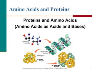
- 1. 1 Amino Acids and Proteins Proteins and Amino Acids (Amino Acids as Acids and Bases) Copyright © 2007 by Pearson Education, Inc. Publishing as Benjamin Cummings
- 2. 2
- 3. Proteins are molecular tools They are a diverse and complex group of macromolecules Protein Diversity From McKee and McKee, Biochemistry, 5th Edition, © 2011 by Oxford University Press Proteins
- 4. 4 Functions of Proteins Proteins perform many different functions in the body. TABLE 19.1
- 5. Functions of Proteins Antibody Antibodies bind to specific foreign particles, such as viruses and bacteria, to help protect the body. Immunoglobulin G (IgG) Enzyme Enzymes carry out almost all of the thousands of chemical reactions that take place in cells. They also assist with the formation of new molecules by reading the genetic information stored in DNA. Phenylalanine hydrox ylase Messenger Messenger proteins, such as some types of hormones, transmit signals to coordinate biological processes between different cells, tissues, and organs. Growth hormone Structural component These proteins provide structure and support for cells. On a larger scale, they also allow the body to move. Actin Transport/storage These proteins bind and carry atoms and small molecules within cells and throughout the body. Ferritin
- 6. 6 Amino Acids Amino acids Are the building blocks of proteins. There are 20 standard amino acids Contain a carboxylic acid group and an amino group on the alpha () carbon. Are ionized in solution. Each contain a different side group (R). R R │ + │ H2N—C —COOH H3N—C —COO− │ │ H H ionized form
- 7. 7 Examples of Amino Acids H + │ H3N—C—COO− │ H glycine CH3 + │ H3N—C—COO− │ H alanine
- 8. 8 Types of Amino Acids Amino acids are classified as Nonpolar (hydrophobic) with hydrocarbon side chains. Polar (hydrophilic) with polar or ionic side chains. Acidic (hydrophilic) with acidic side chains. Basic (hydrophilic) with –NH2 side chains. Nonpolar Polar Acidic Basic Copyright © 2007 by Pearson Education, Inc. Publishing as Benjamin Cummings
- 9. 9 Amino Acids Classification Table
- 10. 10 Nonpolar Amino Acids A nonpolar amino acid has An R group that is H, an alkyl group, or aromatic. Copyright © 2007 by Pearson Education, Inc. Publishing as Benjamin Cummings
- 11. 11 Polar Amino Acids A polar amino acid has An R group that is an alcohol, thiol, or amide. Copyright © 2007 by Pearson Education, Inc. Publishing as Benjamin Cummings
- 12. 12 Acidic and Basic Amino Acids An amino acid is Acidic with a carboxyl R group (COO−). Basic with an amino R group (NH3 +). Copyright © 2007 by Pearson Education, Inc. Publishing as Benjamin Cummings Basic Amino Acids D E
- 13. 13 Learning Check Identify each as (P) polar or (NP) nonpolar. + A. H3N–CH2–COO− (Glycine) CH3 | CH–OH + │ B. H3N–CH–COO− (Threonine)
- 14. 14 Solution Identify each as (P) polar or (NP) nonpolar. + A. H3N–CH2–COO− (Glycine) (NP) nonpolar CH3 | CH–OH + │ B. H3N–CH–COO− (Threonine) (P) polar
- 15. 15 Optical Activity Amino acids Are chiral except for glycine. Have Fischer projections that are stereoisomers. That are L are used in proteins. L-alanine D-alanine L-cysteine D-cysteine CH2SH H2N H COOH CH2SH H NH2 COOH CH3 H NH2 COOH CH3 H2N H COOH Fischer Projections of Amino Acids
- 16. 16 • Amino acids are amphoteric molecules have both basic and acidic group • Zwitterions (dipolar molecules), have charged —NH3 + and COO- groups. • Forms when both the —NH2 and the —COOH groups in an amino acid ionize in water. • Has equal + and − charges at the isoelectric point (pI). O O ║ + ║ NH2—CH2—C—OH H3N—CH2—C—O– Glycine Zwitterion of glycine Amphoteric Properties Zwitterions and Isoelectric Points
- 17. At physiological pH (7.4), a zwitterion forms Both + and – charges Overall neutral Amphoteric Amino group is protonated Carboxyl group is deprotonated Soluble in polar solvents due to ionic character Structure of R also influence solubility Zwitterions
- 18. 18 In solutions more basic than the pI, The —NH3 + in the amino acid donates a proton. + OH– H3N—CH2—COO– H2N—CH2—COO– Zwitterion Negative ion at pI pH > pI Charge: 0 Charge: 1− Amino Acids as Acids
- 19. 19 In solutions more acidic than the pI, The COO− in the amino acid accepts a proton. + H+ + H3N—CH2—COO– H3N—CH2—COOH Zwitterion Positive ion at pI pH< pI Charge: 0 Charge: 1+ Amino Acids as Bases
- 20. 20 pH and Ionization H+ OH− + + H3N–CH2–COOH H3N–CH2–COO– H2N–CH2–COO– positive ion zwitterion negative ion (at low pH) (at pI) (at high pH)
- 21. Aas have multiple pKa’s due to multiple ionizable groups Acid-base Properties pK1 ~ 2.2 (protonated below 2.2) pK2 ~ 9.4 (NH3 + below 9.4) pKR (when applicable)
- 22. Table 3-1 Note 3-letter and 1-letter abbreviations Amino acid organization chart
- 23. Titration of Glycine pK1 [cation] = [zwitterion] pK2 [zwitterion] = [anion] First equivalence point Zwitterion Molecule has no net charge pH = pI (Isoelectric point) pI = average of pKa’s = ½ (pK1 + pK2) pIglycine = ½ (2.34 + 9.60) = 5.97 Animation
- 24. 24 Electrophoresis: Separation of Amino Acids In electrophoresis, an electric current is used to separate a mixture of amino acids, and The positively charged amino acids move toward the negative electrode. The negatively charged amino acids move toward the positive electrode. An amino acid at its pI does not migrate. The amino acids are identified as separate bands on the filter paper or thinlayer plate.
- 25. 25 Electrophoresis With an electric current, a mixture of lysine, aspartate, and valine are separated. Copyright © 2007 by Pearson Education, Inc. Publishing as Benjamin Cummings
- 26. 26 CH3 CH3 + | | H3N—CH—COOH H2N—CH—COO– (1) (2) Which structure represents: A. Alanine at a pH above its pI? B. Alanine at a pH below its pI? Learning Check
- 27. 27 CH3 CH3 + | | H3N—CH—COOH H2N—CH—COO– (1) (2) Which structure represents: A. Alanine at a pH above its pI? (2) B. Alanine at a pH below its pI? (1) Solution
- 28. 28 Essential amino acids Must be obtained from the diet. Are the ten amino acids not synthesized by the body. Are in meat and diary products. Are missing (one or more) in grains and vegetables. Essential Amino Acids TABLE 19.3 Copyright © 2007 by Pearson Education, Inc Publishing as Benjamin Cummings
- 29. 29 Amino Acids and Proteins Formation of Peptides Copyright © 2007 by Pearson Education, Inc Publishing as Benjamin Cummings
- 30. 30 The Peptide Bond A peptide bond Is an amide bond. Forms between the carboxyl group of one amino acid and the amino group of the next amino acid. O CH3 O + || + | || H3N—CH2—C—O– + H3N—CH—C—O– O H CH3 O + || | | || H3N—CH2—C—N—CH—C—O– + H2O peptide bond
- 31. 31 Formation of A Dipeptide Copyright © 2007 by Pearson Education, Inc Publishing as Benjamin Cummings
- 32. 32 Learning Check Write the dipeptide Ser-Thr. OH CH3 | | CH2 O HCOH O + | ║ + | ║ H3N─CH─C─O – + H3N─CH─C─O– Ser Thr
- 33. 33 Solution Write the dipeptide Ser-Thr. OH CH3 | | CH2 O HCOH O + | ║ + | ║ H3N─CH─C─O – + H3N─CH─C─O– Ser peptide Thr OH bond CH3 | | CH2 O H HCOH O + | ║ | | ║ NH3─CH─C─N ─CH─C─O– + H2O Ser-Thr
- 34. 34 Naming Dipeptides A dipeptide is named with A -yl ending for the N-terminal amino acid. The full amino acid name of the free carboxyl group (COO-) at the C-terminal end. Copyright © 2007 by Pearson Education, Inc Publishing as Benjamin Cummings
- 35. 35 Write the three-letter abbreviations and names of the tripeptides that could form from two glycine and one alanine. Learning Check
- 36. 36 Write the names and three-letter abbreviations of the tripeptides that could form from two glycine and one alanine. Glycylglycylalanine Gly-Gly-Ala Glycylalanylglycine Gly-Ala-Gly Alanylglycylglycine Ala-Gly-Gly Solution
- 37. 37 Learning Check What are the possible tripeptides formed from one each of leucine, glycine, and alanine?
- 38. 38 Solution Tripeptides possible from one each of leucine, glycine, and alanine Leu-Gly-Ala Leu-Ala-Gly Ala-Leu-Gly Ala-Gly-Leu Gly-Ala-Leu Gly-Leu-Ala
- 39. 39 Learning Check Write the three-letter abbreviation and name for the following tetrapeptide: CH3 │ CH3 S │ │ CH–CH3 SH CH2 │ │ │ CH3 O H CH2 O H CH2O H CH2 O + │ ║ │ │ ║ │ │ ║ │ │ ║ H3N–CH–C–N–CH–C–N–CH–C–N–CH–CO–
- 40. 40 Solution Ala-Leu-Cys-Met Alanylleucylcysteylmethionine CH3 │ CH3 S │ │ CH–CH3 SH CH2 │ │ │ CH3 O H CH O H CH2O H CH2 O + │ ║ │ │ ║ │ │ ║ │ │ ║ H3N–CH–C–N–CH–C–N–CH–C–N–CH–CO– Ala Leu Cys Met
- 41. 41 Amino Acids and Proteins Protein Structure: Primary and Secondary Levels Copyright © 2007 by Pearson Education, Inc Publishing as Benjamin Cummings
- 42. Protein Diversity From McKee and McKee, Biochemistry, 5th Edition, © 2011 by Oxford University Press Proteins
- 43. 43 Primary Structure of Proteins The primary structure of a protein is The particular sequence of amino acids. The backbone of a peptide chain or protein. Ala─Leu─Cys─Met CH3 SH CH2 CH3 S CH2 CH2CH O O- CCH H N O CCH H N O CCH H N O CCHH3N CH3 CH3CH Copyright © 2007 by Pearson Education, Inc Publishing as Benjamin Cummings
- 44. 44 Primary Structures The nanopeptides oxytocin and vasopressin Have similar primary structures. Differ only in the amino acids at positions 3 and 8. Copyright © 2007 by Pearson Education, Inc Publishing as Benjamin Cummings
- 45. 45 Primary Structure of Insulin Insulin Was the first protein to have its primary structure determined. Has a primary structure of two polypeptide chains linked by disulfide bonds. Has a chain A with 21 amino acids and a chain B with 30 amino acids. Copyright © 2007 by Pearson Education, Inc Publishing as Benjamin Cummings
- 46. 46 Secondary Structure – Alpha Helix The secondary structures of proteins indicate the three-dimensional spatial arrangements of the polypeptide chains. An alpha helix has A coiled shape held in place by hydrogen bonds between the amide groups and the carbonyl groups of the amino acids along the chain. Hydrogen bonds between the H of a –N-H group and the O of C=O of the fourth amino acid down the chain.
- 47. 47 Secondary Structure – Alpha Helix Copyright © 2007 by Pearson Education, Inc Publishing as Benjamin Cummings
- 48. 48 Secondary Structure – Beta Pleated Sheet A beta-pleated sheet is a secondary structure that Consists of polypeptide chains arranged side by side. Has hydrogen bonds between chains. Has R groups above and below the sheet. Is typical of fibrous proteins such as silk.
- 49. 49 Secondary Structure: β-Pleated Sheet Copyright © 2007 by Pearson Education, Inc Publishing as Benjamin Cummings
- 50. 50 Secondary Structure: Triple Helix A triple helix Consists of three alpha helix chains woven together. Contains large amounts glycine, proline, hydroxy proline, and hydroxylysine that contain –OH groups for hydrogen bonding. Is found in collagen, connective tissue, skin, tendons, and cartilage. Copyright © 2007 by Pearson Education, Inc Publishing as Benjamin Cummings
- 51. 51 Indicate the type of protein structure as 1) primary 2) alpha helix 3) beta-pleated sheet 4) triple helix A. Polypeptide chains held side by side by H bonds. B. Sequence of amino acids in a polypeptide chain. C. Corkscrew shape with H bonds between amino acids. D. Three peptide chains woven like a rope. Learning Check
- 52. 52 Indicate the type of protein structure as: 1) primary 2) alpha helix 3) beta-pleated sheet 4) triple helix A. 3 Polypeptide chains held side by side by H bonds. B. 1 Sequence of amino acids in a polypeptide chain. C. 2 Corkscrew shape with H bonds between amino acids. D. 4 Three peptide chains woven like a rope. Solution
- 53. 53 Amino Acids and Proteins Protein Structure: Tertiary and Quaternary Levels Copyright © 2007 by Pearson Education, Inc Publishing as Benjamin Cummings
- 54. 54 The tertiary structure of a protein Gives a specific three dimensional shape to the polypeptide chain. Involves interactions and cross links between different parts of the peptide chain. Is stabilized by Hydrophobic and hydrophilic interactions. Salt bridges. Hydrogen bonds. Disulfide bonds. Tertiary Structure
- 55. 55 Tertiary Structure The interactions of the R groups give a protein its specific three- dimensional tertiary structure. Copyright © 2007 by Pearson Education, Inc Publishing as Benjamin Cummings
- 57. 57 Globular Proteins Globular proteins Have compact, spherical shapes. Carry out synthesis, transport, and metabolism in the cells. Such as myoglobin store and transport oxygen in muscle. Myoglobin Copyright © 2007 by Pearson Education, Inc Publishing as Benjamin Cummings
- 58. 58 Fibrous Proteins Fibrous proteins Consist of long, fiber-like shapes. Such as alpha keratins make up hair, wool, skin, and nails. Such as feathers contain beta keratins with large amounts of beta-pleated sheet structures. Copyright © 2007 by Pearson Education, Inc Publishing as Benjamin Cummings
- 59. 59 Select the type of tertiary interaction 1) disulfide 2) ionic 3) H bonds 4) hydrophobic A. Leucine and valine B. Two cysteines C. Aspartic acid and lysine D. Serine and threonine Learning Check
- 60. 60 Select the type of tertiary interaction as: 1) disulfide 2) ionic 3) H bonds 4) hydrophobic A. 4 Leucine and valine B. 1 Two cysteines C. 2 Aspartic acid and lysine D. 3 Serine and threonine Solution
- 61. 61 Quaternary Structure The quaternary structure Is the combination of two or more tertiary units. Is stabilized by the same interactions found in tertiary structures. Of hemoglobin consists of two alpha chains and two beta chains. The heme group in each subunit picks up oxygen for transport in the blood to the tissues. Copyright © 2007 by Pearson Education, Inc Publishing as Benjamin Cummings hemoglobin
- 62. 62 Summary of Protein Structure
- 63. 63 Summary of Protein Structures Copyright © 2007 by Pearson Education, Inc Publishing as Benjamin Cummings
- 64. 64 Identify the level of protein structure as: 1) Primary 2) Secondary 3) Tertiary 4) Quaternary A. Beta-pleated sheet B. Order of amino acids in a protein C. A protein with two or more peptide chains D. The shape of a globular protein E. Disulfide bonds between R groups Learning Check
- 65. 65 Identify the level of protein structure 1. Primary 2. Secondary 3. Tertiary 4. Quaternary A. 2 Beta-pleated sheet. B. 1 Order of amino acids in a protein. C. 4 A protein with two or more peptide chains. D. 3 The shape of a globular protein. E. 3 Disulfide bonds between R groups. Solution
- 66. 66 Protein Hydrolysis and Denaturation Amino Acids and Proteins Copyright © 2007 by Pearson Education, Inc. Publishing as Benjamin Cummings
- 67. 67 Protein hydrolysis Splits the peptide bonds to give smaller peptides and amino acids. Occurs in the digestion of proteins. Occurs in cells when amino acids are needed to synthesize new proteins and repair tissues. Protein Hydrolysis
- 68. 68 Hydrolysis of a Dipeptide In the lab, the hydrolysis of a peptide requires acid or base, water and heat. In the body, enzymes catalyze the hydrolysis of proteins. + H3N CH COH OCH3 + H2O, H + ++ heat, CH2 OH H3N CH C O N H CH C O OH CH3 CH2 OH CH C O OHH3N
- 69. 69 Denaturation involves The disruption of bonds in the secondary, tertiary and quaternary protein structures. Heat and organic compounds that break apart H bonds and disrupt hydrophobic interactions. Acids and bases that break H bonds between polar R groups and disrupt ionic bonds. Heavy metal ions that react with S-S bonds to form solids. Agitation such as whipping that stretches peptide chains until bonds break. Denaturation
- 70. 70 Denaturation of protein occurs when An egg is cooked. The skin is wiped with alcohol. Heat is used to cauterize blood vessels. Instruments are sterilized in autoclaves. Applications of Denaturation Copyright © 2007 by Pearson Education, Inc Publishing as Benjamin Cummings
- 71. • Ribonuclease is a small protein that contains 8 cysteins linked via four disulfide bonds • Urea in the presence of 2- mercaptoethanol fully denatures ribonuclease • When urea and 2-mercaptoethanol are removed, the protein spontaneously refolds, and the correct disulfide bonds are reformed • The sequence alone determines the native conformation • Quite “simple” experiment, but so important it earned Chris Anfinsen the 1972 Chemistry Nobel Prize Ribonuclease Refolding Experiment
- 72. 72 What are the products of the complete hydrolysis of the peptide Ala-Ser-Val? Learning Check
- 73. 73 The products of the complete hydrolysis of the peptide Ala-Ser-Val are alanine serine valine Solution
- 74. 74 Tannic acid is used to form a scab on a burn. An egg is hard boiled by placing it in boiling water. What is similar about these two events? Learning Check
- 75. 75 Acid and heat cause the denaturation of protein. They both break bonds in the secondary and tertiary structures of proteins. Solution