
Empfohlen
Weitere ähnliche Inhalte
Ähnlich wie Introduction - Cellular Pathology 22-23(1).pptx
Ähnlich wie Introduction - Cellular Pathology 22-23(1).pptx (20)
Kürzlich hochgeladen
Kürzlich hochgeladen (20)
Introduction - Cellular Pathology 22-23(1).pptx
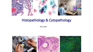
- 1. Histopathology & Cytopathology Gus Lusack
- 2. Histology, Histopathology, Histochemistry, Immunocytochemistry & Cytopathology • What are they? - A mixture of anatomy, biochemistry and physiology. • Histology - the microscopic study of normal tissue and its structure. • Histopathology - as above but looking at diseased tissue. This includes diseases such as infection, inflammation and cancer. • Histochemistry - this is the study of known substances within the structural framework, as opposed to using ‘dyes’ for histology, the chemistry is understood. • Immunocytochemistry - localisation of specific protein or antigen in cells using primary antibodies • Cytopathology – is closely allied to histopathology but distinct as it involves analysis of cell preparations
- 3. What does Histopathology involve? • Patients give consent to have histopathology specimens according to Human Tissue Act 2004 • The results are communicated to the patients by the requesting clinicians • Majority of the diagnoses are based on the assessment of haematoxylin and eosin (H&E)- stained sections of formalin-fixed, paraffin wax-embedded tissue Steps from specimen to report Fixation Specimen Collection, transportation and receipt Tissue selection and description Tissue processing Embedding Microtomy Staining and mounting Quality assurance Reporting Computer entry & report dispatch
- 4. The Main Disease Processes 1 Protective response to foreign agents: Microorganisms: bacteria, fungi, viruses, parasites Physical agents: trauma, chemicals, radiation (UV, Radiotherapy), temperature Inflammation Organ / Tissue Tissues Inflammation Brain Meninges Meningitis Mouth Gum (gingiva) Tonsil Gingivitis Tonsilitis Gastrointestinal tract Oesophagus Stomach Appendix Colon Oesophagitis Gastritis Appendicitis Colitis Lung Bronchi Lung Bronchitis Pneumonitis (pneumonia) Breast Breast Mastitis Liver Liver Hepatitis Bladder Bladder mucosa Cystitis Skin Dermis Dermatitis Musculoskeletal system Tendon Joints Tendonitis Arthritis
- 5. The Main Disease Processes 2 • Neoplasia means new growth o Ability to grow and enlarge unless it is treated o Sometimes referred to as tumour (swelling) o Classified into benign and malignant types Neoplasia Benign Malignant Size Small Large Borders Well defined Ill (poorly) defined Differentiation Resembles tissue of origin Variable Growth rate Slow Rapid Mitotic figures Rare Common Necrosis No Yes Invasion No Yes Metastasis No Yes
- 6. The main disease processes 3 Neoplasia Benign Malignant Epithelial Squamous Squamous cell papilloma Squamous cell carcinoma Transitional Transitional cell papilloma Transitional cell carcinoma Glandular Adenoma Adenocarcinoma Mesenchymal Fibrous tissue Fibroma Fibrosarcoma Adipose tissue Lipoma Liposarcoma Blood vessels Haemangioma Angiosarcoma Cartilage Chondroma Chondrosarcoma Bone Osteoma Osteosarcoma Classification of benign and malignant neoplasms
- 7. Types of biopsy submitted for histopathology
- 8. Dealing with Dead Tissue – Preparation: Whole tissue Fixation Dehydration Clearing Isolated Cells Freezing Decalcification Impregnation Embedding Microtomy Staining Microscopy
- 10. Haematoxylin and Eosin (H & E) Stain • H & E is the most widely used: oHaematoxylin is used as a nuclear stain – shows chromatin pattern in a distinct blue/black oIt requires oxidation to haematein (formalin pigment) oEosin is an acid dye – red colour which complements haematoxylin
- 12. Automated Tissue Processing & Staining XY Stainer Coverslipper Leica Microsystems Thermo enclosed tissue processor Thermo microwave tissue processor
- 13. Non-Melanoma Skin Cancer (NMSC) Squamous Cell Carcinomas (SCCs) H&E stained Normal Skin H&E stained SCC
- 14. Special Stains Alcian blue -acid mucins Periodic acid Schiff (PAS) – Glycogen and neutral mucins Toluidine blue – Heparin Diastase PAS – Neutral mucins Colon mucus secreting glands stomach Cervix mast cells Elastic van Gieson – elastin Lung Masson Trichrome Collagen – blue Muscle, RBC - red
- 15. Immunocytochemistry in Histopathology Indirect immunocytochemistry Direct immunocytochemistry / Fluorescent Tag / Enzyme Tag Colon carcinoma – Anti-Cytokeratin 10 antibody Streptavidin Poly-HRP Conjugate Anti-Cytokeratin antibody – Fluorescent Tag
- 16. Molecular Diagnostics in Histopathology • In situ Hybridisation oPolymerase chain reaction (PCR) oSouthern blotting In situ Hybridisation (ISH) Fluorescence In situ Hybridisation (FISH) Breast cancer cells- multiple copies of HER2 gene (Red) In situ Hybridisation (ISH) Squamous cell carcinoma (SCC) – probes for Human Papillomavirus (HPV) DNA
- 17. Review and Reporting by Histopathologist • Check request form and slides match • H&E stained sections are examined and assessed for adequacy of sectioning and staining • Identify tissue type • Biopsy assessed for adequacy of sampling, size and preservation • A systematic approach to analyse tissue type and disease process – e.g. skin biopsy is assessed by observing the overall architecture of tissue (epidermis, the epidermal-dermal interface, the dermis and subcutaneous tissue) • Assessment of cellular composition and identify pathological changes • Identify the disease process
- 18. Cytopathology •Cervical screening •Cytology of urine •Lower Respiratory tract cytology •Serous effusions •Basic semen analysis (Andrology)
- 19. Cervical Screening • Prevention of carcinoma of the uterine cervix • Cervical carcinomas: oSquamous cell carcinoma derived from ectocervix oAdenocarcinoma derived from glandular epithelial cells of endocervix oThe precursors are termed as intraepithelial- abnormal cells remain within the epithelium oCervical carcinomas are caused by specific types of Human papillomaviruses (HPVs) oMost infections resolve within 6-18 months oSome persist and increase the risk oIt takes about 20 years from HPV infection to invasion of the lesion
- 20. Cervical Screening • The NHS screening programme; • For women aged 25 to 49 - 3 years interval • For women aged 50 to 64 – 5 years interval • Samples (Transformation Zone) usually taken in GP practice • Most popular staining method for wet-fixed preparation is Papanicolaou technique –Pap smear (George Papanicolaou 1940s) • Haematoxylin to stain the nuclei of cells and two cytoplasmic counterstains – orange G and eosin azure
- 21. Cervical Screening Liquid Based Cytology (LBC) • A number of points about the PAP smear: o LBC involves immediately dispersing the cells in a liquid transport medium o Monolayer (thin layer) preparation – facilitating the automated evaluation of individual cells o Membrane filtration/density gradient centrifugation - Reduce the number of lucocytes (WBCs) and erythrocytes (RBCs) o 6% reduction in tests considered inadequate for reporting in 2009 – reduced the need for many repeat tests
- 22. Normal Cervical Cytology • Chromatin pattern and distribution are the most useful features that distinguish normal cells from neoplastic cells • Nuclear shape e.g. round, oval, irregular • Regularity of the nuclear membrane, e.g. smooth, wrinkled, angular • Intensity of nuclear staining, e.g. normochromasia, hyperchromasia (darkly stained) and hypochromasia (pale staining)
- 23. Normal Cervical Cytology Metaplastic cells Endometrial cells
- 24. Abnormal Cervical Cytology Mild dyskaryosis A normal cell undergoing mitosis A abnormal cell undergoing mitosis Normal CIN1 CIN2 CIN3 Tripolar mitosis Moderate dyskaryosis Severe dyskaryosis
- 25. Abnormal Cervical Cytology (a) A ‘tadpole’ cell (the pink cell left of centre) and a fibre cell (right of centre). (b) Cytoplasmic keratinisation in malignant squamous cells (c) A microbiopsy of severe dyskaryotic cells (d) Tumour diathesis o Koilocytosis is caused by HPV o Superficial squamous cells with large o perinuclear clear space o Thickened uneven rim of dense o cytoplasm – wire loop appearance o Binucleation / multinucleation o Dyskeratosis is usually present
- 26. Cytological features suggestive of invasive carcinoma • Numerous dyskeratotic cells • Cellular pleomorphism (bizarre-shaped, fibre-shaped, tadpole shaped) • Very coarse aggregates of nuclear chromatin • Large, irregular, sometimes multiple nucleoli • Cytoplasmic keratinisation – presence of anucleate fragments of keratinsed cytoplasm • Tissue fragments composed of dyskeratotic cells • Tumour diathesis – mixture of necrotic cell debris, inflammatory exudate and blood
- 27. Cytological pitfalls Potential false positives Immature squamous metaplastic cells mimic moderate/severe dyskeratosis Endometrial cells- appear as hyperchromatic cells with high N:C ratio Histiocytes – mimic of dyskaryosis Potential false negatives Small cell dyskaryosis Pale dyskaryosis – less chromatin Intensity compared to adjacent polymorphs A microbiopsy / hyperchromatic crowded cell group
- 28. Cytology of Urine • Sample types: ovoided urine oIleal conduit urine – urinary diversion for bladder cancer patients oCatheter urine oBladder washings – not a common source • Benign changes - bladder could be infected by bacteria, fungi, viruses or parasites • Useful test for identification of high-grade urothelial cancer
- 29. High-Grade Urothelial Carcinoma (a) Increased cellularity (b) Isolated malignant cells (c) Clusters of malignant cell with hyperchromatic nuclei (d) Malignant cells showing pleomorphism (e) Very high nuclear to cytoplasmic ratio (f & g) Marked nuclear hyperchromasia (h) Coarse chromatin pattern (i) These two cells have coarse and regular chromatin pattern (j) Angular nuclear outline (k) Prominent nucleoli seen
- 30. Cytology of the Lower Respiratory Tract • Sample types: oSputum – mixture of mucus and cells oBronchial brushings oBronchial washings oTransbronchial fine needle aspiration oTransthorasic fine needle aspiration • Infections: bacterial (pneumonia), TB (Mycobacterium tuberculosis), Herpes simplex virus • Lung cancer: small cell lung cancer & bronchial carcinoid
- 31. Cytology of the Lower Respiratory Tract • Bacterial infection Pneumonia sputum sample –mixture of inflammatory cells in necrotic debris Actinomyces – filamentous branching bacteria, often found – no significance Bronchial washing sample from a patient confirmed with TB –giant cells (the nuclei are at one pole) Necrotic debris from a TB patient
- 32. Serous effusions
- 33. Serous Effusions - Adenocarcinoma • The most common malignancy in effusions • May present as papillary (nipple –like protrusion) or acina (gland- like) • Multilayered clusters of cells • Nuclei - pleomorphism, enlargement, hyperchromasia (a) Cluster of malignant cells (b) Breast carcinoma showing spherical clusters (c) Ovarian carcinoma showing papillary and acinar clusters; (d) psammoma body; (e) vacuolated malignant cells (f) Lung carcinoma showing signet ring formation
- 34. Basic semen analysis (Andrology) • A number of risk factors associated with poor semen quality • History of reproductive tract infections / sexually transmitted diseases • Exposure to environmental pollutants • Athletes exposed to conditions which may lower sperm counts • Alcohol abuse/ use of tobacco/ recreational drugs • Malignancy • Medicinal drugs, especially steroids • Previous testicular surgery / recent trauma
- 35. Basic Semen Analysis • Two sample types; oPost-vasectomy oInfertility • Equipment : An improved Neubauer Haemocytometer & Microscope Infertility 1. Liquefaction 2. Appearance 3. Volume 4. Viscosity 5. pH 6. Motility 7. Morphology (% of normal sperm) 8. Sperm count and leucocytes count (millions per ml) 9. Anti sperm antibodies – potential cause of infertility 10. Vitality (% of live sperm)
- 36. Basic Semen Analysis Normal Sperm Test Parameter Lower Reference Values WHO Semen volume 1.5 ml or more Total number of sperm per ejaculate 39 million Sperm concentration 15 million sperm per ml Total motility 40% Progressive motility 32% Vitality (live) 58% Morphology (% normal) 4% pH 7.2 or higher White blood cells Fewer than I million per ml Sperm antibodies <50% of sperm reactive Abnormal sperm morphology
- 37. Quality Assurance (QA) 1 Internal Quality Control (IQC): • Ensure measures and procedures employed are performed to expected standards • Use known positive controls in histochemistry or with special stains • e.g. A control slide known to contain tubercle bacilli with every batch of slides stained with a Ziehl Neelson (ZN) stain • Slides from patients are submitted to a Histopathologist for reporting only if the control slide is positive
- 38. Quality Assurance 2 • External Quality Assessment (EQA) • Membership of an external QA scheme is a requirement for UKAS ISO 15189 accreditation • Over 140 schemes operate from 24 centres based at major hospitals, research institutions and universities • Cover qualitative and interpretive investigations in andrology, clinical chemistry, genetics, haematology, histopathology, immunology and microbiology • Participants receive independent, objective and impartial reports – identify weaknesses and take appropriate action • In histopathology; • Covers diagnostic aspects of the service – UK NEQAS Breast Screening Pathology • Covers technical aspects – UK NEQAS Cellular Pathology Technique and UK NEQAS Immunocytochemistry and In situ hybridisation National External Quality Assessment Scheme (NEQAS) www.ukneqas.org.uk
- 39. Exercise 1 • Write short notes on the following: • Carcinoma • Adenocarcinoma • Sarcoma • Leukaemia • Lymphoma • Myeloma • UKAS ISO15189 Accreditation • Human Tissue Act 2004 (Word count:1500)