
Empfohlen
Weitere ähnliche Inhalte
Was ist angesagt?
Was ist angesagt? (20)
Andere mochten auch
Andere mochten auch (20)
Ähnlich wie General-Purpose Ultrasound
Ähnlich wie General-Purpose Ultrasound (20)
General-Purpose Ultrasound
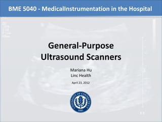
- 1. BME 5040 - MedicalInstrumentation in the Hospital General-Purpose Ultrasound Scanners Mariana Hu Linc Health April 23, 2012
- 2. Outline 1. Introduction 2. System components 3. Imaging principles 4. Types of transducers 5. Imaging modes 6. Conclusion
- 3. Outline 1. Introduction 2. System components 3. Imaging principles 4. Types of transducers 5. Imaging modes 6. Conclusion
- 4. Introduction • Ultrasound imaging utilizes the interaction of high frequency sound waves with living tissue to obtain an image of the tissue or to determine the velocity of a moving tissue • The dynamic, real-time images provide quantitative structural and functional information
- 5. Introduction • General-Purpose • abdominal • obstetric/gynecologic • small parts • vascular Types of Ultrasound • Dedicated Cardiac Scanners •real-time, non-invasive imaging of heart structures andfunctionality • Dedicated Intravascular • 360o cross sectional images of blood vessels for diagnostic and therapeutic purposes • Portable • Compact and light weight to be carried by hand between exams
- 6. Introduction • General-Purpose • abdominal • obstetric/gynecologic • small parts • vascular Types of Ultrasound • Dedicated Cardiac Scanners •real-time, non-invasive imaging of heart structures andfunctionality • Dedicated Intravascular • 360o cross sectional images of blood vessels for diagnostic and therapeutic purposes • Portable • Compact and light weight to be carried by hand between exams
- 7. Introduction • General-Purpose • abdominal • obstetric/gynecologic • small parts • vascular Types of Ultrasound • Dedicated Cardiac Scanners •real-time, non-invasive imaging of heart structures andfunctionality • Dedicated Intravascular • 360o cross sectional images of blood vessels for diagnostic and therapeutic purposes • Portable • Compact and light weight to be carried by hand between exams
- 8. Introduction • General-Purpose • abdominal • obstetric/gynecologic • small parts • vascular Types of Ultrasound • Dedicated Cardiac Scanners •real-time, non-invasive imaging of heart structures andfunctionality • Dedicated Intravascular • 360o cross sectional images of blood vessels for diagnostic and therapeutic purposes • Portable • Compact and light weight to be carried by hand between exams
- 9. Introduction • General-Purpose • abdominal • obstetric/gynecologic • small parts • vascular Types of Ultrasound • Dedicated Cardiac Scanners •real-time, non-invasive imaging of heart structures andfunctionality • Dedicated Intravascular • 360o cross sectional images of blood vessels for diagnostic and therapeutic purposes • Portable • Compact and light weight to be carried by hand between exams
- 10. Outline 1. Introduction 2. System components 3. Imaging principles 4. Types of transducers 5. Imaging modes 6. Conclusion
- 11. System components Keyboard/ Cursor Ultrasound Pulse Control Image Storage System Ultrasound Monitor Probe Ultrasound Echo ________________________ Pulse Pulse Reflecting interface
- 12. System components Keyboard/ Cursor Ultrasound Pulse Control Image Storage System Ultrasound Monitor Probe Ultrasound Echo ________________________ Pulse Pulse Reflecting interface
- 13. System components Keyboard/ Cursor Ultrasound Pulse Control Image Storage System Ultrasound Monitor Probe Ultrasound Echo ________________________ Pulse Pulse Reflecting interface
- 14. System components -Duration -Frequency Keyboard/ Cursor -Scan mode Ultrasound Pulse Control Image Storage System Ultrasound Monitor Probe Ultrasound Echo ________________________ Pulse Pulse Reflecting interface
- 15. System components Keyboard/ Cursor Ultrasound Pulse Control Image Storage System Ultrasound Monitor PACS Probe Ultrasound Echo ________________________ Pulse Pulse Reflecting interface
- 16. System components Keyboard/ Cursor Ultrasound Pulse Control Image Storage System Ultrasound Monitor 2-20 MHz Probe Ultrasound Echo Pulse Pulse ________________________ Reflecting interface
- 17. System components Keyboard/ Cursor Ultrasound Pulse Control Image Storage System Ultrasound Monitor 2-20 MHz Probe Ultrasound Echo Pulse Pulse ________________________ Reflecting interface
- 18. Outline 1. Introduction 2. System components 3. Imaging principles 4. Types of transducers 5. Imaging modes 6. Conclusion
- 19. Imaging Principles • Ultrasound waves generated by a transducer are transmitted through soft tissue relative to their acoustic impedance • Acoustic impedance: Z = vxd
- 20. Imaging Principles • Ultrasound waves generated by a transducer are transmitted through soft tissue relative to their acoustic impedance • Acoustic impedance: Z = vxd Velocity of sound Tissue density transmission
- 21. Imaging Principles • Ultrasound waves generated by a transducer are transmitted through soft tissue relative to their acoustic impedance • Acoustic impedance: Z is primarily a function of the tissue Z = vxd density Velocity of sound Tissue density transmission Constant: 1540 m/s
- 22. Imaging Principles • When two tissues with different densities are located next to each other, the acoustic impedance mismatch causes the sound waves to be reflected • The transducer receives the reflections and converts them into an electronic signal • The image is created based on the signal amplitude and frequency, and the time delay between the transmitted pulse and its echo
- 23. Imaging Principles • When two tissues with different densities are located next to each other, the acoustic impedance mismatch causes the sound waves to be reflected • The transducer receives the reflections and converts them into an electronic signal • The image is created based on the signal amplitudeand frequency, and the time delay between the transmitted pulse and its echo a The time delay is measure of the depth of the tissue interface
- 24. Imaging Principles The transducer continuously switches between transmit and receive modes Probe Probe Ultrasound Echo Pulse Pulse Interface Penetrating Ultrasound Pulse Transmit Mode Receive Mode
- 26. Imaging Principles Ultrasound Image frequency resolution
- 27. Imaging Principles Ultrasound Image frequency resolution Tissue penetration
- 28. Outline 1. Introduction 2. System components 3. Imaging principles 4. Types of transducers 5. Imaging modes 6. Conclusion
- 29. Types of Transducers Piezoelectric effect Electrical energy Mechanical energy
- 30. Types of Transducers The transducer’s physical design and the technique of beam generation determine the direction of the ultrasound beams This, in turn, determines the shape of the image
- 31. Types of transducers Types of Images Rectangular Sector Steered Linear Array Convex Array Phased Array Vector Array Linear Array Transducer Transducer Transducer Transducer Transducer
- 32. Types of transducers Types of Images Rectangular Sector Steered Linear Array Convex Array Phased Array Vector Array Linear Array Transducer Transducer Transducer Transducer Transducer
- 33. Types of transducers Types of Images Rectangular Sector Steered Linear Array Convex Array Phased Array Vector Array Linear Array Transducer Transducer Transducer Transducer Transducer
- 34. Types of transducers Types of Images Rectangular Sector Steered Linear Array Convex Array Phased Array Vector Array Linear Array Transducer Transducer Transducer Transducer Transducer
- 35. Types of transducers Types of Images Rectangular Sector Steered Linear Array Convex Array Phased Array Vector Array Linear Array Transducer Transducer Transducer Transducer Transducer
- 36. Types of transducers Types of Images Rectangular Sector Steered Linear Array Convex Array Phased Array Vector Array Linear Array Transducer Transducer Transducer Transducer Transducer
- 37. Types of transducers Types of Images Rectangular Sector Steered Linear Array Convex Array Phased Array Vector Array Linear Array Transducer Transducer Transducer Transducer Transducer
- 38. Types of transducers Types of • Individual beams travel at the same Images angle, perpendicular to the transducer’s scanning surface Rectangular Sector Steered Linear Array Convex Array Phased Array Vector Array Linear Array Transducer Transducer Transducer Transducer Transducer
- 39. Types of transducers •Externally similar to linear array transducers Types of • Electronic steering directs beams Images at an angle slanted relative to the scanning surface. Rectangular Sector Steered Linear Array Convex Array Phased Array Vector Array Linear Array Transducer Transducer Transducer Transducer Transducer
- 40. Types of transducers Types of Images Rectangular Sector Steered Linear Array Convex Array Phased Array Vector Array Linear Array Transducer Transducer Transducer Transducer Transducer
- 41. Types of transducers Types of Images Rectangular Sector Steered Linear Array Convex Array Phased Array Vector Array Linear Array Transducer Transducer Transducer Transducer Transducer
- 42. Types of transducers Types of Images Rectangular Sector Steered Linear Array Convex Array Phased Array Vector Array Linear Array Transducer Transducer Transducer Transducer Transducer
- 43. Types of transducers Types of Images • Curved surface • Emission of beams at different angles Rectangular Sector Steered Linear Array Convex Array Phased Array Vector Array Linear Array Transducer Transducer Transducer Transducer Transducer
- 44. Types of transducers Types of Images • Smaller footprint • Imaging in tight areas or behind obstructions Rectangular Sector Steered Micro Linear Array Convex Array Phased Array Vector Array Linear Array Transducer Transducer Transducer Transducer Transducer
- 45. Types of transducers • Flat and small • Even narrower in the near field Types of Images • Electronic steering directs beams at different angles Rectangular Sector Steered Linear Array Convex Array Phased Array Vector Array Linear Array Transducer Transducer Transducer Transducer Transducer
- 46. Types of transducers • Electronic steering directs beams at different angles Types of ImagesLarger scanning surface • Rectangular Sector Steered Linear Array Convex Array Phased Array Vector Array Linear Array Transducer Transducer Transducer Transducer Transducer
- 47. Types of transducers Types of Images Rectangular Sector Steered Linear Array Convex Array Phased Array Vector Array Linear Array Transducer Transducer Transducer Transducer Transducer
- 48. Types of transducers Types of Endocavity transducers Images Rectangular Sector Steered Linear Array Convex Array Phased Array Vector Array Linear Array Transducer Transducer Transducer Transducer Transducer
- 49. Types of transducers Endovaginal Types of Endocavity transducers Images Rectangular Sector Steered Linear Array Convex Array Phased Array Vector Array Linear Array Transducer Transducer Transducer Transducer Transducer
- 50. Outline 1. Introduction 2. System components 3. Imaging principles 4. Types of transducers 5. Imaging modes 6. Conclusion
- 51. Imaging modes • B-Mode • M-Mode • Harmonic • Doppler – Spectral – Color Flow Mapping – Color Power
- 52. Imaging modes • B-Mode • M-Mode • Harmonic • Doppler – Spectral – Color Flow Mapping – Color Power
- 53. Imaging modes • B-Mode Brightness Mode
- 54. Imaging modes • B-Mode Brightness Mode – Shows the signal amplitude as the brightness of a point – An array of transducers scan a plane through the body simultaneously
- 55. Imaging modes • B-Mode Brightness Mode – Shows the signal amplitude as the brightness of a point – An array of transducers scan a plane through the body simultaneously – Real-time, 2-D image representing cross-sectional slice
- 56. Imaging modes • B-Mode Brightness Mode – Shows the signal amplitude as the brightness of a point – An array of transducers scan a plane through the body simultaneously – Real-time, 2-D image representing cross-sectional slice
- 57. Imaging modes • B-Mode Brightness Mode – Shows the signal amplitude as the brightness of a point – An array of transducers scan a plane through the body simultaneously – Real-time, 2-D image representing cross-sectional slice B-Mode image of the liver
- 58. Imaging modes • B-Mode • M-Mode • Harmonic • Doppler – Spectral – Color Flow Mapping – Color Power
- 59. Imaging modes • M-Mode Motion Mode
- 60. Imaging modes • M-Mode Motion Mode – Captures returning echoes in only one line of the B-mode image but displays them over a time axis – Pulses are emitted in quick succession
- 61. Imaging modes • M-Mode Motion Mode – Captures returning echoes in only one line of the B-mode image but displays them over a time axis – Pulses are emitted in quick succession – Produces a moving display of a structure over time
- 62. Imaging modes • M-Mode Motion Mode – Captures returning echoes in only one line of the B-mode image but displays them over a time axis – Pulses are emitted in quick succession – Produces a moving display of a structure over time
- 63. Imaging modes • M-Mode Motion Mode – Captures returning echoes in only one line of the B-mode image but displays them over a time axis – Pulses are emitted in quick succession – Produces a moving display of a structure over time M-Mode image of left ventricle
- 64. Imaging modes • B-Mode • M-Mode • Harmonic • Doppler – Spectral – Color Flow Mapping – Color Power
- 65. Imaging modes Harmonics are frequencies that occur at multiple times of the transmitted • Harmonic
- 66. Imaging modes Harmonics are frequencies that occur at multiple times of the transmitted • Harmonic – Ultrasound is transmitted at one frequency and received at twice the transmitted frequency – The returning high-frequency signal is isolated from the fundamental signal by using a filter
- 67. Imaging modes Harmonics are frequencies that occur at multiple times of the transmitted • Harmonic – Ultrasound is transmitted at one frequency and received at twice the transmitted frequency – The returning high-frequency signal is isolated from the fundamental signal by using a filter – Image is produced by high frequency signal
- 68. Imaging modes Harmonics are frequencies that occur at multiple times of the transmitted • Harmonic – Ultrasound is transmitted at one frequency and received at twice the transmitted frequency – The returning high-frequency signal is isolated from the fundamental signal by using a filter – Image is produced by high frequency signal vs
- 69. Imaging modes Harmonics are frequencies that occur at multiple times of the transmitted • Harmonic – Ultrasound is transmitted at one frequency and received at twice the transmitted frequency – The returning high-frequency signal is isolated from the fundamental signal by using a filter – Image is produced by high frequency signal vs
- 70. Imaging modes • B-Mode • M-Mode • Harmonic • Doppler Spectral Color Flow Mapping Color Power
- 72. Imaging modes • Doppler – Utilizes “Doppler shift” principle to determine blood flow velocity and direction
- 73. Imaging modes • Doppler – Utilizes “Doppler shift” principle to determine blood flow velocity and direction Blood cells flowing toward transducer
- 74. Imaging modes • Doppler – Utilizes “Doppler shift” principle to determine blood flow velocity and direction Blood cells flowing toward transducer
- 75. Imaging modes • Doppler – Utilizes “Doppler shift” principle to determine blood flow velocity and direction Blood cells flowing toward transducer Blood cells flowing away from transducer
- 76. Imaging modes • Doppler – Utilizes “Doppler shift” principle to determine blood flow velocity and direction Blood cells flowing toward transducer Blood cells flowing away from transducer
- 77. Imaging modes • Doppler – Utilizes “Doppler shift” principle to determine blood flow velocity and direction Blood cells Doppler shift: the flowing difference in toward transmitted and transducer reflected wave frequency Blood cells flowing away from transducer
- 78. Imaging modes • Doppler – Utilizes “Doppler shift” principle to determine blood flow velocity and direction Blood cells Doppler shift: the flowing difference in toward transmitted and transducer reflected wave frequency Blood cells flowing away Doppler shift from transducer Velocity of flow
- 79. Imaging modes • B-Mode • M-Mode • Harmonic • Doppler Spectral Color Flow Mapping Color Power
- 80. Imaging modes • Spectral Doppler
- 81. Imaging modes • Spectral Doppler – Pulsed Wave (PW): the same transducer is used both for transmitting and receiving • User can select a specific depth • Limited maximum velocity that can be measured
- 82. Imaging modes • Spectral Doppler – Pulsed Wave (PW): the same transducer is used both for transmitting and receiving • User can select a specific depth • Limited maximum velocity that can be measured – Continuous Wave (CW): one transducer transmits and another transducer detects signal • No information about depth of received signal • Can measure very high velocities
- 83. CW Doppler Image across the left PW Doppler Image of left ventricular outflow tract and aortic ventricular outflow tract valve in subject with aortic stenosis
- 84. Imaging modes • B-Mode • M-Mode • Harmonic • Doppler Spectral Color Flow Mapping Color Power
- 85. Imaging modes • Color Flow Mapping (CFM)
- 86. Imaging modes • Color Flow Mapping (CFM) – Color-encoded map of flow velocity and direction superimposed on a real-time 2-D image – Preset color flow map assigns one color to a flow velocity and direction
- 87. Imaging modes • Color Flow Mapping (CFM) – Color-encoded map of flow velocity and direction superimposed on a real-time 2-D image – Preset color flow map assigns one color to a flow velocity and direction
- 88. Imaging modes • Color Flow Mapping (CFM) – Color-encoded map of flow velocity and direction superimposed on a real-time 2-D image – Preset color flow map assigned one color to a flow velocity and direction CFM image of the kidney
- 89. Imaging modes • B-Mode • M-Mode • Harmonic • Doppler Spectral ColorFlow Mapping Color Power
- 90. Imaging modes • Color Power Doppler
- 91. Imaging modes • Color Power Doppler – Images blood flow by displaying the density of RBC rather than their velocity – Does not display flow direction or different velocities – Increased flow sensitivity – Very poor temporal resolution – Susceptible to noise
- 92. Imaging modes • Color Power Doppler – Images blood flow by displaying the density of RBC rather than their velocity – Does not display flow direction or different velocities – Increased flow sensitivity – Very poor temporal resolution – Susceptible to noise
- 93. Imaging modes • Color Power Doppler – Images blood flow by displaying the density of RBC rather than their velocity – Does not display flow direction or different velocities – Increased flow sensitivity – Very poor temporal resolution – Susceptible to noise Power Doppler image of the liver
- 94. Outline 1. Introduction 2. System components 3. Imaging principles 4. Types of transducers 5. Imaging modes 6. Conclusion
- 95. Conclusion • Ultrasound imaging is highly versatile
- 96. Conclusion • Ultrasound imaging is highly versatile • It presents many advantages: – Safety – Portability – Economic feasibility
- 97. Conclusion • Ultrasound imaging is highly versatile • It presents many advantages: – Safety – Portability – Economic feasibility • A limitation: – Inability to image bone- and gas-filled structures
- 98. End Thank you for your attention!
Hinweis der Redaktion
- The CPU controls and processes the majority of the functions of the ultrasound system. Itis responsible for the input to the other components, such as the transducer and monitor. It alsoreceives and analyzes electronic input from the transducer to ultimately construct the image.Nowadays, they have full digital image processing capabilities and the computing speed neededfor high resolution imaging at high frame rates. The CPU can also serve as a temporary storagesystem for images. Rather than requiring purchase of an entirely new system, CPUs can beupgraded easily by installing software as technology advances
- The CPU controls and processes the majority of the functions of the ultrasound system. Itis responsible for the input to the other components, such as the transducer and monitor. It alsoreceives and analyzes electronic input from the transducer to ultimately construct the image.Nowadays, they have full digital image processing capabilities and the computing speed neededfor high resolution imaging at high frame rates. The CPU can also serve as a temporary storagesystem for images. Rather than requiring purchase of an entirely new system, CPUs can beupgraded easily by installing software as technology advances
- The CPU controls and processes the majority of the functions of the ultrasound system.It is responsible for the input to the other components, such as the transducer and monitor.It also receives and analyzes electronic input from the transducer to ultimately construct the image.Nowadays, they have full digital image processing capabilities and the computing speed neededfor high resolution imaging at high frame rates. The CPU can also serve as a temporary storagesystem for images. Rather than requiring purchase of an entirely new system, CPUs can beupgraded easily by installing software as technology advances
- The CPU controls and processes the majority of the functions of the ultrasound system. Itis responsible for the input to the other components, such as the transducer and monitor. It alsoreceives and analyzes electronic input from the transducer to ultimately construct the image.Nowadays, they have full digital image processing capabilities and the computing speed neededfor high resolution imaging at high frame rates. The CPU can also serve as a temporary storagesystem for images. Rather than requiring purchase of an entirely new system, CPUs can beupgraded easily by installing software as technology advances
- The CPU controls and processes the majority of the functions of the ultrasound system. Itis responsible for the input to the other components, such as the transducer and monitor. It alsoreceives and analyzes electronic input from the transducer to ultimately construct the image.Nowadays, they have full digital image processing capabilities and the computing speed neededfor high resolution imaging at high frame rates. The CPU can also serve as a temporary storagesystem for images. Rather than requiring purchase of an entirely new system, CPUs can beupgraded easily by installing software as technology advancesDICOM stands for "Digital imaging & Communications in Medicine" and is a format for passing information between various pieces of digital medical equipment. It is the industry standard format for transferring images and movies from an ultrasound machine to a PACS network, storage device or computer.
- The CPU controls and processes the majority of the functions of the ultrasound system. Itis responsible for the input to the other components, such as the transducer and monitor. It alsoreceives and analyzes electronic input from the transducer to ultimately construct the image.Nowadays, they have full digital image processing capabilities and the computing speed neededfor high resolution imaging at high frame rates. The CPU can also serve as a temporary storagesystem for images. Rather than requiring purchase of an entirely new system, CPUs can beupgraded easily by installing software as technology advancesOn current systems, the operator can store images or transfer them via networks forstorage on picture archiving and communication systems (PACS). Many ultrasound scannersuppliers incorporate the Digital Imaging and Communications in Medicine (DICOM) Standard.Its purpose is to allow digital images produced by any medical device to be stored andtransferred through PACS) regardless of the device supplier 3.
- The CPU controls and processes the majority of the functions of the ultrasound system. Itis responsible for the input to the other components, such as the transducer and monitor. It alsoreceives and analyzes electronic input from the transducer to ultimately construct the image.Nowadays, they have full digital image processing capabilities and the computing speed neededfor high resolution imaging at high frame rates. The CPU can also serve as a temporary storagesystem for images. Rather than requiring purchase of an entirely new system, CPUs can beupgraded easily by installing software as technology advances
- Ultrasound waves travel through the tissue until they reach a boundary between substances (organ and fluid, or muscle and bone) where they are reflected back to the transducer. Using the strength of the signal and the length of time it took the echoes to return, the transducer uses them to turn them into an image
- One limitation of using higher frequencies is that as the frequency increases, the soundwave penetration decreases. Lower sound frequencies provide greater tissue penetration butdecreased resolution. The latter is desirable, for example, in pediatric applications or whenviewing small body parts because the targets are generally within the depth penetration. Thereexist multifrequency (broadband) transducers that have larger frequency ranges than traditionaltransducers. These allow the user to easily select transducer resolution and tissue penetration indifferent imaging procedures
- Ultraosund transducers work on the basis of the piezoelectric effect, which consists of the conversion of electrical into mechanical energy and viceversa. The transducers are made of a piezoelecctric material between 2 electrodes that apply AC voltage and make it vibrate and generate the ultrasound wave. When the sound wave is reflected back and hits the piezoelectric material, this mechanical energy causes it to generate an electric signal. Ultrasound transducers work on the basis of the piezoelectric effect in which an alternating voltage applied to piezoelectric crystal material causes the crystals to become electrically polarized and hence vibrate to produce ultrasound. Such crystals also become electrically polarized when any incident sound wave and hence the generation of an alternating voltage.Thus, an ultrasound transducer consists of a suitable piezoelectric material sandwiched between electrodes that are used to provide a fluctuating electric field when the transducer is required to generate ultrasound. When the transducer is required to detect ultrasound, the electrodes may be used to detect any fluctuating voltages produced as a result of the polarization of the crystals of the piezoelectric material in response to incident sound which generates fluctuating mechanical stresses on the material. Piezoelectric materials include quartz, ferroelectric crystals such as tourmaline and Rochelle salt as well as the group of materials known as the piezoelectric ceramics, which include lead titanate (PbTiO3) and lead zirconate (PbZrO3).To produce an ultrasound, alternating current is applied across a piezoelectric crystal. The piezoelectric crystal grows and shrinks depending on the voltage run through it. Running an alternating current through it causes it to vibrate at a high speed and to produce an ultrasound. This conversion of electrical energy to mechanical energy is known as the piezoelectric effect. The sound then bounces back off the object under investigation. The sound hits the piezoelectric crystal and then has the reverse effect - causing the mechanical energy produced from the sound vibrating the crystal to be converted into electrical energy. By measuring the time between when the sound was sent and received, the amplitude of the sound and the pitch of the sound, a computer can produce images, calculate depths and calculate speeds.
- The transducer’s physical design and the technique of beam generation determine the shape of the image
- Rectangular images are produced by linear array transducers. They are the same width in the near field as they are in the far field. This makes a linear array system ideal for obstetric examinations in which the placenta or fetal skull might be positioned close to the transducer
- Rectangular images are produced by linear array transducers. They are the same width in the near field as they are in the far field. This makes a linear array system ideal for obstetric examinations in which the placenta or fetal skull might be positioned close to the transducer
- AbdominalOb/gynSmall partsvascular
- Peripheral vascular imaging
- Some convex-array, phased-array, vectorarray,and mechanically steered transducers are configuredfor endocavity applications
- Ideally, endocavity transducers arespecially designed for either endovaginal or endorectal studies,but some vendors compromise by offering a single transducerfor both studies. Left: Endovaginal transducers are intended forgynecologic and early-trimester obstetric studies
- Endovaginal: gynecologic and early-trimester obstetric studies
- A B-mode image of the liver. It is the most basic imaging mode. Itproduces a real-time, 2-D image that represents a cross-sectional slice of the area under study.The image is created as the transducer sweeps the pulsed ultrasound beam through the imageplane either mechanically or electronically. The image is updated multiple times to produce amoving image, and the sweep (or frame) rate determines how often the image updating occurs
- A B-mode image of the liver. It is the most basic imaging mode. Itproduces a real-time, 2-D image that represents a cross-sectional slice of the area under study.The image is created as the transducer sweeps the pulsed ultrasound beam through the imageplane either mechanically or electronically. The image is updated multiple times to produce amoving image, and the sweep (or frame) rate determines how often the image updating occurs
- A B-mode image of the liver. It is the most basic imaging mode. Itproduces a real-time, 2-D image that represents a cross-sectional slice of the area under study.The image is created as the transducer sweeps the pulsed ultrasound beam through the imageplane either mechanically or electronically. The image is updated multiple times to produce amoving image, and the sweep (or frame) rate determines how often the image updating occurs
- A B-mode image of the liver. It is the most basic imaging mode. Itproduces a real-time, 2-D image that represents a cross-sectional slice of the area under study.The image is created as the transducer sweeps the pulsed ultrasound beam through the imageplane either mechanically or electronically. The image is updated multiple times to produce amoving image, and the sweep (or frame) rate determines how often the image updating occurs
- A B-mode image of the liver. It is the most basic imaging mode. Itproduces a real-time, 2-D image that represents a cross-sectional slice of the area under study.The image is created as the transducer sweeps the pulsed ultrasound beam through the imageplane either mechanically or electronically. The image is updated multiple times to produce amoving image, and the sweep (or frame) rate determines how often the image updating occurs
- A B-mode image of the liver. It is the most basic imaging mode. Itproduces a real-time, 2-D image that represents a cross-sectional slice of the area under study.The image is created as the transducer sweeps the pulsed ultrasound beam through the imageplane either mechanically or electronically. The image is updated multiple times to produce amoving image, and the sweep (or frame) rate determines how often the image updating occurs
- A B-mode image of the liver. It is the most basic imaging mode. Itproduces a real-time, 2-D image that represents a cross-sectional slice of the area under study.The image is created as the transducer sweeps the pulsed ultrasound beam through the imageplane either mechanically or electronically. The image is updated multiple times to produce amoving image, and the sweep (or frame) rate determines how often the image updating occurs
- A B-mode image of the liver. It is the most basic imaging mode. Itproduces a real-time, 2-D image that represents a cross-sectional slice of the area under study.The image is created as the transducer sweeps the pulsed ultrasound beam through the imageplane either mechanically or electronically. The image is updated multiple times to produce amoving image, and the sweep (or frame) rate determines how often the image updating occurs
- A B-mode image of the liver. It is the most basic imaging mode. Itproduces a real-time, 2-D image that represents a cross-sectional slice of the area under study.The image is created as the transducer sweeps the pulsed ultrasound beam through the imageplane either mechanically or electronically. The image is updated multiple times to produce amoving image, and the sweep (or frame) rate determines how often the image updating occurs
- In contrast harmonics, the harmonic frequency energy is generated on reflection from the microbubble contrast agent. In tissue harmonics, the harmonic frequency energy is generated gradually as the ultrasonic wave propagates through the tissue. Critical to the utility of tissue-generated harmonic frequencies is their origin beyond the chest wall and their nonlinear relation to the fundamental frequency energy strength. These two characteristics of tissue-generated harmonics ensure that the echoes most likely to produce artifact are least likely to produce harmonic waves
- Left: breast cyst undetermined, taken with conventional UsRight: breast cyst image taken with HI
- It utilizes the Doppler shift principle to determine blood flow velocity and direction. Withrespect to ultrasound imaging, the Doppler effect is the apparent shift in sound wave frequencythat occurs when a sound wave is reflected by a moving target, primarily blood cells. If the bloodcells are flowing toward the transducer, the frequency of the reflected waves is higher than theoriginal transmitted waves. If the blood is flowing away from the transducer, the frequency ofthe reflected waves is lower than that of the original transmitted waves. This difference intransmitted and reflected wave frequency is the “Doppler shift”. The greater the Doppler shift,the greater the velocity of blood flow. The Doppler shift is affected by the alignment of theultrasound beam and the flowing blood. As this alignment diverges from parallel, the detectedDoppler shift is attenuated. Therefore, it is important to orient the ultrasound beam as parallel aspossible to the direction of flow or movement. In most situations, completely parallel alignmentis not possible, so it has been recommended that the alignment be no greater than 20 to 30° fromparallel.
- It utilizes the Doppler shift principle to determine blood flow velocity and direction. Withrespect to ultrasound imaging, the Doppler effect is the apparent shift in sound wave frequencythat occurs when a sound wave is reflected by a moving target, primarily blood cells. If the bloodcells are flowing toward the transducer, the frequency of the reflected waves is higher than theoriginal transmitted waves. If the blood is flowing away from the transducer, the frequency ofthe reflected waves is lower than that of the original transmitted waves. This difference intransmitted and reflected wave frequency is the “Doppler shift”. The greater the Doppler shift,the greater the velocity of blood flow. The Doppler shift is affected by the alignment of theultrasound beam and the flowing blood. As this alignment diverges from parallel, the detectedDoppler shift is attenuated. Therefore, it is important to orient the ultrasound beam as parallel aspossible to the direction of flow or movement. In most situations, completely parallel alignmentis not possible, so it has been recommended that the alignment be no greater than 20 to 30° fromparallel.
- It utilizes the Doppler shift principle to determine blood flow velocity and direction. Withrespect to ultrasound imaging, the Doppler effect is the apparent shift in sound wave frequencythat occurs when a sound wave is reflected by a moving target, primarily blood cells. If the bloodcells are flowing toward the transducer, the frequency of the reflected waves is higher than theoriginal transmitted waves. If the blood is flowing away from the transducer, the frequency ofthe reflected waves is lower than that of the original transmitted waves. This difference intransmitted and reflected wave frequency is the “Doppler shift”. The greater the Doppler shift,the greater the velocity of blood flow. The Doppler shift is affected by the alignment of theultrasound beam and the flowing blood. As this alignment diverges from parallel, the detectedDoppler shift is attenuated. Therefore, it is important to orient the ultrasound beam as parallel aspossible to the direction of flow or movement. In most situations, completely parallel alignmentis not possible, so it has been recommended that the alignment be no greater than 20 to 30° fromparallel.
- It utilizes the Doppler shift principle to determine blood flow velocity and direction. Withrespect to ultrasound imaging, the Doppler effect is the apparent shift in sound wave frequencythat occurs when a sound wave is reflected by a moving target, primarily blood cells. If the bloodcells are flowing toward the transducer, the frequency of the reflected waves is higher than theoriginal transmitted waves. If the blood is flowing away from the transducer, the frequency ofthe reflected waves is lower than that of the original transmitted waves. This difference intransmitted and reflected wave frequency is the “Doppler shift”. The greater the Doppler shift,the greater the velocity of blood flow. The Doppler shift is affected by the alignment of theultrasound beam and the flowing blood. As this alignment diverges from parallel, the detectedDoppler shift is attenuated. Therefore, it is important to orient the ultrasound beam as parallel aspossible to the direction of flow or movement. In most situations, completely parallel alignmentis not possible, so it has been recommended that the alignment be no greater than 20 to 30° fromparallel.
- It utilizes the Doppler shift principle to determine blood flow velocity and direction. Withrespect to ultrasound imaging, the Doppler effect is the apparent shift in sound wave frequencythat occurs when a sound wave is reflected by a moving target, primarily blood cells. If the bloodcells are flowing toward the transducer, the frequency of the reflected waves is higher than theoriginal transmitted waves. If the blood is flowing away from the transducer, the frequency ofthe reflected waves is lower than that of the original transmitted waves. This difference intransmitted and reflected wave frequency is the “Doppler shift”. The greater the Doppler shift,the greater the velocity of blood flow. The Doppler shift is affected by the alignment of theultrasound beam and the flowing blood. As this alignment diverges from parallel, the detectedDoppler shift is attenuated. Therefore, it is important to orient the ultrasound beam as parallel aspossible to the direction of flow or movement. In most situations, completely parallel alignmentis not possible, so it has been recommended that the alignment be no greater than 20 to 30° fromparallel.
- It utilizes the Doppler shift principle to determine blood flow velocity and direction. Withrespect to ultrasound imaging, the Doppler effect is the apparent shift in sound wave frequencythat occurs when a sound wave is reflected by a moving target, primarily blood cells. If the bloodcells are flowing toward the transducer, the frequency of the reflected waves is higher than theoriginal transmitted waves. If the blood is flowing away from the transducer, the frequency ofthe reflected waves is lower than that of the original transmitted waves. This difference intransmitted and reflected wave frequency is the “Doppler shift”. The greater the Doppler shift,the greater the velocity of blood flow. The Doppler shift is affected by the alignment of theultrasound beam and the flowing blood. As this alignment diverges from parallel, the detectedDoppler shift is attenuated. Therefore, it is important to orient the ultrasound beam as parallel aspossible to the direction of flow or movement. In most situations, completely parallel alignmentis not possible, so it has been recommended that the alignment be no greater than 20 to 30° fromparallel.
- It utilizes the Doppler shift principle to determine blood flow velocity and direction. Withrespect to ultrasound imaging, the Doppler effect is the apparent shift in sound wave frequencythat occurs when a sound wave is reflected by a moving target, primarily blood cells. If the bloodcells are flowing toward the transducer, the frequency of the reflected waves is higher than theoriginal transmitted waves. If the blood is flowing away from the transducer, the frequency ofthe reflected waves is lower than that of the original transmitted waves. This difference intransmitted and reflected wave frequency is the “Doppler shift”. The greater the Doppler shift,the greater the velocity of blood flow. The Doppler shift is affected by the alignment of theultrasound beam and the flowing blood. As this alignment diverges from parallel, the detectedDoppler shift is attenuated. Therefore, it is important to orient the ultrasound beam as parallel aspossible to the direction of flow or movement. In most situations, completely parallel alignmentis not possible, so it has been recommended that the alignment be no greater than 20 to 30° fromparallel.
- It utilizes the Doppler shift principle to determine blood flow velocity and direction. Withrespect to ultrasound imaging, the Doppler effect is the apparent shift in sound wave frequencythat occurs when a sound wave is reflected by a moving target, primarily blood cells. If the bloodcells are flowing toward the transducer, the frequency of the reflected waves is higher than theoriginal transmitted waves. If the blood is flowing away from the transducer, the frequency ofthe reflected waves is lower than that of the original transmitted waves. This difference intransmitted and reflected wave frequency is the “Doppler shift”. The greater the Doppler shift,the greater the velocity of blood flow. The Doppler shift is affected by the alignment of theultrasound beam and the flowing blood. As this alignment diverges from parallel, the detectedDoppler shift is attenuated. Therefore, it is important to orient the ultrasound beam as parallel aspossible to the direction of flow or movement. In most situations, completely parallel alignmentis not possible, so it has been recommended that the alignment be no greater than 20 to 30° fromparallel.
- Spectral Doppler displays the velocity within a specific region within a vessel over time.The result is a velocity profile. Spectral Doppler can be either pulsed wave (PW) or continuouswave (CW). In PW Doppler (Figure 8), a single transducer produces and transmits the soundwaves and then receives the reflected sound waves, whereas in CW Doppler (Figure 9), onetransducer produces and transmits the sound waves and a second transducer detects the reflectedsound waves. One advantage of PW Doppler is that it allows the operator to select the area ofinterest for flow analysis using cursors superimposed on the 2D image, therefore allowing thedetermination of the velocity profile from a precise location (range resolution). However, themaximum flow velocity that can be accurately detected by PW Doppler is limited. In contrast,CW Doppler can detect higher flow velocities than PW Doppler but is not capable of rangeresolution.A problem in pulsed Doppler is that the Doppler shift is very small compared to the ultrasound frequency. In order to measure velocity at a certain depth, the next pulse cannot be sent out before the signal is returned. The Doppler shift is thus sampled once for every pulse that is transmitted, and the sampling frequency is thus equal to the pulse repetition frequency (PRF).Pulsed wave Doppler is used to provide analysis of the flow at specific sites in the vessel under investigation. PW produces a series of pulses used to study the motion of blood flow at a small region along a desired scan line. The x-axis of the graph represents time while the Y axis represent Doppler frequency shift. The shift can be converted into velocity and flow if an appropriate angle between the beam and blood flow is known Pulsed wave systems suffer from a fundamental limitation.The time interval between sampling pulses must be sufficient for a pulse to make the return journey from the transducer to the reflector and back. If a second pulse is sent before the first is received, the receiver cannot discriminate between the reflected signal from both pulses and ambiguity in the range of the sample volume ensues. As the depth of investigation increases, the journey time of the pulse to and from the reflector is increased, reducing the pulse repetition frequency for unambiguous ranging. The result is that the maximum fd measurable decreases with depth.When pulses are transmitted at a given sampling frequency (known as the pulse repetition frequency), the maximum Doppler frequency fd that can be measured unambiguously is half the pulse repetition frequency. If the blood velocity and beam/flow angle being measured combine to give a fd value greater than half of the pulse repetition frequency, ambiguity in the Doppler signal occurs. This ambiguity is known as aliasing. A similar effect is seen in films where wagon wheels can appear to be going backwards due to the low frame rate of the film causing misinterpretation of the movement of the wheel spokes.Continuous wave systems use continuous transmission and reception of ultrasound. Doppler signals are obtained from all vessels in the path of the ultrasound beam (until the ultrasound beam becomes sufficiently attenuated due to depth). Continuous wave Doppler ultrasound is unable to determine the specific location of velocities within the beam and cannot be used to produce color flow images. CW allows the operator to measure extremely high velocities. CW is not limited like PW with respect to the ability to measure high velocities as it is always sending and receiving signals, it does not have to wait for a signal to return before sending out another.: Doppler information is sampled along a line through the body, and all velocities detected at each time point is presented (on a time line)CW: high velocities, but no depth resolutionPW: depth resolution, but limited max velocity
- Spectral Doppler displays the velocity within a specific region within a vessel over time.The result is a velocity profile. Spectral Doppler can be either pulsed wave (PW) or continuouswave (CW). In PW Doppler (Figure 8), a single transducer produces and transmits the soundwaves and then receives the reflected sound waves, whereas in CW Doppler (Figure 9), onetransducer produces and transmits the sound waves and a second transducer detects the reflectedsound waves. One advantage of PW Doppler is that it allows the operator to select the area ofinterest for flow analysis using cursors superimposed on the 2D image, therefore allowing thedetermination of the velocity profile from a precise location (range resolution). However, themaximum flow velocity that can be accurately detected by PW Doppler is limited. In contrast,CW Doppler can detect higher flow velocities than PW Doppler but is not capable of rangeresolution.A problem in pulsed Doppler is that the Doppler shift is very small compared to the ultrasound frequency. In order to measure velocity at a certain depth, the next pulse cannot be sent out before the signal is returned. The Doppler shift is thus sampled once for every pulse that is transmitted, and the sampling frequency is thus equal to the pulse repetition frequency (PRF).Pulsed wave Doppler is used to provide analysis of the flow at specific sites in the vessel under investigation. PW produces a series of pulses used to study the motion of blood flow at a small region along a desired scan line. The x-axis of the graph represents time while the Y axis represent Doppler frequency shift. The shift can be converted into velocity and flow if an appropriate angle between the beam and blood flow is known Pulsed wave systems suffer from a fundamental limitation.The time interval between sampling pulses must be sufficient for a pulse to make the return journey from the transducer to the reflector and back. If a second pulse is sent before the first is received, the receiver cannot discriminate between the reflected signal from both pulses and ambiguity in the range of the sample volume ensues. As the depth of investigation increases, the journey time of the pulse to and from the reflector is increased, reducing the pulse repetition frequency for unambiguous ranging. The result is that the maximum fd measurable decreases with depth.When pulses are transmitted at a given sampling frequency (known as the pulse repetition frequency), the maximum Doppler frequency fd that can be measured unambiguously is half the pulse repetition frequency. If the blood velocity and beam/flow angle being measured combine to give a fd value greater than half of the pulse repetition frequency, ambiguity in the Doppler signal occurs. This ambiguity is known as aliasing. A similar effect is seen in films where wagon wheels can appear to be going backwards due to the low frame rate of the film causing misinterpretation of the movement of the wheel spokes.Continuous wave systems use continuous transmission and reception of ultrasound. Doppler signals are obtained from all vessels in the path of the ultrasound beam (until the ultrasound beam becomes sufficiently attenuated due to depth). Continuous wave Doppler ultrasound is unable to determine the specific location of velocities within the beam and cannot be used to produce color flow images. CW allows the operator to measure extremely high velocities. CW is not limited like PW with respect to the ability to measure high velocities as it is always sending and receiving signals, it does not have to wait for a signal to return before sending out another.: Doppler information is sampled along a line through the body, and all velocities detected at each time point is presented (on a time line)CW: high velocities, but no depth resolutionPW: depth resolution, but limited max velocity
- Spectral Doppler displays the velocity within a specific region within a vessel over time.The result is a velocity profile. Spectral Doppler can be either pulsed wave (PW) or continuouswave (CW). In PW Doppler (Figure 8), a single transducer produces and transmits the soundwaves and then receives the reflected sound waves, whereas in CW Doppler (Figure 9), onetransducer produces and transmits the sound waves and a second transducer detects the reflectedsound waves. One advantage of PW Doppler is that it allows the operator to select the area ofinterest for flow analysis using cursors superimposed on the 2D image, therefore allowing thedetermination of the velocity profile from a precise location (range resolution). However, themaximum flow velocity that can be accurately detected by PW Doppler is limited. In contrast,CW Doppler can detect higher flow velocities than PW Doppler but is not capable of rangeresolution.Pulsed wave (PW) Doppler systems use a transducer that alternates transmission and reception of ultrasound in a way similar to the M-mode transducer (Fig. 1.18). One main advantage of pulsed Doppler is its ability to provide Doppler shift data selectively from a small segment along the ultrasound beam, referred to as the "sample volume". The location of the sample volume is operator controlled. An ultrasound pulse is transmitted into the tissues travels for a given time (time X) until it is reflected back by a moving red cell. It then returns to the transducer over the same time interval but at a shifted frequency. The total transit time to and from the area is 2X. Since the speed of ultrasound in the tissues is constant, there is a simple relationship between roundtrip travel time and the location of the sample volume relative to the transducer face (i.e., distance to sample volume equals ultrasound speed divided by round trip travel time). This process is alternately repeated through many transmit-receive cycles each second.A problem in pulsed Doppler is that the Doppler shift is very small compared to the ultrasound frequency. In order to measure velocity at a certain depth, the next pulse cannot be sent out before the signal is returned. The Doppler shift is thus sampled once for every pulse that is transmitted, and the sampling frequency is thus equal to the pulse repetition frequency (PRF).Pulsed wave Doppler is used to provide analysis of the flow at specific sites in the vessel under investigation. PW produces a series of pulses used to study the motion of blood flow at a small region along a desired scan line. The x-axis of the graph represents time while the Y axis represent Doppler frequency shift. The shift can be converted into velocity and flow if an appropriate angle between the beam and blood flow is known Pulsed wave systems suffer from a fundamental limitation.The time interval between sampling pulses must be sufficient for a pulse to make the return journey from the transducer to the reflector and back. If a second pulse is sent before the first is received, the receiver cannot discriminate between the reflected signal from both pulses and ambiguity in the range of the sample volume ensues. As the depth of investigation increases, the journey time of the pulse to and from the reflector is increased, reducing the pulse repetition frequency for unambiguous ranging. The result is that the maximum fd measurable decreases with depth.When pulses are transmitted at a given sampling frequency (known as the pulse repetition frequency), the maximum Doppler frequency fd that can be measured unambiguously is half the pulse repetition frequency. If the blood velocity and beam/flow angle being measured combine to give a fd value greater than half of the pulse repetition frequency, ambiguity in the Doppler signal occurs. This ambiguity is known as aliasing. A similar effect is seen in films where wagon wheels can appear to be going backwards due to the low frame rate of the film causing misinterpretation of the movement of the wheel spokes.Continuous wave systems use continuous transmission and reception of ultrasound. Doppler signals are obtained from all vessels in the path of the ultrasound beam (until the ultrasound beam becomes sufficiently attenuated due to depth). Continuous wave Doppler ultrasound is unable to determine the specific location of velocities within the beam and cannot be used to produce color flow images. CW allows the operator to measure extremely high velocities. CW is not limited like PW with respect to the ability to measure high velocities as it is always sending and receiving signals, it does not have to wait for a signal to return before sending out another.: Doppler information is sampled along a line through the body, and all velocities detected at each time point is presented (on a time line)CW: high velocities, but no depth resolutionPW: depth resolution, but limited max velocity
- Spectral Doppler displays the velocity within a specific region within a vessel over time.The result is a velocity profile. Spectral Doppler can be either pulsed wave (PW) or continuous7wave (CW). In PW Doppler (Figure 8), a single transducer produces and transmits the soundwaves and then receives the reflected sound waves, whereas in CW Doppler (Figure 9), onetransducer produces and transmits the sound waves and a second transducer detects the reflectedsound waves. One advantage of PW Doppler is that it allows the operator to select the area ofinterest for flow analysis using cursors superimposed on the 2D image, therefore allowing thedetermination of the velocity profile from a precise location (range resolution). However, themaximum flow velocity that can be accurately detected by PW Doppler is limited. In contrast,CW Doppler can detect higher flow velocities than PW Doppler but is not capable of rangeresolution.
- Color flow mapping (CFM) Doppler represents a color-encoded map of flow velocity anddirection superimposed on a 2-D image. One color from a preset color flow map is assigned to aflow velocity and direction. These images display the velocity and direction of the blood flow inthe specific target structure in real time
- Color flow mapping (CFM) Doppler represents a color-encoded map of flow velocity anddirection superimposed on a 2-D image. One color from a preset color flow map is assigned to aflow velocity and direction. These images display the velocity and direction of the blood flow inthe specific target structure in real time
- Color flow mapping (CFM) Doppler represents a color-encoded map of flow velocity anddirection superimposed on a 2-D image. One color from a preset color flow map is assigned to aflow velocity and direction. These images display the velocity and direction of the blood flow inthe specific target structure in real time
- Color flow mapping (CFM) Doppler represents a color-encoded map of flow velocity anddirection superimposed on a 2-D image. One color from a preset color flow map is assigned to aflow velocity and direction. These images display the velocity and direction of the blood flow inthe specific target structure in real time
- Color power Doppler (Figure 11) can be used as an adjunct to CFM. It displays theintegrated power of the reflected signal in the conventional color-flow Doppler technique. Itincreases the flow sensitivity of color Doppler imaging and provides good results even at anglesperpendicular to the direction of flow, which cannot be visualized at all with standard Doppler.However, power Doppler provides no quantitative data, such as flow rate or direction 1.Power Doppler does not display flow direction or different velocities. Power mode is used to image blood flow by displaying the density of red blood cells, as opposed to their velocity. Power increases the sensitivity range of the soundwaves allowing even small flows of blood through organs to be tracked rather than the faster, more substantial flow in arteries and veins detected by Color Doppler. Power Doppler is:sensitive to low flows,No directional information in some modes,Very poor temporal resolution,Susceptible to noise
- Color power Doppler (Figure 11) can be used as an adjunct to CFM. It displays theintegrated power of the reflected signal in the conventional color-flow Doppler technique. Itincreases the flow sensitivity of color Doppler imaging and provides good results even at anglesperpendicular to the direction of flow, which cannot be visualized at all with standard Doppler.However, power Doppler provides no quantitative data, such as flow rate or direction 1.power Doppler sonography is valuable in low-flow states and when optimal Doppler angles cannot be obtainedPower Doppler does not display flow direction or different velocities. Power mode is used to image blood flow by displaying the density of red blood cells, as opposed to their velocity. Power increases the sensitivity range of the soundwaves allowing even small flows of blood through organs to be tracked rather than the faster, more substantial flow in arteries and veins detected by Color Doppler. Power Doppler is:sensitive to low flows,No directional information in some modes,Very poor temporal resolution,Susceptible to noise
- Color power Doppler (Figure 11) can be used as an adjunct to CFM. It displays theintegrated power of the reflected signal in the conventional color-flow Doppler technique. Itincreases the flow sensitivity of color Doppler imaging and provides good results even at anglesperpendicular to the direction of flow, which cannot be visualized at all with standard Doppler.However, power Doppler provides no quantitative data, such as flow rate or direction 1.Power Doppler does not display flow direction or different velocities. Power mode is used to image blood flow by displaying the density of red blood cells, as opposed to their velocity. Power increases the sensitivity range of the soundwaves allowing even small flows of blood through organs to be tracked rather than the faster, more substantial flow in arteries and veins detected by Color Doppler. Power Doppler is:sensitive to low flows,No directional information in some modes,Very poor temporal resolution,Susceptible to noise
- Color power Doppler (Figure 11) can be used as an adjunct to CFM. It displays theintegrated power of the reflected signal in the conventional color-flow Doppler technique. Itincreases the flow sensitivity of color Doppler imaging and provides good results even at anglesperpendicular to the direction of flow, which cannot be visualized at all with standard Doppler.However, power Doppler provides no quantitative data, such as flow rate or direction 1.Power Doppler does not display flow direction or different velocities. Power mode is used to image blood flow by displaying the density of red blood cells, as opposed to their velocity. Power increases the sensitivity range of the soundwaves allowing even small flows of blood through organs to be tracked rather than the faster, more substantial flow in arteries and veins detected by Color Doppler. Power Doppler is:sensitive to low flows,No directional information in some modes,Very poor temporal resolution,Susceptible to noise