
Recommended
More Related Content
What's hot
What's hot (20)
Viewers also liked
Viewers also liked (20)
Similar to Saliva and salivary gland
Similar to Saliva and salivary gland (20)
More from Priyanka Makkar
Recently uploaded
Recently uploaded (20)
Saliva and salivary gland
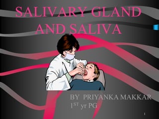
- 1. SALIVARY GLAND AND SALIVA BY PRIYANKA MAKKAR 1ST yr PG 1
- 2. It is critical to the preservation and maintenance of oral health, yet it receives little attention until quantity or quality is diminished. Saliva also has become useful as a noninvasive systemic sampling measure for medical diagnosis and research. 2
- 3. Saliva is a clear, slightly acidic mucoserous exocrine secretion. Whole saliva is a complex mix of fluids from major and minor salivary glands and from gingival crevicular fluid, which contains oral bacteria and food debris. 3
- 4. Its status in the oral cavity is at par with that of blood i.e. to remove waste,supply nutrients and protect the cells 4
- 5. Classification of salivary gland Salivary Gland is any cell or organ discharging a secretion into the oral cavity. • Major and minor Salivary Glands Major (Paired) Collection of secretory cells aggregated into large bilaterally paired extra oral glands with extended duct system through which the gland secretions reach the mouth. - Parotid - Submandibular - Sublingual 5
- 6. Minor salivary glands Collection of secretory cells scattered throughout the mucosa & submucosa of the oral cavity with short ducts opening directly onto mucosal surface. - Serous glands of Von Ebner. - Anterior lingual glands. - Lingual, buccal, labial, palatal glands, glossopalatine and retromolar glands 6
- 7. 2. Based on type of secretory cells 1. Serous : Parotid 2. Mixed (seromucous): Submandibular 3. Mucous: Minor salivary glands. 7
- 8. 8
- 9. Salivary gland anatomy Parotid gland: Largest of all the salivary glands Purely serous gland that produce thin,watery amylase rich saliva Superficial portion lies in front of external ear & deeper portion lies behind the ramus of mandible Stensen's Duct (Parotid Papilla) Opens out adjacent to maxillary second molar 9
- 10. Superficial portion of gland is located subcutaneously, in front of the external ear & deeper portion lies behind ramus of mandible. Associated with facial nerve 10
- 11. Submandibular gland Second largest salivary gland Mixed gland Located in the posterior part of floor of mouth,adjacent to medial aspect of mandible & wrapping around the posterior border of mylohyoid muscle Wharton's Duct Opens beneath the tongue at sub-lingual caruncle lateral to the lingual frenum 11
- 12. 12
- 13. Sublingual gland: Smallest salivary gland Mixed gland but mucous secretory cells predominate Located in anterior part of floor of mouth between the mucosa and mylohyoid muscle Opens through series of small ducts (ducts of rivinus) opening along the sub-lingual fold & often through a larger duct(bartholin’s duct) that opens with the wharton’s duct at the sub-lingual caruncle 13
- 14. Minor Salivary gland: No. between 600 and 1000. Exist as aggregates of secretory tissue present in submucosa throughout most of the oral cavity. Not seen in gingiva & anterior part of hard plate. 14
- 15. Rich in mucin, antibacterial proteins and secretory immunoglobulin. Continuous slow secreting glands, thus have a important role in protecting and moistening oral mucosa, especially when major salivary glands are mostly inactive. 15
- 16. Von Ebners’s Lingual serous gland Located in tongue and open into the troughs surrounding circumvallate papillae on the dorsum of tongue and at the foliate papillae on the side of tongue. Secrete digestive enzymes & proteins that are thought to play role in taste process Fluid of their secretion cleanse the trough & prepare the taste receptors for a new stimulus. 16
- 17. DEVELOPMENT OF GLANDS All salivary glands show a similar pattern of development. MESENCHYME ORAL EPITHELIAL BUDS ECTODERMAL ENDODERMAL PAROTID GLAND AND MINOR SALIVORY GLANDS SUBMANDIBULAR AND SUBLINGUAL GLAND 17
- 18. The Primordia of the glands of humans appear during sixth week whereas the primordium of sublingual glands appear after 7 to 8 weeks of fetal life. The minor salivary glands begin their development during the third month. The epithelial bud grows into an extensively branched system of cords of cell that are first solid but gradually develop a lumen and become ducts. 18
- 19. DEVELOPMENT OF SALIVARY GLANDS Bud formation Formation and growth of epithelial chord. Initiation of branching in terminal parts of epithelial chord. Branching of epithelial chord and lobule formation Canalization Cytodifferentiation Stage I Stage II Stage III Stage IV Stage V Stage VI 19
- 20. Epithelium protrudes into underlying ectomesenchyme & as the epithelium invaginates , it forms a small bud connected to the surface by trailing cord of epithelium - Initial bud stage. At the same time , ectomesenchyme cells condense around this bud. The bud undergoes branching to produce a cluster of branches & buds, -known as Pseudoglandular Stage 20
- 21. Since salivary glands are formed from an initially solid core of epithelial cells –for the proper functioning of the gland the duct needs to undergo cavitation -to allow free access between the saliva producing acini and oral cavity.- known as Canilicular Stage. 21
- 22. 22
- 23. Minor salivary glands are the most important because of their protective components. Major glands do produce more saliva than minor glands, but the quality of content and thus the type of protection varies. 23
- 24. The parotid glands normally contribute about 25% of the total volume of unstimulated whole saliva, while the submandibular glands contribute 60%, the sublingual 7–8%, and the minor mucous glands 7–8%. At very high stimulated flow rates, the parotid becomes the dominant gland, contributing about 50% of the whole saliva. 24
- 25. Flow rate (ml/min) WHOLE PAROTID SUBMANDIBULAR RESTING 0.2-0.4 0.04 0.1 STIMULATED 2.0-5.0 1.0-2.0 0.8 pH 6.7-7.4 6.0-7.8 25
- 26. 0.0 0.1 0.2 0.3 0.4 0.5 20-39 yr 40-59 yr > 60 yr ml/min Age Flow Rate of Saliva unstimulated stimulated 26
- 27. The average daily flow of whole saliva varies in health between 1 and 1.5 L. 27
- 28. ANATOMY OF GLANDS Secretory units are composed of serous, mucous and myoepithelial cells arranged into secretory tubules called-acini. Serous cell specialised for the synthesis, storage and secretion produce protein & glycoprotein ( typically N linked ) 28
- 29. Mucous cell produce mucin , glycoprotein ( typically O linked ) & function mainly to lubricate & form barrier against micro- organism. 29
- 30. THE SECRETORY UNIT The basic building block of all salivary glands ACINI - water and ions derived from plasma Saliva formed in acini flows down DUCTS to empty into the oral cavity. 30
- 32. SEROUS CELLS Typically spherical in shape . 8-12 cells . Cells are pyramidal in shape, with its broad base resting on a thin basal lamina and its narrow apex bordering on the lumen of end piece. The spherical nucleus is located in the basal region of the cell. 32
- 33. secrete a watery fluid, essentially devoid of mucous contain zymogen granules containing a precursor of ptyalin enzyme for digesting starches 33
- 34. MUCOUS CELLS Polyhedral & Contain mucinogen granules. (1) they have little or on enzymatic activity and serve for lubrication and protection of the oral tissues (2) the ratio of carbohydrate to protein is greater, and larger amounts of sialic acid and sulphated sugar residues are present. 34
- 35. DUCTS 35
- 36. 36
- 37. Intercalated ducts The small ducts are intercalated ducts; they are thin branching tubes of variable length that connect to the terminal secretory units to the next larger ducts. Primary saliva produced by secretory end piece passes first thorough intercalated ducts. Diameter of these ducts are smaller 37
- 38. The intercalated ducts cells often contain secretory granules in their apical cytoplasm, and two of the antibacterial proteins in saliva, lysozyme and lactoferrin, have been localized to these ducts. 38
- 39. STRIATED DUCTS Constitute largest portion of ductal system located within lobules of Salivary glands . The striated duct cells contain kallikrein, an enzyme found in saliva, and synthesize secretory glycoproteins, which are stored in the apical granules. 39
- 40. EXCRETORY DUCTS Located in connective tissue septa between lobules of gland Larger in diameter then striated duct . 40
- 41. Regulation of saliva secretion Afferent signals from sensory receptors in mouth Trigeminal,facial,glossopharyngeal nerves Salivary nuclei in the medulla oblongata of brain Parasympathetic nerve bundle sympathetic nerve bundle salivary glands 41
- 42. Innervation o Parasympathetic innervation to major salivary glands • Otic ganglion fibers supply Parotid Gland • Submandibular ganglion supplies other major glands o Sympathetic innervation promotes saliva flow • Stimulates muscle contractions at salivary ducts 42
- 43. Saliva secretion is also controlled by the conditioned reflexes(by sight,smell or thought of food). Besides receiving impulses from the afferents,the salivary nuclei also receives impulses from higher centers of brain which leads to release of variety of neurotransmitters resulting in facilatory or inhibitory effects As a result of such control,unstimulated salivation is inhibited during sleep,fear & mental depression Stress may increase or decrease salivary flow 43
- 44. Salivary secretion:two step model Formation of primary saliva: • Initiated by binding of neurotransmitters in the acinar cell membranes • Acinar cell loses K⁺ to the interstitium & Cl⁻ to the lumen • Gain of Cl⁻ creates negative potential in the lumen,driving interstitial Na⁺ into lumen thereby restoring electroneutrality • Water flux follws the movement of salt into the lumen for osmotic reasons,resulting in acinar cell shrinkage • Outcome is the formation of isotonic primary saliva 44
- 45. • At high flow rates,saliva is in contact with the ductal epithelium for shorter time & Na⁺ & Cl⁻ concentration increase & K⁺ concentration decrease • At low flow rates,the electrolyte concentration change in the opposite direction • The HCO₃⁻ concentration increases with increased flow rates,reflecting the increased secretion of HCO₃⁻ by the acinar cells to drive fluid secretion acini ducts Na+ K+ Cl- HCO3 - 45
- 46. saliva Water -99% solids 1% Organic substance Inorganic substance Gases Enzymes Other org. substance 1.amylase 2maltase 3lingual lipase 4lysozyme 5phosphatase 6carbonic anhydrase 7kalikrein 1.Proteins- mucin & albumin 2.Blood group antigen 3.Free amino acids 4.Non protein nitrogenous substances-urea,uric acid,creatinine,xanthine hypoxanthine 1.Sodium 2.Calcium 3.Potassium 4.Biocarbonate 5.Bromide 6.Chlorine 7.Fluoride 8.phosphate 1.Oxygen 2.Carbon dioxide 3.Nitrogen 46
- 49. Calcium and Phosphate Hydrogen bicarbonate Other ions are:- Fluoride Thiocynate Sodium,Potassium,Chloride Lead,Cadmium,Copper 49
- 51. Organic components of saliva Mucins Proline-rich proteins Amylase Lipase Peroxidase Lysozyme Lactoferrin Secretory IgA Histatins Statherin Blood group substances, sugars, steroid hormones, amino acids, ammonia, urea 51
- 52. Multifunctionality Salivary Functions Anti- Bacterial Buffering Digestion Mineral- ization Lubricat- ion &Visco- elasticity Tissue Coating Anti- Fungal Anti- Viral Carbonic anhydrases, Histatins Amylases, Mucins, Lipase Cystatins, Histatins, Proline- rich proteins, Statherins Mucins, Statherins Amylases, Cystatins, Mucins, Proline-rich proteins, Statherins Histatins Cystatins, Mucins Amylases, Cystatins, Histatins, Mucins, Peroxidases 52
- 53. MAJOR FUNCTIONS OF SALIVA 1.PROTECTION: Mechanical washing action: - Flushes away non-adherent bacterial and acellular debris from the mouth. -Clearance of sugars from the mouth limits their availability to acidogenic plaque microorganisms. Lubricant: (mucin) - Protects the lining mucosa by forming a barrier against noxious stimuli, microbial toxins and minor trauma. - Allows the oral surface to move one another with minimal friction during function. 2.BUFFERING: Bicarbonate ,phosphate, some salivary proteins. Metabolism of salivary proteins and peptides produce ammonia and urea which help in increase of pH. Buffers the acid produced by plaque microorganism. 53
- 54. 3.Pellicle formation: Salivary proteins bind to surface of the teeth and oral mucosa, thus forming salivary pellicle which behaves as a protective membrane . Binding site for bacteria – plaque. 4.MAINTAINENCE OF TOOTH INTEGRITY: Saliva is saturated with Ca+ & PO4- : acidic prolein rich proteins & statherin High concentration of these ions ensures that ionic exchange with the tooth surface results in post eruptive enamel maturation, increase in surface hardness, decrease in permeability and increase in resistance to demineralization. Remineralization of initial caries. 54
- 55. 5.ANTIMICROBIAL ACTION: Lysozyme is an enzyme that can hydrolyse the cell wall of some bacteria. Lactoferrin binds free iron & in doing so deprives bacteria of this essential element. Antibodies present in saliva (IgA) -has the capacity to agglutinate microorganisms that are swallowed. -prevent their agglutination to oral tissue. Mucin and specific agglutins: aggregate microorganisms. Histatin and peroxidase 55
- 56. 6.ROLE OF SALIVA IN TISSUE REPAIR: Bleeding time of oral tissues is shorter than other tissues. Experiment have shown that wound healing is faster & wound contraction is also increased in the presence of saliva. 7.DIGESTION: Forms the food bolus-preparation of the ingested food for deglutition. Breaks down starch (Amylase). Lipase Dilutes gastric chyme. 8.TASTE: Saliva is required to dissolve substance to be tasted & carry them to the taste buds. It also contains a protein called gustin that is thought to be necessary for growth & maturation of the taste buds. It is thought to be secreted by circumvallate papillae. 56
- 57. 8.Salivary anticaries activity: Carbohydrate & microbial clearance from the oral cavity. Buffering action - Salivary bicarbonates. Remineralization of incipient carious lesion-Ca+, PO4 +, Fl- Increase enamel resistance to acid decalcification- Fl- Salivary urea and bicarbonate can increase rate of glycolysis, thus leading to faster carbohydrate metabolization, which inturn leads to reduced duration of the enamel exposure to critical pH levels. 57
- 58. Functions in detail according to its composition Calcium and phosphate Help to prevent dissolution of dental enamel Calcium • 1.4 mmol/lt. (1.7 mmol/lt. in stimulated saliva) • 50% in ionic form • sublingual > submandibular > parotid Phosphate • 6 mmol/lt. (4 mmol/lt. in stimulated saliva) • 90% in ionic form pH around 6 - hydroxyapatite is unlikely to dissolve Increase of pH - precipitation of calcium salts => dental calculus 58
- 59. Hydrogen Bicarbonate Buffer Low in unstimulated saliva, increases with flow rate Pushes pH of stimulated saliva up to 8 pH 5.6 critical for dissolution of enamel Defence against acids produced by cariogenic bacteria Derived actively from CO2 by carbonic anhydrase Helps to protect the teeth from demineralization caused by bacterial acids produced during sugar metabolism. 59
- 60. Mucins Products of acinar cells from submandibular,sublingual and some minor salivary glands. Asymmetrical molecule with open, randomly organized structure Glycoproteins - protein core with many oligosaccharide side chains attached by glycosidic bond Hydrophillic Unique rheological properties (e.g., high elasticity, adhesiveness, and low solubility) 60
- 61. Major salivary mucins are: MG1-adsorbs tightly to the tooth surface contributing to the enamel pellicle formation, thereby protecting the tooth surface from chemical & physical attack including acidic challenges MG2-also binds to the tooth surface but is easily displaced, however it promotes clearance of oral bacteria by aggregation 61
- 62. Amylases Produced by acinar cells of major salivary glands Metabolizes starch and other polysaccharides into glucose & maltose Calcium metalloenzyme Parotid; 30% of total protein in parotid saliva “Appears” to have digestive function - inactivated in stomach, provides disaccharides for acid-producing bacteria It is also present in tears, serum, bronchial, and male and female urogenital secretions A role in modulating bacterial adherence 62
- 63. Lingual Lipase Secreted by sublingual gland and parotid gland Involved in first phase of fat digestion Hydrolyzes medium to long chain triglycerides Important in digestion of milk fat in newborn Unlike other mammalian lipases, it is highly hydrophobic and readily enters fat globules 63
- 64. Statherins Produced by acinar cells in salivary glands Acidic peptide containing relatively high levels of proline,tyrosine and phosphoserine Inhibits spontaneous precipitation of calcium phosphate salts from supersaturated saliva & favours remineralization Calcium phosphate salts of dental enamel are soluble under typical conditions of pH and ionic strength 64
- 65. Supersaturation of calcium phosphates maintain enamel integrity Also an effective lubricant for the tooth surface thus protecting it from physical forces 65
- 66. Proline-rich Proteins (PRPs) Like statherin, PRPs are also highly asymmetrical Present in the initially formed enamel pellicle and in “mature” pellicles 2 types: Basic Acidic Both are secretory products of major salivary glands 66
- 67. Lactoferrin Iron-binding protein Prevents iron from being used by microorganism that require it for metabolism Nutritional immunity (iron starvation) Some microorganisms (e.g., E. coli) have adapted to this mechanism by producing enterochelins. • bind iron more effectively than lactoferrin • iron-rich enterochelins are then reabsorbed by bacteria Lactoferrin, with or without iron, can be degraded by some bacterial proteases. 67
- 68. CHEMICAL BENEFITS OF SALIVA STIMULATION • Stimulating the flow of saliva alters its composition. Increases the concentration of protein, sodium, chloride and bicarbonate and decreases the concentration of magnesium and phosphorus. • Perhaps of greatest importance is the increase in the concentration of bicarbonate, which increases progressively with the duration of stimulation. The increased concentration of bicarbonate diffuses into the plaque, neutralizes plaque acids, increases the pH of the plaque and favors the remineralization of damaged enamel and dentin 68
- 69. Studies have shown that chewing sugar-free gum after meals results in a significant decrease in the incidence of dental caries and that the benefit is due to stimulating salivary flow rather than any chewing gum ingredient. Stimulating salivary flow after meals reduces the incidence of dental caries so,its practical measures should be considered in caries prevention programs 69
- 70. Saliva:A Diagnostic Fluid ADVANTAGES: • non-invasive • limited training • no special equipment • potentially valuable for children and older adults • cost-effective • eliminates the risk of infection • screening of large populations 70
- 71. Testing of saliva production Unstimulated production – collection of saliva into container during 15 min Stimulated production – collection of saliva during 5 min of chewing 1g paraffin Unstimulated whole saliva flow rates of <0.1 ml/min. and stimulated whole saliva flow rate’s of <1.0 ml/min. are considered abnormally low& indicative of marked salivary hypofunction. 71
- 72. Recent work in Sjogren syndrome is beginning to identify changes in salivary cytokine & other protein levels that may have diagnostic significance . Saliva may play a greater diagnostic role in monitoring for the presence and concentrations of drugs of abuse and therapeutic agents. 72
- 74. 74
- 75. SUBMANDIBULR /SUBLINGUAL Alternatively , for et al have described a simpler method for the collection of submandibular-sublingual. After blocking the parotid saliva secretion by placing a gauze pad at the orifice of the parotid ducts. Saliva can be collected from the floor of the mouth with a micropipette. 75
- 76. MINOR GLANDS Minor gland secretions can be collected by micropipette,absorbent filter paper or strips from the inner surface of lips,palate,or buccal mucosa and quantitated by weight differences or using a Peritron device. 76
- 77. Pathology related to salivary glands 77
- 78. Hypofunction of salivary glands Clinical evaluation Patient’s complaints: • Oral dryness and soreness • Burning sensation of oral mucosa and tongue • Difficulties in speech • Difficulty in chewing dry food • Taste impairment and disturbances • Difficulties in wearing removable dentures • Dry lips • Acid reflux,nausea,heart burn • Sensation of thirst • The oral symptoms are often associated with other symptoms such as dry skin,dry nose,dry eyes,dry vaginal mucosa,dry throat,dry cough and constipation 78
- 79. Signs: • Mucosal dryness:dry glazed and red oral mucosa • Lobulation and fissuring of the dorsal part of tongue • Atrophy of filiform papillae • Dry lips,angular cheilitis • Increased caries experience • Oral candidiasis 79
- 80. Xerostomia It is a clinical manifestation of salivary gland dysfunction and it does not represent a disease entity .Dry mouth varies from minimal viscous appearance of saliva to complete absence of any salivary flow. More prevalent in women. Can cause significant morbidity and a reduction in a patient’s perception of quality of life. When unstimulated salivary flow is less than 0.12 to 0.16 ml/minute, a diagnosis of hypofunction is established. 80
- 81. Etiology Aging Foods:alcohol, coffee, coco cola, smoke Drugs: • Anti-depressants • Anti-histamine Cimitidine • Anti-cholinergic • Anti-HTN (sympathomimetic drugs) • Anti-inflammatory Systemic factors • Emotions: nervousness , excitation, depression, stress.. • Encephalitis, brain tumors, stroke, Parkinson’s disease • Dehydration: diarrhea, vomiting, polyuria of diabetes … • Anemia, nutrition deficiency. 81
- 82. Etiology Radiotherapy • Acini atrophy fibrosis or replaced by fatty tissue • Serous acini: more sensitive to R/T • Saliva: thickened, altered electrolytes, pH↓, secretion of immunoglobulins↓ • >1000rad (2-3wk): felt oral dryness • >4000rad: irreversible change Sjogren’s syndrome Other salivary gland diseases 82
- 83. Antidepressants As used to treat depression but blocked histaminic,cholinergic and alpha-1- adrenergic receptors sites,causes the unwanted side effects called dry mouth. Eg:-citalopram (celexa) escitalopram (laxapro) 83
- 84. Antipsychotics Relative anticholinergic potential of selected antipsychotics: Eg:- Promazine,triflupromazine,clozapine. Chlorpromazine,pimozide. 84
- 85. Antihistaminic drugs Relative potencies for anticholinergic effects of these drugs are: Eg:-Diphenhydramine,promethazine Chlorphenarimine,hydroxyzin 85
- 86. Analgesics Immediate release of oxycodone causes more oral dryness than sustained release. 86
- 88. Trauma or surgery Trauma to the mouth or throat Nerve damage to the head and neck 88
- 89. Need of sialogogues Required in case of reduced salivary secretion,commonly that condition is known as dry mouth or xerostomia. Seen in all age groups but common in elders,30%of people beyond 60 yrs experience this disorder. 89
- 90. Symptoms & Signs Symptoms: • Oral dryness (most common) • Halitosis • Burning sensation • Loss of sense of taste or bizarre taste • Difficulty in swallowing • Tongue tends to stick to the palate • Decreased retention of denture 90
- 91. Signs: • Saliva pool disappear • Mucosa: dry or glossy • Duct orifices: viscous and opaque saliva • Tongue: glossitis fissured red with papilla atrophy • Angular cheilitis • Rampant caries: cervical or cusp tip • Periodontitis • Candidiasis 91
- 92. Clinical Appearance: • Oral mucosa appears dry, pale, or atrophic. • Tongue may be devoid of papillae with fissured and inflamed appearance. • New and recurrent dental caries. • Difficulty with chewing, swallowing, and tasting may occur. • Fungal infections are common. 92
- 93. Pale Fisured Tongue Due To Severe Dry Mouth 93
- 95. Severe Dry Mouth (Strawberry Tongue) 95
- 96. One of the major problems associated with xerostomic patients is the poor tolerance and retention of removable dental prostheses because of thin dry atrophic mucosa and lack of a saliva film. Combined with oral discomfort and loss of taste acuity it causes increase caries incidence, burning sensation and problem during speech Problem 96
- 97. The use of salivary substitutes can improve lubrication, provide irrigation for dry mucosa, provide significant relief from symptoms, and also improve the retention of removable prostheses. They are three types-glycerine and lemon based, carboxymethylcellulose, based on mucin even milk can be used as saliva substitute. Management should be directed toward relief of symptoms. These substitutes can be delivered over prolonged periods by using saliva reservoirs in the prosthesis itself as it is difficult to carry salivary substitutes all the time. Intervention 97
- 99. Retention Of Denture Salivary wetting mechanics are necessary to create adhesion,cohesion and surface tension that helps in retention of denture ADHESION:is the bond created by saliva between oral mucosal epithelium and denture base.Thin film of saliva hold the denture to mucosa. Amount of adhesion present is directly proportional to denture base area. 99
- 100. COHESION is the bond between salivary components that helps in retention of prosthesis.Watery serous saliva forms a thinner film and is more cohesive than thick serous saliva. 100
- 101. Viscosity of saliva It also determines denture retention. Serous saliva has a moderate flow favorably contributes to retention. Thick ropy saliva often forces the denture out of their position thus providing a detrimental factor for retention. 101
- 102. Saliva allows for the vaccum pressure formation that contributes to the fit of prosthesis. Adequate amount,flow and consistency of saliva ensures the optimal denture functioning by providing the fit,support and stabilization of the denture 102
- 103. In CD patients Saliva is required to make a intimate surface between denture and soft tissues Keeps the denture in stable position Helps in providing the retention Tissue contacting denture will become irritated in absence of adequate amount of saliva. Denture sores can occur in case of hyposalivation 103
- 104. Dry mouth is managed in following ways By treating the drug induced xerostomia. Salivary substitutes. Sialagogue herbs. 104
- 105. How to treat drug induced xerostomia Following ways can be used to modify patient intake of xerogenic drugs: 1.elimination and reduction of select drugs. 2.modification of drug intake schedule. 3.substitution of one drug with another with less noxious effects. 105
- 106. For antidepressants: Newer multiple receptors antidepressants are less xerogenic than tricyclics: Eg.nefazodone,mirtazapine. Anticholinergics/antispasmodics: Tolterodine tartarate is less xerogenic than oxybutynin chloride. 106
- 107. When drug therapies are not enough…. When it is impossible to determine or eliminate the cause of oral dryness or drugs will do minimally help,then attempts are made to stimulate the patient saliva by Use of SIALAGOGUES or to moisturize the oral mucosa by Use of SALIVARY SUBSTITUTES/ ORAL MOISTURIZERS. 107
- 108. Salivary stimulants…. A) mechanical B) electrical C) pharmacological D) chemical 108
- 109. a) Mechanical stimulants… Sugarless Gums like Biotene,Eclipse,Extra,Orbit has Xylitol,Sorbitol,mannitol Acesulfame,Aspartame etc… 109
- 110. b) Electrical stimulants.. Salitron:- Proponents of electro stimulation as a treatment option postulate that stimulating the tongue and the roof of the mouth simultaneously will result in impulses to all residual salivary tissues, major and minor, in the oral and pharyngeal regions, thus causing salivation. 110
- 111. Pharmacological stimulants… Cholinergic drugs like PILOCARPINE HCL(salagen) and CEVIMELINE HCL (evoxac) 111
- 112. Pilocarpine is also used to treat xerostomia which can occur, for example, as a side effect of radiationtherapy for head and neck cancers, and in Sjogren's syndrome. Pilocarpine stimulates the secretion of large amounts of saliva and sweat. Dose : 5 mg four times a day 112
- 113. Pilocarpine has been known to cause excessive sweating, excessive salivation, bronchospasm, increased bronchial mucus secretion, bradycardia, vasodilati on, and diarrhea. 113
- 114. Oral moisturizer… •Like:- WATER, Salivart ,Oralube, Xerolube, Plax 114
- 115. To fabricate maxillary prosthesis using resilient liner in the floor of reservoir for management of hyposalivation problem in xerostomic patients PURPOSE OF STUDY 115
- 116. Complete all the steps for complete denture fabrication in a conventional manner up to the trial placement appointment At the trial placement appointment, add modeling wax to the palatal surface of the denture base and evaluate speech, adjusting palatal contours as necessary. If needed make a palatogram . Technique 116
- 117. 117
- 118. After the addition of wax, make an index of the palatal surface with Type III dental stone . Note that this index serves as a guide when fabricating the floor of the reservoir. Remove the modeling wax and process the dentures. 118
- 119. Adapt a sheet of modeling wax over the surface of the stone index. Flask and process it in clear heat-polymerized acrylic resin to form the floor of the reservoir . 119
- 120. To make it functional, make a hole with roughened borders in the anterior part of the floor and line it with a resilient liner . This completes the fabrication of the floor of the reservoir. 120
- 121. Attach the floor to the palatal surface of the reservoir with autopolymerizing acrylic resin . 121
- 122. Drill a 1 mm diameter hole in the most anterior part of the floor. This will be the lowest point of the reservoir floor. Demonstrate to the patient how to inject saliva substitute through the hole by using a 5 mL disposable syringe and needle. Select a needle with a diameter slightly smaller than that of the hole. Have the patient practice this procedure until they are able to inject the saliva substitute easily. 122
- 123. artificial salivas normally contain a mixture of buffering agents, cellulose derivatives (to increase stickiness and moistening ability) and flavoring agents (such as sorbitol). 123
- 124. The technique given for the construction of reservoir using resilient liner the tongue presses against the liner during swallowing ,resulting in flow of saliva substitute through a hole in the anterior part of floor of the reservoir. Discussion 124
- 125. Advantages of this functional saliva reservoir design include the swallowing cycle controlling the flow of saliva, less outlet clogging, ease of fabrication, and low cost. The reservoir does not contain any metal components, which could increase prosthesis weight . Advantages 125
- 126. Limitations It cannot be used for patients with shallow palatal forms, there is a loss of resiliency of liner over the period of time Increased weight of the denture is also an issue Disadvantages 126
- 127. Remove all acrylic resin undercuts from the internal surface of the denture, reestablish the borders and the posterior palatal seal with impression compound, and place an impression adhesive on the appropriate surfaces. Make a final impression with a material of choice. Zinc oxide-eugenol impression pastes should not be used in patients with xerostomia because of its irritating effect on the dry, vulnerable oral mucosa Construction of an artificial saliva reservoir in an existing denture V. ink, M. C. HuismanConstruction of an artificial saliva reservoir in an existing maxillary denture, j Pros dent.1986;5:56;1:70-75 127
- 128. Thicken the palatal surface of the denture with soft beeswax by approximately 9 mm. Mold the wax so that oral function, especially speech, is not disturbed. Normal speech should be acceptable, although rapid speech will be difficult . 128
- 129. Cover the beeswax with a thin film of modeling wax to prevent it from sticking to the mold. Pour a cast in plaster and key it Lightly lubricate the superior surface of the cast with waterglass and cover it with a layer of plaster to form the nonfitting surface of the mold . Separate the nonfitting surface of the mold and remove the beeswax. 129
- 130. Fill the space created by the beeswax with Optosil and replace the nonfitting surface of the mold. Remove the excess Optosil . 130
- 131. Shorten the borders of the denture and remove the palatal part . With sticky wax secure the teeth and the remaining base material to the nonfitting surface of the mold . 131
- 132. Construct a chrome-cobalt palatal plate on a duplicate cast. The metal base should cover the palate and terminate 5 mm anterior to the posterior extension.The metal base should be 0.45 mm thick at the center and 1 mm thick where it joins the acrylic resin base Leakage at the junction does not occur because of the border seal and the rheological properties of the saliva substitute. Place two filling holes (1.5 mm in diameter) in the metal base, one anterior and one posterior at the midline. Adapt a shellac baseplate to the upper half of the flask that holds the teeth and glue the metal palate to the flasked cast. 132
- 133. Cover both halves of the flask with polythene foil and fill the remaining space with Optosil . The amount of Optosil is less than that used because the shellac baseplate allows for the acrylic resin covering of the oral side of the reservoir. Remove excess Optosil so that 2 mm of the outline of the chrome-cobalt baseplate is free. Glue the Optosil to the metal base and glue a small round piece of tinfoil to the Optosil and The tinfoil creates an area for the valve. Pack acrylic resin into the flask and cure in the usual manner. 133
- 134. Unsnap the metal base and remove the filler and the tinfoil from the denture Place a hole in the middle of the acrylic resin that covered the tinfoil. Cover the opening with a latex membrane . The membrane is secured to the acrylic resin with cyanoacrylate glue. Punch a hole in the membrane. Snap the metal base to position . The reservoir can be filled with a syringe. The patient can moisten the oral cavity by sucking some artificial saliva out of the reservoir. 134
- 135. 135
- 136. 136
- 137. Burning Mouth Syndrome Painful,frustating condition often described as a scalding sensation in tongue,lips,palate or throughout the mouth Can affect anyone but occurs most commonly in middle-aged or older women occurs with a range of medical and dental conditions, from nutritional deficiencies and menopause to dry mouth and allergies the exact cause of burning mouth syndrome cannot always be identified with certainty. 137
- 138. SIGNS AND SYMPTOMS Moderate to severe burning in the mouth is the main symptom of BMS and can persist for months or years the burning sensation begins in late morning, builds to a peak by evening, and often subsides at night Some feel constant pain; for others, pain comes and goes Anxiety and depression are common in people with burning mouth syndrome and may result from their chronic pain. 138
- 139. Other symptoms of BMS include: tingling or numbness on the tip of the tongue or in the mouth bitter or metallic changes in taste dry or sore mouth 139
- 140. Causes: damage to nerves that control pain and taste hormonal changes dry mouth, which can be caused by many medicines and disorders such as Sjögren’s syndrome or diabetes nutritional deficiencies oral candidiasis, a fungal infection in the mouth acid reflux poorly-fitting dentures or allergies to denture materials anxiety and depression. In some people, burning mouth syndrome may have more than one cause. But for many, the exact cause of their symptoms cannot be found 140
- 141. Diagnosis: A review of medical history, a thorough oral examination, and a general medical examination may help identify the source of burning mouth. Tests may include: • blood work to look for infection, nutritional deficiencies, and disorders associated with BMS such as diabetes or thyroid problems • oral swab to check for oral candidiasis • allergy testing for denture materials, certain foods, or other substances that may be causing symptoms. 141
- 142. Treatment: Treatment should be tailored to individual needs. Depending on the cause of BMS symptoms, possible treatments may include: adjusting or replacing irritating dentures treating existing disorders such as diabetes, Sjögren’s syndrome, or a thyroid problem to improve burning mouth symptoms recommending supplements for nutritional deficiencies 142
- 143. switching medicine, where possible, if a drug is causing burning mouth • prescribing medications to — relieve dry mouth — treat oral candidiasis — help control pain from nerve damage — relieve anxiety and depression. When no underlying cause can be found, treatment is aimed at the symptoms to try to reduce the pain associated with burning mouth syndrome. 143
- 144. Helpful tips: Sip water frequently. Suck on ice chips. Avoid irritating substances like hot, spicy foods; mouthwashes that contain alcohol; and products high in acid, like citrus fruits and juices. Chew sugarless gum. Brush teeth/dentures with baking soda and water. Avoid alcohol and tobacco products. 144
- 145. Clinical implications: Difficulties in chewing tasting swallowing speaking Increased chances of developing dental decay & other infections in mouth Mouth sores Difficult for operator to work when saliva pools in mouth (in case of sialorrhea) uncoordinated swallowing poorly synchronized lip closure abnormal increase in tone of the muscles that open the mouth 145
- 146. 2. HYPERSALIVATION The excess secretion of saliva is known as hypersalivation . Hypersalivation in pathological condition is known as ptyalism , sialorrhea , sialism or sialosis. Hypersalivation occurs in the following conditions :- 1) Decay of tooth or neoplasm of mouth or tongue due to continuous irritation of nerve endings in the mouth 2) Disease of esophagus , stomach & intestine 3) Neurological disorder such as cerebral palsy & mental retardation 4) Cerebral stroke 5) Parkinsonism 6) Some psychological & psychiatric conditions 7) Nausea & vomiting 146
- 147. DROOLING Uncontrolled flow of saliva outside the mouth is called drooling . It is often called ptyalism. Drooling occurs because of excess production of saliva in association with inability to retain saliva within the mouth. Drooling in small children is a normal part of development. Teeth are coming in, they put everything in their little mouths, and they haven’t developed the habit of keeping the lips together. While child is teething ,their gums will produce excessive saliva 147
- 148. The saliva which is produce during drooling is designed to moisten and lubricate babys tender gums. Drooling serves to help make teething process more bearable for child. 148
- 149. Antisialogogue… Antisialagogues are substances that decrease the production of saliva and their effect is opposite to that of sialagogues. Their origin may be both natural and synthetic. Classic antisialagogues include: atropine, opium, alkalies, belladonna, tobacco in excess, 149
- 150. Atropine dilates the pupils, increases heart rate, and reduces salivation and other secretions. Atropine is an effective salivary-secretion suppressant and, therefore, useful in dentistry. Its value depends upon a careful selection of proper dosage in order to achieve maximum antisialogogic effects with a minimum of side actions. This dose would range between 0.4 mg. and 0.8 mg. of atropine sulfate (total dose) for an average adult. Brand names : atrophen,atrisolon,diafort,lomophen contain atropine sulphate. 150
- 151. Saliva & Age: With age, a generalized loss of salivary gland parenchymal tissue occurs. The lost salivary cells often are replaced by adipose tissue. Although decreased production of saliva often is produced in older persons,whether this is related directly to the decrease in parenchymal tissue is not clear. Some studies of healthy older individuals,in which the use of medication were carefully controlled,revealed little or no loss of salivary function. Other studies suggest that although resting salivary secretion is in the normal range,the volume of saliva produced during stimulated secretion is less than normal. 151
- 152. DIAGNOSTIC IMAGING FOR SALIVARY GLAND The methods employed are: - Plain Film Radiography - IntraOral Radiography - ExtraOral Radiography - Conventional Sialography - Computed Tomography (CT) - Magnetic Resonance Imaging - Scintigraphy - UltraSonography 152
- 153. 153