anatomy and physiology of the eye.pptx
•Download as PPTX, PDF•
0 likes•37 views

Anatomy part 2

Recommended
More Related Content
Similar to anatomy and physiology of the eye.pptx
Similar to anatomy and physiology of the eye.pptx (20)
More from mahamed adam
More from mahamed adam (20)
Recently uploaded
Recently uploaded (20)
anatomy and physiology of the eye.pptx
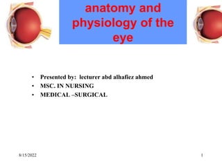
- 1. anatomy and physiology of the eye • Presented by: lecturer abd alhafiez ahmed • MSC. IN NURSING • MEDICAL –SURGICAL 1 8/15/2022
- 2. • Eye ball العين مقلة : is a set in a bony structure ( eye orbit عين )مدار surrounded by sinuses , move by muscle , innervated by cranial nerve 111, 1v, v1, interiorly protected by eye lids . 2 anatomy and physiology of the eye 8/15/2022
- 3. 3 Special Senses • Sensory receptors are within large, complex sensory organs in the head • Smell in olfactory organs • Taste in taste buds • Hearing and equilibrium in ears • Sight in eyes 8/15/2022
- 4. process of vision • The human eye is the organ which gives us the sense of sight, allowing us to learn more about the surrounding world than we do with any of the other four senses. • The eye allows us to see and interpret the shapes, colors, and dimensions of objects in the world by processing the light they reflect or emit. 4 8/15/2022
- 5. 5 Sense of Sight • Visual accessory organs • Eyelids جفون • Lacrimal apparatus الدمعي الجهاز • Extrinsic eye muscles 8/15/2022
- 6. Eyelids جفون •Orbicularis oculi – closes eyelid • Levator palpebrae superioris – opens eyelid • Tarsal glands – secrete oil onto eyelashes الرموش • Conjunctiva – mucous membrane; lines eyelid and covers portion of eyeball Eyelash Cornea Conjunctiva Eyelid Tendon of levator palpebrae superioris Superior rectus Orbicularis oculi Inferior rectus Tarsal glands Copyright © The McGraw-Hill Companies, Inc. Permission required for reproduction or display. 6 8/15/2022
- 7. Lacrimal Apparatus • Lacrimal gland • Lateral to eye • Secretes tears • Canaliculi • Collect tears • Lacrimal sac • Collects from canaliculi • Nasolacrimal duct • Collects from lacrimal sac • Empties tears into nasal cavity Lacrimal gland Lacrimal sac Superior and inferior canaliculi Nasolacrimal duct Copyright © The McGraw-Hill Companies, Inc. Permission required for reproduction or display. 7 8/15/2022
- 8. Extrinsic Eye Muscles Inferior rectus Inferior oblique Medial rectus Superior rectus Superior oblique Lateral rectus (cut) Copyright © The McGraw-Hill Companies, Inc. Permission required for reproduction or display. • Superior rectus • Rotates eye up and medially • Inferior rectus • Rotates eye down and medially • Medial rectus • Rotates eye medially 8 8/15/2022
- 9. Extrinsic Eye Muscles Inferior rectus Inferior oblique Medial rectus Superior rectus Superior oblique Lateral rectus (cut) Copyright © The McGraw-Hill Companies, Inc. Permission required for reproduction or display. • Lateral rectus • Rotates eye laterally • Superior oblique • Rotates eye down and laterally • Inferior oblique • Rotates eye up and laterally 9 8/15/2022
- 10. 10 Structure of the Eye • Wall has three (3) layers: • Outer fibrous tunic • Middle vascular tunic • Inner nervous tunic الداخلية العصبية السترة Ciliary body Retina Choroid coat Sclera Fovea centralis Optic nerve Lens Iris Pupil Cornea Lateral rectus Medial rectus Optic disc Posterior cavity Vitreous humor Posterior chamber Anterior chamber Aqueous humor Suspensory ligaments Copyright © The McGraw-Hill Companies, Inc. Permission required for reproduction or display. Anterior cavity 8/15/2022
- 11. 11 Outer Tunic • Corneaالقرنية • Anterior portion • Transparent • Light transmission • Light refraction • Sclera الصلبة •: is the white of the eye , maintain the shape of the eye and protect it from trauma . • Posterior portion • Opaque • Protection Ciliary body Retina Choroid coat Sclera Fovea centralis Optic nerve Lens Iris Pupil Cornea Lateral rectus Medial rectus Optic disc Posterior cavity Vitreous humor Posterior chamber Anterior chamber Aqueous humor Suspensory ligaments Copyright © The McGraw-Hill Companies, Inc. Permission required for reproduction or display. Anterior cavity 8/15/2022
- 12. 12 Middle Tunic • Iris آيريس •is the color part of the eye • vascular • Anterior portion • Pigmented • Controls light intensity • Ciliary body • Anterior portion • Pigmented • Holds lens • Moves lens for focusing • Choroid coat • Provides blood supply • Pigments absorb extra light Ciliary body Retina Choroid coat Sclera Fovea centralis Optic nerve Lens Iris Pupil Cornea Lateral rectus Medial rectus Optic disc Posterior cavity Vitreous humor Posterior chamber Anterior chamber Aqueous humor Suspensory ligaments Copyright © The McGraw-Hill Companies, Inc. Permission required for reproduction or display. Anterior cavity 8/15/2022
- 13. 13 Anterior Portion of Eye •Is lies behind the cornea • Filled with aqueous humor Conjunctiva Iris Lens Ciliary process Ciliary muscles Sclera Cornea Anterior chamber Vitreous humor Suspensory ligaments Posterior chamber Ciliary body Copyright © The McGraw-Hill Companies, Inc. Permission required for reproduction or display. 8/15/2022
- 14. 14 Lens • Transparent • Biconvex • Lies behind iris • Largely composed of lens fibers • Elastic • Held in place by suspensory ligaments of ciliary body Conjunctiva Iris Lens Ciliary process Ciliary muscles Sclera Cornea Anterior chamber Vitreous humor Suspensory ligaments Posterior chamber Copyright © The McGraw-Hill Companies, Inc. Permission required for reproduction or display. Ciliary body 8/15/2022
- 15. 15 Ciliary Body • Forms internal ring around the front of the eye • Ciliary processes – radiating folds • Ciliary muscles – contract and relax to move lens Retina Choroid coat Sclera Lens Ciliary processes of ciliary body Suspensory ligaments Copyright © The McGraw-Hill Companies, Inc. Permission required for reproduction or display. 8/15/2022
- 16. 16 Accommodation اإلسكان • Changing of lens shape to view objects (a) Lens thick Lens thin (b) Ciliary muscle fibers contracted Suspensory ligaments relaxed Ciliary muscle fibers relaxed Suspensory ligaments taut Copyright © The McGraw-Hill Companies, Inc. Permission required for reproduction or display. 8/15/2022
- 17. 17 Iris Sympathetic motor nerve fiber In dim light Copyright © The McGraw-Hill Companies, Inc. Permission required for reproduction or display. Radially arranged Smooth muscle fibers of the iris Circularly arranged smooth muscle fibers of the iris Pupil In normal light In bright light Parasympathetic ganglion Parasympathetic motor nerve fiber • Composed of connective tissue and smooth muscle • Pupil is hole in iris • Dim light stimulates radial muscles and pupil dilates • Bright light stimulates circular muscles and pupil constricts 8/15/2022
- 18. 18 Aqueous Humor • Fluid in anterior cavity of eye • Secreted by epithelium on inner surface of the ciliary body • Provides nutrients • Maintains shape of anterior portion of eye • Leaves cavity through Canal of Schlemm Sclera Iris Lens Aqueous humor Cornea Vitreous humor Ciliary process Ciliary muscles Copyright © The McGraw-Hill Companies, Inc. Permission required for reproduction or display. Posterior chamber Ciliary body Scleral venous sinus (canal of Schlemm) Anterior chamber 8/15/2022
- 19. 19 Inner Tunic • Retina العين ّةيشبك • Contains visual receptors • Continuous with optic nerve • Ends just behind margin of the ciliary body • Composed of several layers • Macula lutea – yellowish spot in retina • Fovea centralis – center of macula lutea; produces sharpest vision • Optic disc البصري القرص – blind spot المخفية ;النقطة contains no visual receptors • Vitreous humor الزجاجي المرح – thick gel that holds retina flat against choroid coat 8/15/2022
- 20. 20 Posterior Cavity • Contains vitreous humor – thick gel that holds retina flat against choroid coat Ciliary body Retina Choroid coat Sclera Fovea centralis Optic nerve Lens Iris Pupil Cornea Lateral rectus Medial rectus Optic disc Posterior cavity Vitreous humor Posterior chamber Anterior chamber Aqueous humor Suspensory ligaments Copyright © The McGraw-Hill Companies, Inc. Permission required for reproduction or display. Anterior cavity 8/15/2022
- 21. 21 Light Refraction • Refraction • Bending of light • Occurs when light waves pass at an oblique angle into mediums of different densities Light wave Perpendicular line Air Glass Refracted light wave Copyright © The McGraw-Hill Companies, Inc. Permission required for reproduction or display. 8/15/2022
- 22. 22 Types of Lenses • Convex بّدمح lenses cause light waves to converge • Concave رّعمق lenses cause light waves to diverge Air Glass (a) (b) Diverging light waves Convex surface Light wave Converging light waves Concave surface Copyright © The McGraw-Hill Companies, Inc. Permission required for reproduction or display. 8/15/2022
- 23. 23 Focusing On Retina • As light enters eye, it is refracted by: • Convex surface of cornea • Convex surface of lens • Image focused on retina is upside down and reversed from left to right Light waves Object Cornea Image Retina Copyright © The McGraw-Hill Companies, Inc. Permission required for reproduction or display. 8/15/2022
- 24. 24 Stereoscopic Vision • Provides perception of distance and depth • Results from formation of two slightly different retinal images Light waves Right eye Left eye Copyright © The McGraw-Hill Companies, Inc. Permission required for reproduction or display. 8/15/2022
- 25. 25 Lifespan Changes • Age-related visual problems include: • Dry eyes • Floaters (crystals in vitreous humor) • Loss of elasticity of lens • Glaucoma • Cataracts • Macular degeneration 8/15/2022