
Empfohlen
Weitere ähnliche Inhalte
Was ist angesagt?
Was ist angesagt? (20)
Ähnlich wie Amino acids
Ähnlich wie Amino acids (20)
Kürzlich hochgeladen
Kürzlich hochgeladen (20)
Amino acids
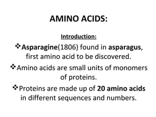
- 1. AMINO ACIDS: Introduction: Asparagine(1806) found in asparagus, first amino acid to be discovered. Amino acids are small units of monomers of proteins. Proteins are made up of 20 amino acids in different sequences and numbers.
- 2. Structure: All the standard amino acids except (glycine) the α carbon is asymmetric. α carbon is bonded to four different substituent groups: carboxyl group amino group R group hydrogen atom.
- 3. R (in red) represents the side chain( different in each amino acid). GLYCINE the α-carbon atom has four different substituent groups.
- 4. Stereoisomerism: Proteins Contain L-Amino Acids. L-Amino acids are those with the αamino group on the left. The naming of configurations of both simple sugars and amino acids is based on the absolute configuration of glyceraldehyde.
- 6. CLASSIFICATION: Classified based on the properties of their R groups. Tendency to interact with water at biological pH (near pH 7.0).
- 7. ;those whose R groups are: 1)nonpolar and aliphatic 2)aromatic (generally nonpolar) 3) polar but uncharged 4) negatively charged 5) positively charged. Within each class, there are gradations of polarity, size, and shape of the R groups.
- 8. 1) Nonpolar Aliphatic R group: The hydrocarbon R groups are nonpolar and hydrophobic. The bulky side chains of alanine, valine, leucine, and isoleucine, promote hydrophobic interactions within protein structures.
- 9. Glycine : Simplest amino acid structure. Side chain( optically inactive) allows much more structural flexibility than the other amino acids. Proline: The secondary amino (imino) group is held in a rigid conformation that reduces the structural flexibility of the protein at that point.
- 11. 2) Aromatic R Groups: Phenylalanine (Phe), tyrosine(Tyr), and tryptophan (Trp), are relatively nonpolar. The OH group of tyrosine can form hydrogen bonds, hence important functional group in the activity of some enzymes. Tyrosine and tryptophan are more polar than phenylalanine because of the tyrosine hydroxyl group and the nitrogen of the tryptophan indole ring. Tryptophan and tyrosine, and phenylalanine(lesser), absorb ultraviolet light.
- 13. 3) Polar, Uncharged R Groups : Are more soluble in water than nonpolar amino acids, because they contain functional groups that form hydrogen bonds with water. Includes: I. Serine(OH) II. Threonine(OH) III. cysteine( S) IV.Methionine(S, sulphur atom) V. Asparagine(NH2,amide group) VI.glutamine. (NH2)
- 15. Asparagine and Glutamine are easily hydrolyzed by acid or base. Cysteine has an R group (a thiol group) that is acidic . It is readily oxidized to form cystine, in which two cysteine molecules are joined by a disulfide bridge which stabilize the protein structure.
- 17. 4) Negatively Charged (Acidic) R Groups: Include: 1) aspartate 2) glutamate Each have a second carboxyl group. These are the parent compounds of asparagine and glutamine.
- 19. 5) Positively Charged (Basic) R Groups: Includes: lysine, has a second amino group at the e position on its aliphatic chain; arginine, which has a positively charged guanidino group; and Histidine, has an imidazole group. standard amino acid having a side chain with a pKa near neutrality.
- 21. GENERAL PROPERTIES OF AMINO ACIDS: 1) Isomerism: Two types: 1) L form 2) D forms. a) stereo-isomerism: all amino acids except glycine exist as D and L- isomers. - In D-amino acids, -NH2 group (right). In animals and plants. - In L-amino acids (left) ,occur in bacteria.
- 22. b) Optical isomerism: Dextro-rotatory and Laevo-rotatory
- 23. 2) Ionization: Amino acids in aqueous solution are ionized and can act as acids or bases. Those having a single amino group and a single carboxyl group crystallize from neutral aqueous solutions as fully ionized species known as zwitterions (German for "hybrid ions"), each having both a positive and a negative charge.
- 24. Zwitterions: are electrically neutral and remain stationary in an electric field. The crystal lattice of amino acids is held together by strong electrostatic forces between positively and negatively charged functional groups of neighboring molecules, resembling the stable ionic crystal lattice of NaCI.
- 26. 2)Amphoteric nature: Amino Acids Can Act as Acids and as Bases. When a crystalline amino acid, such as alanine, is dissolved in water, it exists in solution as the dipolar ion, or zwitterion, which can act either as an acid (proton donor),
- 28. or as a base (proton acceptor):
- 29. Substances having this dual nature are amphoteric and are often called ampholytes.
- 30. 3) Physical properties: Colorless, crystalline substances. soluble in water(tyrosine in soluble in hot water) Melting point is higher than 200degrees Celsius.
- 31. 4) Undergo Characteristic Chemical Reactions: Ninhydrin reaction: When amino acids are heated with excess ninhydrin, all those having a free α-amino group yield a purple product. Intensity of color produced proportional to the amino acid concentration. Comparing the absorbance to that of appropriate standard solutions is an accurate and technically simple method for measuring amino acid concentration.
- 33. PEPTIDES: polymers of amino acids. Two amino acid molecules can be covalently joined through a peptide bond, to yield a dipeptide. linkage is formed by removal of the elements of water from the α-carboxyl group of one amino acid and the αamino group of another.
- 35. When a few amino acids are joined in this fashion, the structure is called an oligopeptide. When many amino acids are joined, the product is called a polypeptide. The peptide bond is the most important covalent bond linking amino acids in peptides and proteins.
- 36. Structure of the pentapeptide serylglycyltyrosinylalanylleucine,
- 38. PROTEINS: Classification according to their functions: 1) Enzymes :The most varied and highly specialized proteins are those with catalytic activity. They catalyze all the chemical reactions in cells. Many thousands of different enzymes have been discovered in different organisms.
- 39. The light produced by fireflies is the result of a light-producing reaction involving luciferin and ATP that is catalyzed by the enzyme luciferase .
- 40. 2) Transport Proteins: Hemoglobin of erythrocytes binds oxygen as the blood passes through the lungs, carries it to the peripheral tissues, and there releases it to participate in the energyyielding oxidation of nutrients. lipoproteins, which carry lipids from the liver to other organs.
- 42. 3) Nutrient and Storage Proteins Ovalbumin, the major protein of egg white. casein, the major protein of milk, is a nutrient protein. The ferritin found in some bacteria and in plant and animal tissues stores iron.
- 43. 4) Contractile or Motile Proteins Actin and myosin function in the contractile system of skeletal muscle and also in many non-muscle cells. Tubulin is the protein from which microtubules are built. Microtubules act in concert with the protein dynein in flagella and cilia to propel cells.
- 44. The movement of cilia in protozoans depends on the action of the protein dynein.
- 45. 5) Structural Proteins Give biological structures strength or protection. collagen(tendons and cartilage), has very high tensile strength. Ligaments contain elastin, capable of stretching in two dimensions. Hair, fingernails, and feathers consist largely of the tough, insoluble protein keratin. The major component of silk fibers and spider webs is fibroin .
- 47. 6) Defense Proteins: The Immunoglobulins/antibodies, made by the lymphocytes of vertebrates, can recognize and neutralize invading bacteria, viruses. Fibrinogen and thrombin are bloodclotting proteins that prevent loss of blood when the vascular system is injured. toxic plant proteins( ricin), also appear to have defensive functions.
- 48. Castor beans contain a highly toxic protein called ricin.
- 49. 6) Regulatory Proteins: regulate cellular or physiological activity E.g. insulin, which regulates sugar metabolism.
- 50. Growth hormones (Somatotropin) Pituitary hormone(prolactin)
- 51. Cancerous tumors are often made up of cells that have defects involving one or more of the proteins that regulate cell division.
- 52. Protein separation: • properties such as charge, size, and solubility. • binding properties. • cells must be broken open and the protein released into a solution called a crude extract. • differential centrifugation( sucrose)
- 53. Methods used: 1. Paper chromatography: The physicochemical factors involved in chromatography are adsorption, partition, ion exchange, and molecular sieving. There are two phases in all types of chromatography (i) stationary phase and the (ii) mobile phase. In paper chromatography, paper is the stationary phase and organic solvent is the mobile phase.
- 54. what Mann 1 or 3 filter paper is used as supporting material for the stationary phase. An organic solvent layer of mixture of butanol: acetic acid: water in the ratio of 4:1:5 is used as solvent or mobile phase.
- 55. Retention value (Rf value): the ratio of distance moved by a compound (protein) to the distance moved by the solvent front. When another solvent system in a perpendicular direction is employed, it is a two dimensional paper chromatography that separates the proteins more distinctively.
- 56. 1) Size-exclusion chromatography/gel filtration. Separates proteins according to size. column contains a cross-linked polymer with pores of selected size. Larger proteins migrate faster than smaller ones, because they are too large to enter the pores in the beads. The smaller proteins enter the pores and are slowed by the path they take through the column.
- 58. 3) Affinity chromatography Separates proteins by their binding specificities. The proteins retained on the column are those that bind specifically to a ligand(a group or molecule that is bound) cross-linked to the beads. After nonspecific proteins are washed through the column, the bound protein of particular interest is eluted by a solution containing free ligand.
- 61. 4) Electrophoresis Separation based on the migration of charged proteins in an electric field. Different samples are loaded in wells at the top of the polyacrylamide gel. The proteins move into the gel when an electric field is applied. The gel minimizes convection currents caused by small temperature gradients, and it minimizes protein movements other than those induced by the electric field.
- 62. (b) Proteins can be visualized by treating the gel with a stain such as Coomassie blue, which binds to the proteins. Each band on the gel represents a different protein: smaller proteins are found nearer the bottom of the gel. The first lane shows the proteins present in the crude cellular extract. Successive lanes show the proteins present after each purification step. The purified protein contains four subunits, as seen in the last lane on the right.
- 66. 1) Primary structure: Is the linear sequence of amino acids held together by peptide bonds. Peptide bond form the backbone and side chains of amino acid residues project outside the peptide backbone. The free –NH2 group is called N-terminal end and –COOH group is C-terminal end.
- 67. 2) Secondary Structure regular, recurring arrangements in space of adjacent amino acid residues in a polypeptide chain. Types : α helix and the β conformation. makes up hair and wool (the fibrous protein α-keratin) has a regular structure that repeats every 0.54 nm.
- 68. α helix Is a helical structure (polypeptide chain with its rigid peptide bonds but with the other single bonds free to rotate) Polypeptide backbone is tightly wound around the long axis of the molecule, and the R groups of the amino acid residues protrude outward from the helical backbone. The repeating unit is a single turn of the helix, which extends about 0.56 nm along the long axis.
- 70. 3) Protein Tertiary Structure Formed as a result of further folding, super folding or twisting of Secondary structure. Occur in native protein. Amino acids located far apart are brought closer. Bonds responsible are: a)hydrophobic interactions: occur between nonpolar side chains of amino acids such as alanine, leucine etc.
- 71. b) Hydrogen bonds( polar side) c) Ionic bonds( between oppositely charged polar side chains of amino acids) d) Vander Waals forces( occur between non polar side chains) e) Disulphide bond(are S-S bonds between –SH group of distance cysteine residues.
- 72. heme group, present in myoglobin (protoporphyrin, to which is bound an iron atom in its ferrous (Fe2+ ) state)
- 73. 4) Protein Quaternary Structure Constitute arrangement of proteins and protein subunits in three-dimensional complexes. multiple noncovalent interactions stabilize the structure The assembly is called oligomer. Each constituent peptide chain is called monomer.
- 74. Structure of Hemoglobin contains four polypeptide chains and four heme groups, in which the iron atoms are in the ferrous (Fe2+) state. Globin(protein portion), consists of two α chains (141 residues each) and two β chains (146 residues). hemoglobin molecule is roughly spherical, with a diameter of about 5.5
- 75. α subunits(white and light blue); the β subunits( pink and purple). heme groups( red), are far apart.