

Empfohlen
Weitere ähnliche Inhalte
Was ist angesagt?
Was ist angesagt? (20)
Ähnlich wie Pelvic c clamp
Ähnlich wie Pelvic c clamp (20)
Kürzlich hochgeladen
Kürzlich hochgeladen (20)
Pelvic c clamp
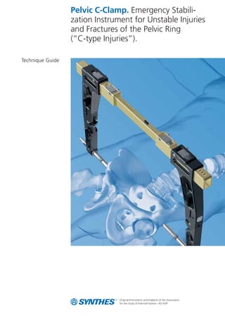
- 1. Pelvic C-Clamp. Emergency Stabili- zation Instrument for Unstable Injuries and Fractures of the Pelvic Ring (“C-type Injuries”). Technique Guide
- 2. Synthes 1 Features and Benefits 2 Indications and Contraindications 4 Implants and Instruments 5 Technique Guide 6 Bibliography 15 Table of Contents Image intensifier control Warning This guide is not sufficient for an immediate application of the Pelvic C-Clamp. Instruction by an experienced surgeon in hand- ling the Pelvic C-Clamp is highly recommended.
- 3. 2 Synthes Pelvic C-Clamp Technique Guide Features and Benefits Description The Pelvic C-Clamp is an emergency stablization instrument for unstable injuries and fractures of the pelvic ring. Unstable pelvic ring fractures can be associated with massive blood loss that can cause terminal shock. The Pelvic C-Clamp allows rapid reduc- tion and stabilization of these unstable pelvic ring fractures. It is therefore an invaluable tool for gaining control of the shock reaction, whilst neither delaying nor hindering subsequent diagnosis and therapy. The Pelvic C-Clamp does not control the rare cases of life threatening arterial bleeding. However, by mechanical stabilisa- tion alone, or enhanced by additional pelvic packing, concomi- tant venous bleeding can be managed, thereby gaining enough time for definitive hemmorage or arterial embolisation control. The Pelvic C-Clamp is comprised of rails and arms with a locking mechanism. This allows a linear movement of the arms. The Pelvic C-Clamp should always be used in combination with the designated cannulated nails. Features and Benefits – Permits quick and efficient compression and stabilization of fractures or luxations, and thus control of haemorrhaging in the unstable posterior pelvic ring. – Gains time for subsequent diagnostic or therapeutic procedures. – The patient can be passed through a CT-gantry without removing the device. – Provides unrestricted access to the abdomen, pelvis or proximal femur. – The Pelvic C-Clamp can be applied quickly outside the operating room, e.g. in the emergency room or on the X-ray table. – MR-safe: made entirely of non-ferrous materials.
- 4. Synthes 3 In cases of sacral fractures or disruptions of the sacroiliac joint, there is often concomitant blood loss from the fracture surface and from the veins of the presacral plexus. The use of the Pelvic C-Clamp in those settings can save lives. Direct transverse compression of the sacroiliac joint controls bleeding from the fracture surfaces and from the presacral plexus. With the Pelvic C-Clamp applied, there is still adequate access for a laporotomy or tamponade. Inner rail Outer rail Upper arm Lower arm Threaded tube Cannulated nail (190 mm or 210 mm) Side buttons Upper buttons Locking mechanism
- 5. Indications and Contraindications Indications – Emergency stabilization of sacroiliac disruptions and fractures of the sacrum in type C pelvic injuries with associated circula- tory instability Contraindications Absolute contraindications are: – Fracture lines within the illium (“transiliac instabilities”; “transiliac fracture dislocation”) as it bears the risk of pin perforation through the fracture line – Hemodynamic stability in A-type Pelvic fractures Relative contraindications are: – B-type injuries (sufficient stability provided by external fixator) – Hemodynamic stability of the patient after B and C-type injuries – Comminuted sacral fractures with the risk of compression of the sacral nerve plexus Note: In life threatening situations hemorrhage control takes priority over the potential risk of nerve root compression. 4 Synthes Pelvic C-Clamp Technique Guide
- 6. Synthes 5 02.306.006 Nail for Pelvic C-Clamp, cannulated, short, length 190 mm 02.306.007 Nail for Pelvic C-Clamp, cannulated, long, length 210 mm 391.930 Wire Cutter, large, with multiplication, length 220 mm 321.200 Ratchet Wrench for Nut, hexagonal, 11mm 398.320 Socket Wrench л 11 mm with Hammer 03.306.009 Guide Handle, for Kirschner Wire л 2.5 mm Implants and Instruments 292.260 Kirschner Wire л 2.5 mm with trocar tip, length 280 mm Sets and Cases 01.306.000 Set Pelvic C-Clamp in Vario Case 68.306.000 Vario Case for Pelvic C-Clamp, no contents 1 03.306.010 Pelvic C-Clamp, complete 1 02.306.006 Nail for Pelvic C-Clamp, cannulated, short, length 190 mm 2 02.306.007 Nail for Pelvic C-Clamp, cannulated, long, length 210 mm 2 321.200 Ratchet Wrench for Nut, hexagonal, 11 mm 2 398.320 Socket Wrench л 11 mm with Hammer 1 391.930 Wire Cutter, large, with multiplication, length 220 mm 1 03.306.009 Guide Handle, for Kirschner Wire л 2.5 mm 1 292.260 Kirschner Wire л 2.5 mm with trocar tip, length 280 mm 10 Optional instrument 359.204 Pliers, flat-nosed Spare Parts 03.306.000 Inner Rail for Pelvic C-Clamp 1 03.306.001 Outer Rail for Pelvic C-Clamp 2 03.306.002 Upper Side Arm for Pelvic C-Clamp 2 03.306.003 Lower Side Arm for Pelvic C-Clamp 2 03.306.008 Threaded Tube for Pelvic C-Clamp 2
- 7. 1 Pre-operative preparation Required instruments Set Pelvic C-Clamp 01.306.000 The Pelvic C-Clamp set consists of the instruments and implants for the emergency treatment of the indicated fracture types. The bottom layer of the Vario Case houses the Pelvic C-Clamp and the optional pliers. The upper layer houses the cannulated nails in two lengths and the remaining instruments. The complete sterile Pelvic C-Clamp set should be kept ready for use in the resuscitation room. Depending on the type of injury, the orientation points in the pelvic region of the injured person may be unclear. Should there be doubts about the anatomic references, use an image intensifier during application of the Pelvic-C-Clamp. Pre-operative preparation – Anteroposterior (AP) plain pelvic radiograph if necessary, oblique views (Inlet and Outlet) or CT. – Patient positioning must allow for intraoperative fluoroscopic controls in AP, Inlet and Outlet Projections. – Have an image intensifier available. Positioning – Place the patient in a supine position. – To facilitate reduction, ensure free draping of the leg on the injured side. Strive for good draping coverage of the genital region. – To prepare the patient, disinfect the proximal femur and the buttocks and cover with sterile sheets. Technique Guide 6 Synthes Pelvic C-Clamp Technique Guide
- 8. Synthes 7 (a) (b) (c) 1 Unlock 2 Lock Preparation of the Pelvic C-Clamp Open the lower side arms by depressing the buttons on the arms to prepare the Pelvic C-Clamp for use. (a) Note: Hold upper and lower arms with both hands and ensure that the lower arm is locked when fully extended. Extend the upper bars by depressing the buttons on the upper rails whilst simultaneously pulling on the side arms. (b) Note: Maximal extension of the Pelvic C-Clamp is advantageous for easy and safe positioning. With a light twist, place the cannulated nails of preferred length into the threaded tubes. (c) The little teeth on the tip of the nail allow a better grip onto the bone. The buttons on the top of the Pelvic C-Clamp can be locked. Ensure the buttons are not locked when applying the Pelvic C-Clamp to the patient, otherwise no or insufficient com- pression can be achieved.
- 9. 2 Identifying nail insertion point Required instrument Guide Handle, for Kirschner Wire л 2.5 mm 03.306.009 Make an incision at the intersection between the extension of the line of the femoral axis over the tip of the greater trochanter, and a vertical line from the anterior superior iliac spine in the dorsal direction (see illustration). If orientation is difficult, use an image intensifier. The surface reference point of the outer side of the ilium changes at the level of the sacroiliac joints. In emergency situations, the resulting “fossa” can be used as a relatively secure point of refer- ence aid. For secure anchoring, the Pelvic C-Clamp must be placed at the level of the sacroiliac joints. Palpation with a blunt instrument, such as the Guide Handle for Kirschner Wire, allows for easy identification of this site, even with severe soft-tissue swelling. Note: If the nails are placed too ventrally to the correct insertion point, there is a risk of perforation of the ilium, which can result in organ injury. Placement of the pins in an excessively dorsal position may result in injury to gluteal nerves and vessels. Inserting the nail too distally endangers the sciatic nerve and the gluteal vessels in the sciatic notch. Malpositioning of the nail in osteoporotic bone, combined with excessive compression, can result in unwanted nail penetration. 8 Synthes Pelvic C-Clamp Technique Guide
- 10. Synthes 9 3 Kirschner Wire placement Required instruments Guide Handle, for Kirschner Wire л 2.5 mm 03.306.009 Kirschner Wire л 2.5 mm with trocar tip, length 280 mm 292.260 Socket Wrench л 11 mm with Hammer 398.320 After having identified the insertion point, a Kirschner Wire can be placed through the Guide Handle (only on the uninjured side). Gently hammer the Kirschner Wire into the bone with the Socket Wrench with Hammer. This Kirschner Wire will ensure an exact placement of the cannulated nail and prevents the nail from slipping. Note: Malpositioned Kirschner wires can be removed with the optional pliers or the wire cutter.
- 11. 4 Placement of the Pelvic C-Clamp Required instruments Pelvic C-Clamp, complete 03.306.010 Nail for Pelvic C-Clamp, cannulated, short, length 190 mm 02.306.006 Nail for Pelvic C-Clamp, cannulated, long, length 210 mm 02.306.007 Ratchet Wrench for Nut, hexagonal, 11 mm 321.200 Wire Cutter, large, with multiplication, length 220 mm 391.930 After inserting the Kirschner Wire on the uninjured side, slide the clamp with cannulated nails over the wire and ensure that the tip of the nail grips the bone securely. Then place the second nail on the injured side (no Kirschner wire is necessary on this side). Note: In cases of severe dislocations of the pelvis, pulling on the leg, internal rotation and even lateral compression may improve reduction and facilitate application of the Pelvic C-Clamp. 10 Synthes Pelvic C-Clamp Technique Guide
- 12. Synthes 11 (a) (b) Alternative: Both nails can be placed at the same time. To do this take off one side arm. After both nails have been seated properly the arm can be placed over the rail again and compres- sion can be achieved as described below. When both nails are correctly seated, manually compress the upper side arms (a) and ensure final fixation by tightening the threaded tubes with the Ratchet Wrench (b). The Kirschner Wire may now be cut off with the Wire Cutter or may be removed. After complete application of the Pelvic C-Clamp, verify fixation with an image intensifier or X-ray (pelvic AP view) and pad the nails. Note: The locking mechanism locks the upper buttons, thus preventing unintended loss of compression during movement of the Pelvic C-Clamp. Once mounted, the Pelvic C-Clamp can be swung caudally and cranially, e.g. for a laparotomy or an angiography. Notes It is recommended to place a drape cloth or lap sponges as a cushion between the Pelvic C-Clamp and the patient. Do not use the Pelvic C-Clamp to lift the patient.
- 13. 5 Postoperative Management – AP plain radiograph, CT if required, rarely oblique view films after application of Pelvic C-Clamp and during follow-up. – Do not use the Pelvic C-Clamp to lift the patient. – Wound closure; extended incisions may require a coapting skin suture. – Continuing injury management according to polytrauma pro- tocols. – The nail insertion sites must be meticulously disinfected and dressed. – Should the patient need to be moved, he/she should on no account be placed on his/her side as this could cause one of the nails to penetrate the bone excessively. 6 Removal The Pelvic C-Clamp is removed prior to definitive treatment of the posterior pelvic ring injury. 12 Synthes Pelvic C-Clamp Technique Guide
- 14. Synthes 13 7 Disassembly of the Pelvic C-Clamp The lower arms can be raised by pressing the side buttons (1). To remove the lower arms completely, keep pushing the buttons and slide the arms out. The upper arms can be removed from the upper rails by pressing the top buttons (2) whilst simultaneously pulling on the arms. Be sure to hold the rails during this procedure to prevent the rails from falling. The outer rails can be separated from the inner rail by pressing the buttons on the outer rails and pulling the two rails apart. The threaded tubes can be unscrewed. 8 Cleaning For cleaning, the Pelvic C-Clamp should be taken apart as described above. Clean the rails, arms and threaded tubes manually, e.g. by using a brush. The springs in the upper arm should be cleaned through the little hole. If necessary, broken or damaged parts should be exchanged. After cleaning, oil the thread of the threaded tube (Oil Dispenser with Synthes special oil, Art.No. 519.970) and reassemble the device. The Pelvic C-Clamp should be checked after every use/ cleaning/ sterilization to confirm correct function. The complete sterile Pelvic C-Clamp should be kept ready for use in the resuscitation room.
- 15. 14 Synthes Pelvic C-Clamp Technique Guide
- 16. Synthes 15 Pohlemann T, Braune C, et al. (2004) Pelvic emergency clamps: anatomic landmarks for a safe primary application. J Orthop Trauma 18(2):102-5 Heini PF, Witt J, Ganz R (1996) The Pelvic C-Clamp for the emer- gency treatment of unstable pelvic ring injuries. A report on clin- ical experience of 30 cases. Injury Vol. 27, Suppl. 1 Schütz M, Stöckle U, Hoffmann R, Südkamp N, Haas N (1996) Clinical experience with two types of Pelvic C-Clamps for unsta- ble pelvic ring injuries. Injury Vol. 27, Suppl. 1 Witschger P, Heini P, Ganz R (1992) Beckenzwinge zur Schock- bekämpfung bei hinteren Beckenringverletzungen. Orthopäde 21:393-399 Pohlemann T, Culemann U, Gänsslen A, Tscherne H (1996) Die schwere Beckenverletzung mit pelviner Massenblutung: Ermitt- lung der Blutungsschwere und klinische Erfahrung mit der Not- fallstabilisierung. Unfallchirurg 99:734-743 Pohlemann T, Gänsslen A, Hartung S (1997) Beckenverletzun- gen/Pelvic Injuries, Ergebnisse der multizentrischen Studie der Arbeitsgruppe Becken der AO und DGU/Results of a German Multicentre Study Group. In: Schweiberer L and Tscherne H (ed.) Hefte zu der Unfallchirurgie, Berlin, Heidelberg, New York: Springer Tscherne H, Pohlemann T (1998) Tscherne Unfallchirurgie Becken und Acetabulum. Berlin, Heidelberg, New York: Springer Buckle R, Browner BD, Morandi M (1995) Emergency Reduction for Pelvic Ring Disruptions and Control of Associated Hemor- rhage Using the Pelvic Stabilizer. Techniques in Orthopaedics 9 (4):258-266 Pohlemann T, Krettek C, Hoffmann R, Culemann U, Gänsslen (1994) Biomechanischer Vergleich verschiedener Notfallstabi- lisierungsmassnahmen am Beckenring. Unfallchirurg 97:503-510 Gänsslen A, Krettek C, Pohlemann T (2004) Emergency Stabi- lization with the Pelvic C-Clamp. Eur J Trauma 30:412-9 Rüedi Tp, Murphy WM (2000) AO Principles of Fracture Man- agement. Berlin Heidelberg New York: Springer Bibliography
- 17. 16 Synthes Pelvic C-Clamp Technique Guide