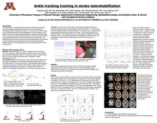
1. Huiqiong Deng, MD, MS; Will Durfee, PhD; David Nuckley, PhD; Brandon Rheude, SPT; Amy Severson, SPT; Katie Skluzacek, SPT; Kristen Spindler, SPT; Cynthia Davey, MS; James Carey, PhD, PT University of Minnesota, Program in Physical Therapy, Department of Mechanical Engineering, Biostatistical Design and Analysis Center, & Clinical and Translational Science Institute F unded by the NIH/NICHD R03HD051615 and NIH/NCRR P41 RR008079 and M01-RR00400. Introduction: Telerehabilitation is emerging that may allow rehabilitative training to continue remotely following discharge from acute care amidst an increasingly difficult health care economy . One aim of the current study was to determine whether stroke rehabilitation could be effective in the subject’s own home using telerehabilitation to improve ankle dorsiflexion (DF). A second aim was to determine whether recovery of ankle function could be influenced more by forced learning than by forced use. We hypothesized that tracking movements would yield greater improvements in ankle dorsiflexion during gait than simple movements and that a different pattern of brain reorganization would emerge between the two training forms. * ** * * Ankle tracking training in stroke telerehabilitation Methods and Procedures (Fig 1): Subjects- Sixteen subjects (11 males, 5 females) with mean age of 54.7±12.5 years and mean post stroke duration of 42.8 ±39.3 months were randomly assigned to either a Track group (N=8) or a Move group (N=8). Inclusion criteria included at least 10 degrees of ankle plantarflexion (PF)-DF. Pre-test/Post-test- Cortical activation was measured during fMRI while the subject completed a 7-minute task alternating ankle tracking of a sine wave target (.4 Hz) with rest (Fig 2). Gait kinematics were captured in the gait lab using an 8 camera Vicon Motion Capture System (Fig 3). Subjects also performed two trials of a 10-meter walk at their self-selected (“comfortable”) speed and then two trials at maximum speed. Analysis: The Wilcoxon signed-rank test was used to evaluate within-group median differences and the Wilcoxon rank-sum test to evaluate between-group. Differences in frequency of the direction of change of the fMRI variables were evaluated using Fisher’s exact test. Results: The primary outcome measure, paretic ankle DF during gait, showed a significant within-group median (Q1, Q3) increase from pretest at 6.75 (4.84, 8.99) degrees to posttest at 12.86 (8.20, 14.15) degrees for the Track group (p=0.008) and a trend toward an increase from pretest at 6.61 (5.80, 10.31) degrees to posttest at 8.99 (5.61, 10.95) degrees in the Move group (p=0.055) (Fig. 6A). Importantly, the average variance of toe clearance in the Track group decreased from 0.48 at pretest to 0.29 mm at posttest (p=0.014), whereas in the Move group it decreased from 0.47 to 0.34 mm (p=0.082). For the 10-meter walk tests, all within- and between-group differences were NS. Conclusions: (1) Telerehabilitation was feasible for stroke groups; (2) Forced learning (Track group) produced greater ankle DF during gait than forced use (Move group) Fig 4 Various postures Fig 6 Median (Q1, Q3, min, max) values of gait variables. GTSR= gait temporal symmetry ratio. Training- From their home (Fig 5 right), both groups completed 180 ankle-movement trials each day, five days/week, totally 3600 trials. The Track group used their affected ankle to trace a waveform on a screen (Fig 1, 5) which forced temporospatial processing to achieve accuracy. The Move group performed repetitive ankle PF-DF movements (Fig 1). Both groups completed trials in four positions (Fig 4). The subjects experienced a therapeutic relationship with the investigator through phone conferencing and web camera sessions 2-3 times/week. Fig 5 (Left) Various waveforms, frequency, amplitude and duration of training trials. (Right) Training set-up at subject’s own home. Fig 3 (Left) Vicon system camera set-up. (Right) Dynamic Plug-in-Gait Helen Hayes marker placement and model. None of the ROIs showed a significant median change in these measures (Fig 7). Inspection of Fig 7 reveals that, for the Track group, the direction of change for the median values of the fMRI variables generally showed decreases from pretest to posttest, whereas the Move group generally showed increases . Of the 8 volume and the 8 percent volume measures (2 sides by 4 ROIs), 7 showed a median increase in the Move group, while all values showed a median decrease in the Track group (Fisher’s exact test p=0.0014). For the 8 intensity measures, 7 of 8 showed a median increase in the Move group, while 7 of 8 showed a median decrease in the Track group (Fisher’s exact test p=0.01). Fig. 8 contrasts the change in the cortical activation maps from pretest to posttest for one subject from each group. Fig 7 Median change (Q1, Q3, min, max) values from pretest to posttest of volume, % volume and BOLD intensity of activation. Fig 8 Cortical activation at one coronal slice and three transverse slices for one subject in Move group with right hemisphere stroke tracking with paretic left ankle and one subject in Track group with left hemisphere stroke tracking with paretic right ankle. Change from pretest to posttest shows reduction in activation with greater focus to the ipsilesional hemisphere in the Track subject compared to the Move subject. Fig 2 Set-up for ankle movement test during fMRI. Fig 1 Procedure. (Upper) Track subject’s tracking performance; blue: target; red: response. (Lower) Screen in training session for Move group.