SlideShare a Scribd company logo
1 of 15
Download to read offline
Nanomedicine Opportunities
in Cardiology
GREGORY LANZA,a PATRICK WINTER,a TILLMANN CYRUS,a
SHELTON CARUTHERS,a,b JON MARSH,a MICHAEL HUGHES,a
AND SAMUEL WICKLINEa
a Washington    University School of Medicine, St. Louis, Missouri 63110, USA
b Philips   Medical Systems, Cleveland, Ohio 44143, USA


     ABSTRACT: Despite myriad advances, cardiovascular-related diseases
     continue to remain our greatest health problem. In more than half of pa-
     tients with atherosclerotic disease, their first presentation to medical at-
     tention becomes their last. Patients often survive their first cardiac event
     through acute revascularization and placement of drug-eluting stents
     (DES), but only select coronary lesions are amenable to DES placement,
     resulting in the use of bare metal or no stent, both of which lack the bene-
     fit of antirestenotic therapy. In other patients, transient ischemic attacks
     (TIAs) and stroke constitute the initial presentation of disease. In these
     patients, the diagnostic and therapeutic options are woefully inadequate.
     Nanomedicine offers options to each of these challenges. Antiangiogenic
     paramagnetic nanoparticles may be used to serially assess the severity of
     atherosclerotic disease in asymptomatic, high-risk patients by detecting
     the development of plaque neovasculature, which reflects the underlying
     lesion activity and vulnerability to rupture. The nanoparticles can locally
     deliver antiangiogenic therapy, which may acutely retard plaque progres-
     sion, allowing aggressive statin therapy to become effective. Moreover,
     these agents may be useful as a quantitative marker to guide atheroscle-
     rotic management in an asymptomatic patient. In those cases proceeding
     to the catheterization laboratory for revascularization, nanoparticles in-
     corporating antirestenotic drugs can be delivered directly into the wall
     of lesions not amenable to DES placement. Targeted nanoparticles could
     help ensure that antirestenotic drugs are available for all lesions. More-
     over, displacement of antiproliferative agents from the intimal surface
     into the vascular wall is likely to improve rehealing of the endothelium,
     improving postprocedural management of these patients.

     KEYWORDS: nanoparticle; angiogenesis; restenosis; thrombolysis


 Address for correspondence: Prof. Gregory M. Lanza, M.D., Ph.D., Med and Biomed Engineering,
WUSTL, 4003 Kingshighway Bldg., St. Louis, MO 63130. Voice: 314-454-8813; fax: 314-454-5265.
 e-mail: greg@cvu.wustl.edu

  Ann. N.Y. Acad. Sci. 1080: 451–465 (2006).   C   2006 New York Academy of Sciences.
  doi: 10.1196/annals.1380.034

                                                   451
452                                  ANNALS NEW YORK ACADEMY OF SCIENCES


                             INTRODUCTION
   Cardiovascular disease (CVD), principally heart disease and stroke, contin-
ues to be the nation’s leading killer for both men and women across all racial
and ethnic groups. Nearly 1 million or 42% of all American deaths are due to
CVD, and these victims were not simply the elderly. Approximately 160,000
individuals between the ages of 35 and 64 years died.1 Current techniques
for early medical detection and treatment are limited and their effectiveness
in actually preventing heart attacks is debatable. In one retrospective study,
86 of 326 individuals received physical examinations within a 7-day period
prior to death from heart attack, and their physicians predicted none to have a
myocardial infarction. As tragic as this death toll is, even more grievous are
the 57 million American survivors who daily struggle with the complications
of CVD. Moreover, the direct medical and lost productivity costs to society
are staggering, approximately $274 billion each year and growing annually.
Although changes in environmental exposures, reduction in tobacco use, ad-
justments in diet, and increased physical activity can all improve patient health,
the progression of CVD is relentless in Western societies. New paradigms to
detect and treat CVD in asymptomatic patients are needed in order to prevent
the first presentation of symptoms from being the last. Improved and safer
approaches to coronary and intracranial revascularization are still required,
despite the myriad of advances in the last 10 years.
   No single technology offers a solution for all problems. However, rapid
evolution of molecular biology, cell biology, genomics, and proteomics com-
bined with discoveries in material sciences and bioengineering have created
many new cadres of “nanotools” to address these challenges. Pharmaceutical
nanoparticles have emerged as multifaceted systems capable of identifying
and characterizing early disease before the gross anatomical manifestations
are easily apparent with a variety of clinically relevant imaging modalities.
Moreover, targeted particles can deliver therapeutics preferentially to sites of
pathologic disease by recognizing and binding to unique biochemical signa-
tures. The synergy of biomarker imaging and therapy is a powerful adjunctive
paradigm to current medical practice, which offers a rich palette of approaches
to address cardiovascular problems from a new perspective.

LIGAND-DIRECTED PERFLUOROCARBON NANOPARTICLES
   Perfluorocarbon (PFC) nanoparticles are unusual lipid-encapsulated col-
loidal emulsions with nominal sizes between 200 nm and 250 nm. The core
of the emulsion particle (98 vol%) comprises perfluorochemicals, which
have twice the specific gravity of water and offer excellent safety pro-
files in pharmaceutical formulations. 2 The fluorine-carbon bonds of these
compounds render them both chemically and biologically inert. Chemically
stable, nonmetabolizable, and intrinsically nontoxic, perfluorochemicals have
seen use in varied human applications including blood replacement, liquid
LANZA et al.: NANOMEDICINE OPPORTUNITIES IN CARDIOLOGY                         453


breathing, ocular fluid replacement, MR imaging, CT imaging, ultrasound
imaging, and percutaneous transluminal cardiac angioplasty (PTCA), with
many products approved or in development.
   For imaging, the perfluorocarbon core of the nanoparticles provides inherent
acoustic contrast relative to blood and tissues due primarily to a speed-of-sound
that is one-half to one-third that of water.2–4 Moreover, this echo contrast ef-
fect can be augmented by further decreases in the speed-of-sound imparted by
heating.5 For traditional proton MR imaging, the high surface area of nanopar-
ticles increases the ionic relaxivity of each atom of gadolinium by three- to
sixfold due to the slowed rotational effects, while increasing the payloads of
paramagnetic metals from a few to 100,000 per particle greatly amplifies the
signal, that is, the molecular or particular relaxivity.6–8 As with ultrasound, the
perfluorocarbon core of the particle can contribute to the MR signal through
19
   F imaging and spectroscopy.9–11 The high concentration of 19 F at sites tar-
geted with nanoparticles in combination with the negligible amount of fluorine
in the surrounding tissues creates a unique and inherent second marker. In ad-
dition, the fluorine signal provides a confirmation of nanoparticle delivery as
well as the quantity of particles delivered within a voxel or region independent
of the local tissue environment.
   As site-targeted agents for medical applications, in vivo stability and pro-
longed circulatory clearance offers many advantages. Liquid PFC nanoparti-
cles minimize rapid systemic destruction, clearance, and coalescence without
the addition of surface polyethylene glycol groups or surfactant cross-linking,
which frequently complicate targeting efforts, interfere with drug transport, or
mask surface components such as metal chelates or bioactive agents.


     ASSESSING AND TREATING ATHEROSCLEROSIS IN
   ASYMPTOMATIC PATIENTS WITH PERFLUOROCARBON
                   NANOPARTICLES

   Perhaps one of the most active areas of cardiovascular research of immediate
clinical significance is the quest to identify, quantify, and treat vulnerable and
unstable plaque. For some time it has been recognized that thrombosis asso-
ciated with plaque rupture is the principal cause of acute coronary syndromes
and strokes, and that these events occur more often than not in asymptomatic
vascular regions with approximately 50% diameter stenosis. Until recently, the
dogma has been that a single complex lesion was responsible for the clinical
event. But the diffuse nature of arterial tree inflammation renders many le-
sions within a vascular bed equally susceptible to extrinsic mechanical forces
modulated by sympathetic tone or direct proteolytic degradation of the fibrous
cap. Although multiple sites of rupture are uncommon as a cause of sudden
coronary death, luminal fibrin from multiple ruptures are frequent and asso-
ciated with plaque hemorrhage and superficial macrophages.12–14 These sites
of intimal fissuring, demarcated by accumulated surface fibrin, are suggested
454                                  ANNALS NEW YORK ACADEMY OF SCIENCES


to be responsible for the rapid angiographic progression of vascular steno-
sis in patients.15 In fact, accumulated surface fibrin may be a critical hall-
mark of lesion instability, and the sensitive and specific detection of fibrin
by nanoparticle technology may define important strategies for the preven-
tion of plaque progression and its sequellae. We7 have previously reported and
demonstrated the use of fibrin-specific paramagnetic nanoparticles for detect-
ing fibrin with MRI, while others have used small paramagnetic peptides.16,17
However, plaque rupture is a late manifestation of atherosclerotic plaque pro-
gression and further techniques are required to assess and treat the disease
earlier in its natural progression in order to achieve any meaningful clinical
impact.
   One signature of atherosclerosis is the proliferation of an angiogenic vas-
culature, which frequently develops disproportionately from the vasa vasorum
in response to the metabolic activity of plaque cellular constituents.18–22 Ex-
tensive neovascular proliferation has been spatially localized to atherosclerotic
plaque, and in particular, to “culprit” lesions clinically associated with unstable
angina, myocardial infarction, and stroke. In addition, plaque angiogenesis has
been suggested to promote plaque growth, intraplaque hemorrhage, and lesion
instability. The interplay between angiogenesis and plaque development was
explored by Moulton et al.23 in Apo E −/− mice treated with antiangiogenic
therapy for 4 months (20 to 36 weeks): TNP-470, a water-soluble fumagillin
analogue, or endostatin (30 mg/kg every other day, 1.68 g/kg total dose).23 Re-
duction in plaque angiogenesis and diminished atheroma growth were noted
despite persistent elevation of total cholesterol levels. TNP-470 and its parent
compound, fumagillin, directly inhibit endothelial cell proliferation by cova-
lently binding to methionine aminopeptidase 2 specifically, which catalyzes
the cleavage of N-terminal methionine from nascent polypeptides.24–26 Un-
fortunately, chronic, high doses of TNP-470 administered systemically have
caused neurocognitive side effects in humans.27,28
   Site-targeted nanoparticles offer the opportunity for local drug delivery in
combination with molecular imaging, which can provide noninvasive confir-
mation of targeting, spatial localization of drug distribution, and quantifica-
tion of therapeutic payload accumulated at the site. This concept was initially
demonstrated in vitro using doxorubicin and paclitaxel nanoparticles to inhibit
the proliferation of vascular smooth muscle cells.29 At that time, we proposed
that targeted perfluorocarbon nanoparticles could deliver chemotherapeutic
agents through a novel mechanism we called “contact facilitated lipid ex-
change.” Subsequent studies using confocal microscopy have illustrated the
exchange of fluorescent-labeled phospholipids from the outer surfactant layer
of the particle to the target cell membrane.30
   Using a hyperlipidemic New Zealand White rabbit model, we initially
demonstrated the antiangiogenic effectiveness of v 3 -targeted fumagillin
nanoparticles administered as a single dose,31,32 which was several orders
of magnitude less than used systemically in the ApoE model.23 In that study,
LANZA et al.: NANOMEDICINE OPPORTUNITIES IN CARDIOLOGY                        455


hyperlipidemic rabbits (∼80 days on diet) were injected via the ear vein with
  v 3 -targeted fumagillin nanoparticles (n = 5), v 3 -targeted nanoparticles
without fumagillin (n = 6), or nontargeted fumagillin nanoparticles (n = 6)
at 1.0 mL/kg. Four hours after nanoparticle injection, rabbits were reimaged
to assess the magnitude and distribution of signal enhancement. Multislice,
T 1 -weighted, spin-echo, fat-suppressed, black-blood images of the entire ab-
dominal aorta from the renal arteries to the diaphragm (TR = 380 msec, TE =
11 msec, 250 × 250 m inplane resolution, 5 mm slice thickness, number
of signals averaged = 8) were acquired. After treatment, all rabbits were
converted to normal rabbit chow (Purina Mills). One week later, the extent
of v 3 -integrin expression in each animal was reassessed by injection of
integrin-targeted paramagnetic nanoparticles (1.0 mL/kg; no drug) and nonin-
vasive imaging as described earlier. MRI signal enhancement from the aortic
wall was averaged over all imaged slices using a custom, semiautomated seg-
mentation program previously described.33 Signal enhancement in the aortic
wall was measured for each individual animal using all properly segmented
slices. The percentage enhancement in MRI signal was calculated slice-by-
slice in the 4-h postinjection images relative to the average preinjection MRI
signal.
   Consistent with the early stage of atherosclerosis in this animal model,
T 1 -weighted, black-blood images showed no gross evidence of plaque devel-
opment in terms of luminal narrowing or wall thickening when compared to
previous experiments using age-matched, nonatherosclerotic rabbits.33 MRI
signal enhancement in the aortic wall following injection of           3 -targeted
nanoparticles, both with and without fumagillin, displayed a patchy distribu-
tion, with typically higher levels of angiogenesis occurring near the diaphragm.
Nontargeted nanoparticles produced less extensive MRI enhancement of the
neovasculature at much lower levels with a similar heterogeneous distribution,
consistent with previous reports. The average MRI signal enhancement per
slice integrated across the entire aortic wall was identical for       3 -targeted
nanoparticles with (16.7 ± 1.1%) and without (16.7 ± 1.6%) fumagillin. Non-
targeted nanoparticles, however, provided less signal enhancement, presum-
ably representing nonspecific accumulation and/or delayed washout within the
tortuous microvasculature. 34
   One week after nanoparticle treatment, the residual expression of            3-
integrin was assessed as a marker of angiogenic activity within the aortic
wall. Preinjection scans were collected, followed by injection of      3 -targeted
paramagnetic nanoparticles (no drug) and contrast enhancement imaging 4 h
post injection. The preinjection aortic wall signal intensities for all groups
at treatment and at the 1-week follow-up were identical, confirming that the
paramagnetic nanoparticles administered 1 week prior were no longer de-
tectable. MRI aortic wall signal enhancement 1 week following          3 -targeted
fumagillin nanoparticle treatment was markedly reduced (2.9 ± 1.6%; P <
0.05) in both spatial distribution as well as intensity. By comparison, MRI
456                                    ANNALS NEW YORK ACADEMY OF SCIENCES


   FIGURE 1. MRI aortic wall signal enhancement with           3 -targeted paramagnetic
nanoparticle (no drug) 1 week following treatment with 3 -targeted fumagillin or control
(no drug) nanoparticles.


signal enhancement 1 week after treatment with               3 -targeted nanoparti-
cles lacking fumagillin was undiminished (18.1 ± 2.1%) (FIG. 1). Treatment
with nontargeted fumagillin nanoparticles did not significantly diminish            3-
integrin levels as determined by MRI signal enhancement 1 week after treat-
ment, although a numerical decrease was observed (12.4 ± 0.9%).
   In this study, the total dose of fumagillin administered as a single injection in
     3 -targeted nanoparticles was >10,000 times lower than the cumulative oral
dose of TNP-470 reported by Moulton et al. Reduced            3 -integrin expression
as determined by MRI molecular imaging and corroborated by decreased                3-
integrin positive vessel density supported the potential reduction in dosage and
increase in efficacy of chemotherapeutic agents using targeted nanomedicine
approaches. Incorporation of fumagillin into paramagnetic nanoparticles al-
lowed both aortic expression of          3 -integrin and local drug delivery to be
assessed and quantified concomitantly and noninvasively. Moreover, the initial
magnitude of the aortic signal response among hyperlipidemic rabbits receiv-
ing       3 -targeted paramagnetic nanoparticles was correlated with the degree
of change in MRI enhancement measured 7 days later with                    3 -targeted
paramagnetic nanoparticles (without drug). These data illustrate the concept
of “rational drug dosing,” which provides noninvasive measures of treatment
dosimetry and allows follow-up of response.
   For asymptomatic patients, nanomedicine approaches offer the ability to
quantitatively interrogate the severity and distribution of disease more directly,
to locally treat pathology with minimal doses, and to follow up response to
LANZA et al.: NANOMEDICINE OPPORTUNITIES IN CARDIOLOGY                        457


treatment noninvasively when there are no clinical symptoms or meaningful
measures with which to titrate treatment efficacy.

           NANOMEDICINE APPROACH TO CORONARY
                  REVASCULARIZATION

   Far too often the progression of atherosclerosis to acute coronary syndromes
presents the need for acute revascularization. Fortunately, continuous advances
from balloon angioplasty, bare-metal stents, and drug-covered stents such as
heparin-coated stents, to the more recent, new class of drug-eluting stents
(DES), have expanded our armamentarium for reopening stenotic vessels while
preventing vascular reocclusion. Within the last few years, the use of conven-
tional balloon angioplasty and bare-metal stent implantation, which were asso-
ciated with clinical restenosis rates of 32–42% and 19–30%, respectively,35–37
have been improved with the local deposition of a pharmacologic agent to
suppress neointimal proliferation. Current DES have reduced the rate of an-
giographic restenosis to below 9% and diminished the frequency for repeat
revascularization to below 5%.38,39 Unfortunately, DES cannot be routinely
used for all lesions. In some situations, vessel tortuosity or the distal location
of lesions prevents manipulation of the relatively inflexible DES. In other cases,
the vessel diameter at the culprit lesion is too small for stent placement. As
a result many lesions, in whole or part, do not receive the local antirestenotic
therapy after revascularization.
   Moreover, despite the clear success of DES, the incidence of late instent
thrombosis has arisen as an infrequent but serious complication of delayed
endothelial healing.40 To avoid acute thrombosis, aggressive dual (and occa-
sionally triple) antiplatelet therapy is employed for 6 months to a year. We
now recognize that some patients are nonresponders to one or more of the
drugs.41–43 In other instances, thrombosis presents when antithrombotic drugs
are withheld secondary to bleeding complications or the need for emergent
surgery. Late instent thrombosis has been linked to fatal outcomes,40 and the
risk can persist up to 30 months after DES implantation.42,43 We anticipate that
targeted local delivery of antirestenotic drugs such as paclitaxel or rapamycin
into the stretch-injured arterial wall rather than the intimal surface will permit
better healing and recovery of the endothelium. More rapid endothelial repair
of the injured wall should substantially diminish the incidence of thrombosis
and reduce the long-term requirement for aggressive antiplatelet therapy.
   Moreover, DES are now known to elicit unwanted effects on vessel healing
and local endothelium-dependent vasomotor responses 6 months after implan-
tation in the vessel distal to the intervened segment.44 Although still limited,
data on the effect of sirolimus on vasomotor response are accumulating in
animal models and patients alike. Swine coronary artery segments exposed
to sirolimus for 48 h showed severe impairment of endothelial function.45
In patients, exercise-induced coronary vasoconstriction was noted in vessel
458                                 ANNALS NEW YORK ACADEMY OF SCIENCES


segments adjacent to DES but not bare-metal stents.46 The mechanism for
this effect is unclear. Higher rates of restenosis proximal to stent placement
compared with the distal edge support an asymmetric downstream effect.
   We use a nanomedicine approach to address restenosis in lesions not
amenable to current DES stent technology by intramural targeting and an-
choring of rapamycin nanoparticles to the v 3 -integrin, present on smooth
muscle and other plaque components (e.g., macrophages, T cells). In previous
studies, we demonstrated that ligand-directed perfluorocarbon nanoparticles
could penetrate balloon-injured vessel walls and target intramural biomark-
ers, including tissue factor,47 collagen III, and integrins.48 We have recently
reported that integrin-targeted PFC nanoparticles can provide effective intra-
mural delivery of rapamycin and inhibit vascular stenosis following balloon
overstretch injury. In these studies, femoral arteries of 12 rabbits on athero-
genic diets for 3 weeks were subjected to balloon stretch injury via a catheter
approach from the left common carotid artery. Using a double-balloon tech-
nique, paramagnetic         3 -nanoparticles with rapamycin were administered to
one artery while the contralateral vessel received targeted nanoparticles with-
out drug or saline. Two weeks after nanoparticle treatment, plaque development
was determined by MR angiography and by microscopic morphometric quan-
tification. Routine MR angiograms were indistinguishable between control
and targeted-vessel segments. Microscopic analysis of serial vascular sections
2 weeks after injury revealed that the intimal plaque to lumen area ratio of
vessels treated with       3 -targeted rapamycin nanoparticles were significantly
(P < 0.05) less (∼50%) than arteries receiving targeted nanoparticles without
drug or saline (FIG. 2). Scanning electron microscopy of the intima performed
24 h after injury and treatment with          3 -targeted rapamycin nanoparticles
demonstrated their binding to the underlying matrix and cells. Immunofluo-
rescent imaging of the vessel wall ∼2 h after treatment demonstrated that the
nanoparticles had penetrated into the media and adventitia, consistent with
the MR images previously obtained. Although early, the results suggest that
     3 -integrin-targeted nanoparticles can provide effective intramural therapy
and may be a tool to extend the use of antirestenotic drugs to all revascularized
sites with or without adjunctive stent placement.

       NANOMEDICINE APPROACH TO THROMBOLYSIS
   Stroke is the third leading cause of death in the United States and often
results in functional impairment and long-term disability among survivors.49
Clinical trials have demonstrated that thrombolytic treatment such as tissue
plasminogen activator (t-PA) can reduce or reverse ischemia in patients treated
within the first 3 h of onset.50 However, intravenously administered throm-
bolytic agents are associated with an increased incidence of intracerebral hem-
orrhage and expanding stroke. This serious risk frequently delays the receipt
of aggressive thrombolytic therapy until intracranial bleeding can be ruled out
LANZA et al.: NANOMEDICINE OPPORTUNITIES IN CARDIOLOGY                                  459


   FIGURE 2. Microscopic analysis of serial vascular sections 2 weeks after injury re-
vealed that the intimal plaque to lumen area ratio of vessels treated with       3 -targeted
rapamycin nanoparticles were significantly (P < 0.05) less than arteries receiving targeted
nanoparticles without drug or saline.


by CT study of the head. As a result of these delays, the window of opportunity
to ameliorate neural damage is lost, and the personal and societal losses are
magnified.
   The advent of perfluorocarbon nanoparticles to specifically deliver drug
payloads to intravascular sites of interest presents a unique opportunity to tar-
get clot-dissolving therapeutics to cerebral sites of embolism while decreas-
ing the risk of hemorrhagic complications and increasing the effectiveness
of thrombolytic therapy. We have previously demonstrated targeting of liquid
perfluorocarbon nanoparticle emulsions to thrombi in vitro and in vivo,51 with
concomitant enhancement of acoustic reflectivity from the targeted surfaces.
Acoustic reflectivity enhancement of surfaces targeted with the nanoparticles
arises because of the acoustic impedance mismatch between the adherent layer
of nanoparticles and the surrounding media. We have recently demonstrated
that nanoparticles modified with thrombolytic enzyme (streptokinase) can be
targeted onto plasma clots and effect rapid dissolution in the presence of plas-
minogen. In this series of experiments, acellular thrombi were produced from
citrated human plasma combined with 500 mM calcium chloride and thrombin.
Since this study was conducted in vitro, targeting of the nanoparticles to fibrin
was accomplished using a three-step process in which biotinylated antifibrin
antibody (NIB 1H10 52 ), avidin, and biotinylated nanoparticle emulsions (with
or without streptokinase, depending on treatment group) were combined se-
quentially with interval washings of unbound reagents. Acoustic microscopy
was performed on targeted and control samples using a broadband, 25-MHz
immersion transducer (Panametrics V324, Waltham, MA, USA) operated in
460                                  ANNALS NEW YORK ACADEMY OF SCIENCES


pulse-echo mode. A computer-controlled pulser receiver was used to generate
insonifying pulses and amplify the received echoes. The transducer was affixed
to a three-axis, computer-controlled, motorized gantry. Radiofrequency (RF)
data were acquired, digitized to 8 bits at 500 MHz for 2,048 point records, and
stored to disk at every site as the transducer was scanned over each sample in
a rectangular grid with 100- m resolution. In preparation for scanning, each
sample was sealed within a chamber having a cellophane acoustic window, and
which was filled with phosphate buffered saline (PBS). The sample chamber
was submerged within a 37◦ C water bath, and the sample was scanned to yield
a baseline measurement. The chamber was then emptied of PBS through an
injection port and refilled with either plasminogen in PBS buffer (3 U/mL) or
PBS alone. Scans were then performed at 15-min intervals for 3 h, and spatial
registration was maintained at all times.
   RF data were analyzed to assess temporal changes in clot morphology and
backscatter. A sliding Hamming window (0.2 sec duration) was applied and
moved over the data in 2-nsec steps. The sum of the squared values within each
segment, a quantity proportional to the reflected energy, was used as input to
a peak-detection algorithm (implemented in LabVIEW, National Instruments
Corp., Austin, TX, USA) and used to determine the arrival time of the echo
from the thrombus surface. A similar technique was used to detect the echo
from the nitrocellulose substrate in the same waveform. The difference between
the echo arrival times of the clot surface and substrate determined the profile of
the clot, and these values were used to generate surface plots for visualization of
the sample volume. Backscatter was quantified by first applying a rectangular
window to each waveform to isolate the reflection from the targeted surface,
and then by calculating the log spectral difference with respect to the reflection
from a steel plate. The average value within the usable bandwidth (10–30 MHz)
was recorded in dB for each point in the scan, and this value was used to generate
a C-scan image of the integrated backscatter from the sample surface.
   The detected clot volume was dramatically decreased (P < 0.05) for
clots treated with fibrin-targeted streptokinase nanoparticles and exposed to
plasminogen in buffer (FIG. 3). Treatment with fibrin-targeted streptokinase
nanoparticles incubated in saline or fibrin-targeted nanoparticles without strep-
tokinase incubated with plasminogen in buffer had no thrombolytic effect. The
time to complete lysis varied with small changes in the synthesis process of
the streptokinase nanoparticle formulations. Initial conjugates had to be left
overnight for complete dissolution while more optimized agents formed later
completely dissolved clots in vitro within an hour, often less than 15 min.
Fibrin clot dissolution occurred from inside to outside. In some replicates, the
clot measured at 1 h was a hollow fibrin shell, which immediately collapsed
with slight motion. None of the control clots revealed morphologic or acoustic
changes.
   The measurements presented here suggest that fibrin-targeted streptokinase
nanoparticles could be used to promote local thrombolysis of plasma clots
in vivo. We have previously shown that fibrin-targeted nanoparticles can
LANZA et al.: NANOMEDICINE OPPORTUNITIES IN CARDIOLOGY                             461


  FIGURE 3. Mean normalized clot volume following 2-h treatment with fibrin-targeted
nanoparticles streptokinase-modified or control perfluorocarbon nanoparticles incubated
with plasminogen or saline.


penetrate and acoustically enhance acute intravascular thromboses in dogs.
Moreover, we have found that perfluorocarbon nanoparticles are constrained
to the vasculature due to their nominal size, even in “leaky” vascular beds
such as tumor neovasculature. Collectively, those results suggest that fibrin-
targeted streptokinase nanoparticles could be used early in acute stroke or
unstable angina with limited extravascular effects.


                                   SUMMARY

   Nanomedicine is a new evolving field referred to by many names, which
promises to significantly enhance the tools available to clinicians to address
some of the serious challenges responsible for profound mortality, morbidity,
and numerous societal consequences. Unlike the simple pharmaceutics of the
past, nanomedicine agents are typically three-dimensional, multicomponent
systems, which require interdisciplinary expertise to produce and use. In this
review, we have briefly introduced the opportunities associated with targeted
perfluorocarbon nanoparticles in early atherosclerosis, in acute revasculariza-
tion, and in thrombolytic therapy. The potential impact of these three concepts
is enormous but pales in comparison with the advancements likely to evolve
in this field over the coming decades.

                           ACKNOWLEDGMENTS
 This research was supported by the NIH grants (HL-42950, HL-59865,
HL-78631, NO1-CO-37007, and EB-01704), SCAI/Bracco Diagnostics, Inc.-
ACIST Fellowship Program, and the American Heart Association. Philips
462                                    ANNALS NEW YORK ACADEMY OF SCIENCES


Medical Systems (Cleveland, OH, USA) provided valuable equipment, soft-
ware, and engineering support.

                                   REFERENCES

 1. ANONYMOUS. 2005. American Heart Association, Vol. 2005.
 2. LANZA, G., K. WALLACE, M. SCOTT, et al. 1996. A novel site-targeted ultra-
     sonic contrast agent with broad biomedical application. Circulation 94: 3334–
     3340.
 3. MATTREY, R.F. 1994. The potential role of perfluorochemicals (pfcs) in diagnostic
     imaging. Artif. Cells Blood Substit. Immobil. Biotechnol. 22: 295–313.
 4. HALL, C.S., G.M. LANZA, J.H. ROSE, et al. 2000. Experimental determination of
     phase velocity of perfluorocarbons: applications to targeted contrast agents. IEEE
     Trans. Ultrason. Ferroelec. Freq. Contr. 47: 75–84.
 5. HALL, C., J. MARSH, M. SCOTT, et al. 2001. Temperature dependence of ultrasonic
     enhancement with a site-targeted contrast agent. J. Acous. Soc. AM. 110: 1677–
     1684.
 6. LANZA, G., C. LORENZ, S. FISCHER, et al. 1998. Enhanced detection of thrombi
     with a novel fibrin-targeted magnetic resonance imaging agent. Acad. Radiol.
     5(Suppl 1): s173–s176.
 7. FLACKE, S., S. FISCHER, M. SCOTT, et al. 2001. A novel MRI contrast agent for
     molecular imaging of fibrin: implications for detecting vulnerable plaques. Cir-
     culation 104: 1280–1285.
 8. WINTER, P., S. CARUTHERS, X. YU, et al. 2003. Improved molecular imaging
     contrast agent for detection of human thrombus. Mag. Reson. Med. 50: 411–
     416.
 9. YU, X., S.-K. SONG, J. CHEN, et al. 2000. High-resolution MRI characterization of
     human thrombus using a novel fibrin-targeted paramagnetic nanoparticle con-
     trast agent. Mag. Reson. Med. 44: 867–872.
10. LANZA, G.M., P.M. WINTER, A.M. NEUBAUER, et al. 2005. 1 H/19 Fmagnetic reso-
     nance molecular imaging with perfluorocarbon nanoparticles. Curr. Top. Dev.
     Biol. 70: 57–76.
11. CARUTHERS, S.D., A.M. NEUBAUER, F.D. HOCKETT, et al. 2006. In vitro demon-
     stration using 19f magnetic resonance to augment molecular imaging with para-
     magnetic perfluorocarbon nanoparticles at 1.5 tesla. Invest. Radiol. 41: 305–
     312.
12. KOLODGIE, F.D., R. VIRMANI, A.P. BURKE, et al. 2004. Pathologic assessment of
     the vulnerable human coronary plaque. Heart 90: 1385–1391.
13. VIRMANI, R., F.D. KOLODGIE, A.P. BURKE, et al. 2005. Atherosclerotic plaque pro-
     gression and vulnerability to rupture: angiogenesis as a source of intraplaque
     hemorrhage. Arterioscler. Thromb. Vasc. Biol. 25: 2054–2061.
14. SCHAAR, J.A., J.E. MULLER, E. FALK, et al. 2004. Terminology for high-risk
     and vulnerable coronary artery plaques. Report of a meeting on the vulnera-
     ble plaque, June 17 and 18, 2003, Santorini. Greece. Eur. Heart J. 25: 1077–
     1082.
15. OJIO, S., H. TAKATSU, T. TANAKA, et al. 2000. Considerable time from the onset
     of plaque rupture and/or thrombi until the onset of acute myocardial infarction
     in humans: coronary angiographic findings within 1 week before the onset of
     infarction. Circulation 102: 2063–2069.
LANZA et al.: NANOMEDICINE OPPORTUNITIES IN CARDIOLOGY                             463


16. BOTNAR, R.M., A. BUECKER, A.J. WIETHOFF, et al. 2004. In vivo magnetic reso-
     nance imaging of coronary thrombosis using a fibrin-binding molecular magnetic
     resonance contrast agent. Circulation 110: 1463–1466.
17. BOTNAR, R.M., A.S. PEREZ, S. WITTE, et al. 2004. In vivo molecular imaging
     of acute and subacute thrombosis using a fibrin-binding magnetic resonance
     imaging contrast agent. Circulation 109: 2023–2029.
18. KAHLON, R., J. SHAPERO & A.I. GOTLIEB. 1992. Angiogenesis in atherosclerosis.
     Can. J. Cardiol. 8: 60–64.
19. FAN, T.P., R. JAGGAR & R. BICKNELL. 1995. Controlling the vasculature: angiogen-
     esis, anti-angiogenesis and vascular targeting of gene therapy. Trends Pharmacol.
     Sci. 16: 57–66.
20. SUEISHI, K., Y. YONEMITSU, K. NAKAGAWA, et al. 1997. Atherosclerosis and angio-
     genesis. Its pathophysiological significance in humans as well as in an animal
     model induced by the gene transfer of vascular endothelial growth factor. Ann.
     N. Y. Acad. Sci. 811: 311–322; 322–324.
21. MOULTON, K. 2002. Plaque angiogenesis: its functions and regulation. Cold Spring
     Harb. Symp. Quant. Biol. 67: 471–482.
22. MOULTON, K.S. 2001. Plaque angiogenesis and atherosclerosis. Curr. Atheroscler.
     Rep. 3: 225–233.
23. MOULTON, K.S., E. HELLER, M.A. KONERDING, et al. 1999. Angiogenesis inhibitors
     endostatin or tnp-470 reduce intimal neovascularization and plaque growth in
     apolipoprotein e-deficient mice. Circulation 99: 1653–1655.
24. KLEIN, C.D. & G. FOLKERS. 2003. Understanding the selectivity of fumagillin for
     the methionine aminopeptidase type II. Oncol. Res. 13: 513–520.
25. RODRIGUEZ-NIETO, S., M.A. MEDINA & A.R. QUESADA. 2001. A re-evaluation of
     fumagillin selectivity towards endothelial cells. Anticancer Res. 21: 3457–3460.
26. LIU, S., J. WIDOM, C.W. KEMP, et al. 1998. Structure of human methionine
     aminopeptidase-2 complexed with fumagillin. Science 282: 1324–1327.
27. TRAN, H.T., G.R. BLUMENSCHEIN JR., C. LU, et al. 2004. Clinical and pharmacoki-
     netic study of TNP-470, an angiogenesis inhibitor, in combination with paclitaxel
     and carboplatin in patients with solid tumors. Cancer Chemother. Pharmacol. 54:
     308–314.
28. HERBST, R.S., T.L. MADDEN, H.T. TRAN, et al. 2002. Safety and pharmacokinetic
     effects of TNP-470, an angiogenesis inhibitor, combined with paclitaxel in pa-
     tients with solid tumors: evidence for activity in non-small-cell lung cancer. J.
     Clin. Oncol. 20: 4440–4447.
29. LANZA, G.M., X. YU, P.M. WINTER, et al. 2002. Targeted antiproliferative drug
     delivery to vascular smooth muscle cells with a magnetic resonance imaging
     nanoparticle contrast agent: implications for rational therapy of restenosis. Cir-
     culation 106: 2842–2847.
30. CROWDER, K.C., M.S. HUGHES, J.N. MARSH, et al. 2005. Sonic activation of
     molecularly-targeted nanoparticles accelerates transmembrane lipid delivery to
     cancer cells through contact-mediated mechanisms: implications for enhanced
     local drug delivery. Ultrasound Med. Biol. 31: 1693–1700.
31. LANZA, G.M., D.R. ABENDSCHEIN, X. YU, et al. 2002. Molecular imaging and
     targeted drug delivery with a novel, ligand-directed paramagnetic nanoparticle
     technology. Acad. Radiol. 9(Suppl 2): S330–S331.
32. LANZA, G.M., P. WINTER, S. CARUTHERS, et al. 2004. Novel paramagnetic contrast
     agents for molecular imaging and targeted drug delivery. Curr. Pharm. Biotech-
     nol. 5: 495–507.
464                                     ANNALS NEW YORK ACADEMY OF SCIENCES


33. WINTER, P.M., A.M. MORAWSKI, S.D. CARUTHERS, et al. 2003. Molecular imag-
      ing of angiogenesis in early-stage atherosclerosis with alpha(v)beta3-integrin-
      targeted nanoparticles. Circulation 108: 2270–2274.
34. WILSON, M.W., J.M. LABERGE, R.K. KERLAN, et al. 2002. MR portal venography:
      preliminary results of fast acquisition without contrast material or breath holding.
      Acad. Radiol. 9: 1179–1184.
35. JAEGERE PD, P., R. DOMBURG RV, H. NATHOE, et al. 1999. Long-term clinical
      outcome after stent implantation in coronary arteries. Int. J. Cardiovasc. Interv.
      2: 27–34.
36. KIMURA, T., T. TAMURA, H. YOKOI, et al. 1994. Long-term clinical and angiographic
      follow-up after placement of palmaz-schatz coronary stent: a single center ex-
      perience. J. Interv. Cardiol. 7: 129–139.
37. KEANE, D., A.J. AZAR, P. DE JAEGERE, et al. 1996. Clinical and angiographic out-
      come of elective stent implantation in small coronary vessels: an analysis of the
      benestent trial. Semin. Interv. Cardiol. 1: 255–262.
38. SERRUYS, P.W., P.A. LEMOS & B.A. VAN HOUT. 2004. Sirolimus eluting stent im-
      plantation for patients with multivessel disease: rationale for the arterial revas-
      cularisation therapies study part II (ARTS II). Heart 90: 995–998.
39. SERRUYS, P.W., E. REGAR & A.J. CARTER. 2002. Rapamycin eluting stent: the onset
      of a new era in interventional cardiology. Heart 87: 305–307.
40. ONG, A.T., E.P. MCFADDEN, E. REGAR, et al. 2005. Late angiographic stent throm-
      bosis (last) events with drug-eluting stents. J. Am. Coll. Cardiol. 45: 2088–2092.
41. WENAWESER, P. & O. HESS. 2005. Stent thrombosis is associated with an impaired
      response to antiplatelet therapy. J. Am. Coll. Cardiol. 46: CS5–CS6.
42. MCFADDEN, E.P., E. STABILE, E. REGAR, et al. 2004. Late thrombosis in drug-
      eluting coronary stents after discontinuation of antiplatelet therapy. Lancet 364:
      1519–1521.
43. RODRIGUEZ, A.E., J. MIERES, C. FERNANDEZ-PEREIRA, et al. 2006. Coronary stent
      thrombosis in the current drug-eluting stent era: insights from the eraci iii trial.
      J. Am. Col. Cardiol. 47: 205–207.
44. HOFMA, S.H., W.J. VAN DER GIESSEN, B.M. VAN DALEN, et al. 2006. Indication
      of long-term endothelial dysfunction after sirolimus-eluting stent implantation.
      Eur. Heart J. 27: 166–170.
45. JEANMART, H., O. MALO, M. CARRIER, et al. 2002. Comparative study of cy-
      closporine and tacrolimus on coronary endothelial function. J. Heart Lung Trans-
      plant. 21: 990–998.
46. TOGNI, M., S. WINDECKER, R. COCCHIA, et al. 2005. Sirolimus-eluting stents as-
      sociated with paradoxic coronary vasoconstriction. J. Am. Coll. Cardiol. 46:
      231–236.
47. LANZA, G., D. ABENDSCHEIN, C. HALL, et al. 2000. Molecular imaging of stretch-
      induced tissue factor expression in carotid arteries with intravascular ultrasound.
      Invest. Radiol. 35: 227–234.
48. CYRUS, T., D.R. ABENDSCHEIN, S.D. CARUTHERS, et al. 2006. MR three-dimensional
      molecular imaging of intramural biomarkers with targeted nanoparticles. J. Car-
      diovasc. Magn. Reson. 8: 535–541.
49. HINCHEY, J.A. & C. BENESCH. 2000. Thrombolytic therapy in patients with acute
      ischemic stroke. Arch. Neurol. 57: 1430–1436.
50. GROUP, T.N.I.O.N.D.A.S.R.-P.S.S. 1995. Tissue plasminogen activator for acute
      ischemic stroke. The National Institute of Neurological Disorders and Stroke
      rt-PA Stroke Study Group. N. Engl. J. Med. 333: 1581–1587.
LANZA et al.: NANOMEDICINE OPPORTUNITIES IN CARDIOLOGY                             465


51. LANZA, G.M., K.D. WALLACE, M.J. SCOTT, et al. 1996. A novel site-targeted ultra-
     sonic contrast agent with broad biomedical application. Circulation 95: 3334–
     3340.
52. RAUT, S. & P. GAFFNEY. 1996. Evaluation of fibrin binding profile of two antifibrin
     monoclonal antibodies. Thromb. Haemost. 76: 56–64.

More Related Content

What's hot

AKI in COVID 19 infection.
AKI in COVID 19 infection.AKI in COVID 19 infection.
AKI in COVID 19 infection.Dr. Manoj Deepak
 
Magnani2006 myocarditis
Magnani2006 myocarditisMagnani2006 myocarditis
Magnani2006 myocarditistommy8998
 
Pleural thickening 2009 janvol7issue1
Pleural thickening 2009 janvol7issue1Pleural thickening 2009 janvol7issue1
Pleural thickening 2009 janvol7issue1Dr Amolkumar W Diwan
 
Bioabsorbable Drug-Eluting Cardiac Stent Analysis
Bioabsorbable Drug-Eluting Cardiac Stent AnalysisBioabsorbable Drug-Eluting Cardiac Stent Analysis
Bioabsorbable Drug-Eluting Cardiac Stent AnalysisUniversity of Toronto
 
In stent neoatherosclerosis
In stent neoatherosclerosis In stent neoatherosclerosis
In stent neoatherosclerosis Kunal Mahajan
 
Bioabsorbable Scaffolds
Bioabsorbable ScaffoldsBioabsorbable Scaffolds
Bioabsorbable ScaffoldsAnkur Batra
 
An approach to myocardial biopsy interpretation
An approach to myocardial biopsy interpretationAn approach to myocardial biopsy interpretation
An approach to myocardial biopsy interpretationSaurav Singh
 
Bacterial septic arthritis adult
Bacterial septic arthritis adultBacterial septic arthritis adult
Bacterial septic arthritis adultAnderson David
 
Septic arthritis adult
Septic arthritis adultSeptic arthritis adult
Septic arthritis adultAnderson David
 
Calcifilaxia iv
Calcifilaxia ivCalcifilaxia iv
Calcifilaxia ivHOME
 

What's hot (20)

AKI in COVID 19 infection.
AKI in COVID 19 infection.AKI in COVID 19 infection.
AKI in COVID 19 infection.
 
Magnani2006 myocarditis
Magnani2006 myocarditisMagnani2006 myocarditis
Magnani2006 myocarditis
 
Visualization of atherosclerotic vulnerable plaque
Visualization of atherosclerotic vulnerable plaqueVisualization of atherosclerotic vulnerable plaque
Visualization of atherosclerotic vulnerable plaque
 
3
33
3
 
Pleural thickening 2009 janvol7issue1
Pleural thickening 2009 janvol7issue1Pleural thickening 2009 janvol7issue1
Pleural thickening 2009 janvol7issue1
 
Bioabsorbable Drug-Eluting Cardiac Stent Analysis
Bioabsorbable Drug-Eluting Cardiac Stent AnalysisBioabsorbable Drug-Eluting Cardiac Stent Analysis
Bioabsorbable Drug-Eluting Cardiac Stent Analysis
 
IN STENT RESTENOSIS
IN STENT RESTENOSISIN STENT RESTENOSIS
IN STENT RESTENOSIS
 
Instent restenosis
Instent restenosis Instent restenosis
Instent restenosis
 
Singapore conference 2015 jj
Singapore conference  2015 jjSingapore conference  2015 jj
Singapore conference 2015 jj
 
In stent neoatherosclerosis
In stent neoatherosclerosis In stent neoatherosclerosis
In stent neoatherosclerosis
 
Bioabsorbable Scaffolds
Bioabsorbable ScaffoldsBioabsorbable Scaffolds
Bioabsorbable Scaffolds
 
Vulnerable plaque overview
Vulnerable plaque overviewVulnerable plaque overview
Vulnerable plaque overview
 
An approach to myocardial biopsy interpretation
An approach to myocardial biopsy interpretationAn approach to myocardial biopsy interpretation
An approach to myocardial biopsy interpretation
 
Vulnerable patient mar 04
Vulnerable patient mar 04Vulnerable patient mar 04
Vulnerable patient mar 04
 
Tct recognition of vulnerable plaque guidelines
Tct recognition of vulnerable plaque guidelines Tct recognition of vulnerable plaque guidelines
Tct recognition of vulnerable plaque guidelines
 
Emf final present
Emf final presentEmf final present
Emf final present
 
Bacterial septic arthritis adult
Bacterial septic arthritis adultBacterial septic arthritis adult
Bacterial septic arthritis adult
 
International Journal of Orthopedics: Research & Therapy
International Journal of Orthopedics: Research & TherapyInternational Journal of Orthopedics: Research & Therapy
International Journal of Orthopedics: Research & Therapy
 
Septic arthritis adult
Septic arthritis adultSeptic arthritis adult
Septic arthritis adult
 
Calcifilaxia iv
Calcifilaxia ivCalcifilaxia iv
Calcifilaxia iv
 

Viewers also liked

Copy of Cover Letter (Student Theme)
Copy of Cover Letter (Student Theme)Copy of Cover Letter (Student Theme)
Copy of Cover Letter (Student Theme)Nilton Domingues
 
Nasa Langley VCEW 2009 Jones
Nasa Langley VCEW 2009 JonesNasa Langley VCEW 2009 Jones
Nasa Langley VCEW 2009 JonesGreg Jones
 
Pixel Envy
Pixel EnvyPixel Envy
Pixel EnvySimoReid
 

Viewers also liked (7)

Nanomedicina4
Nanomedicina4Nanomedicina4
Nanomedicina4
 
Copy of Cover Letter (Student Theme)
Copy of Cover Letter (Student Theme)Copy of Cover Letter (Student Theme)
Copy of Cover Letter (Student Theme)
 
Nanomedicina3
Nanomedicina3Nanomedicina3
Nanomedicina3
 
Nasa Langley VCEW 2009 Jones
Nasa Langley VCEW 2009 JonesNasa Langley VCEW 2009 Jones
Nasa Langley VCEW 2009 Jones
 
Nanomedicina8
Nanomedicina8Nanomedicina8
Nanomedicina8
 
Pixel Envy
Pixel EnvyPixel Envy
Pixel Envy
 
Nanomedicina5
Nanomedicina5Nanomedicina5
Nanomedicina5
 

Similar to Nanomedicina1

Radiological pathology of cerebrovascular disorders
Radiological pathology of cerebrovascular disordersRadiological pathology of cerebrovascular disorders
Radiological pathology of cerebrovascular disordersProfessor Yasser Metwally
 
Calcifilaxiaiv 100615192457-phpapp02
Calcifilaxiaiv 100615192457-phpapp02Calcifilaxiaiv 100615192457-phpapp02
Calcifilaxiaiv 100615192457-phpapp02UNICA-Publicidad
 
Calcifilaxiaiv 100615192457-phpapp02 (1)
Calcifilaxiaiv 100615192457-phpapp02 (1)Calcifilaxiaiv 100615192457-phpapp02 (1)
Calcifilaxiaiv 100615192457-phpapp02 (1)UNICA-Publicidad
 
Pediatric vasculitides
Pediatric vasculitidesPediatric vasculitides
Pediatric vasculitidesdattasrisaila
 
1Central Venous Catheter UsageCentral Venous Catheter .docx
1Central Venous Catheter UsageCentral Venous Catheter .docx1Central Venous Catheter UsageCentral Venous Catheter .docx
1Central Venous Catheter UsageCentral Venous Catheter .docxfelicidaddinwoodie
 
ClinicalOutcomesFollowingCoronary BifurcationPCITechniques.pdf
ClinicalOutcomesFollowingCoronary BifurcationPCITechniques.pdfClinicalOutcomesFollowingCoronary BifurcationPCITechniques.pdf
ClinicalOutcomesFollowingCoronary BifurcationPCITechniques.pdfIrving Torres Lopez
 
Ccsvi and ms ameds centrum
Ccsvi and ms ameds centrumCcsvi and ms ameds centrum
Ccsvi and ms ameds centrumAmeds Centrum
 
034 everybody has atherosclerosis
034 everybody has atherosclerosis034 everybody has atherosclerosis
034 everybody has atherosclerosisSHAPE Society
 
International Journal of Pharmaceutical Science Invention (IJPSI)
International Journal of Pharmaceutical Science Invention (IJPSI)International Journal of Pharmaceutical Science Invention (IJPSI)
International Journal of Pharmaceutical Science Invention (IJPSI)inventionjournals
 
1 enfermedad cv ateroesclerótica figura 1 (1)
1 enfermedad cv ateroesclerótica figura 1 (1)1 enfermedad cv ateroesclerótica figura 1 (1)
1 enfermedad cv ateroesclerótica figura 1 (1)sagita28
 
Miocardites circulation
Miocardites circulationMiocardites circulation
Miocardites circulationgisa_legal
 
Coagulacion diseminada intravascular
Coagulacion diseminada intravascularCoagulacion diseminada intravascular
Coagulacion diseminada intravascularmrpienda
 

Similar to Nanomedicina1 (20)

205832087 cc-2
205832087 cc-2205832087 cc-2
205832087 cc-2
 
Radiological pathology of cerebrovascular disorders
Radiological pathology of cerebrovascular disordersRadiological pathology of cerebrovascular disorders
Radiological pathology of cerebrovascular disorders
 
Issues in radiological pathology
Issues in radiological pathologyIssues in radiological pathology
Issues in radiological pathology
 
Nephrology & Therapeutics
Nephrology & TherapeuticsNephrology & Therapeutics
Nephrology & Therapeutics
 
Calcifilaxiaiv 100615192457-phpapp02
Calcifilaxiaiv 100615192457-phpapp02Calcifilaxiaiv 100615192457-phpapp02
Calcifilaxiaiv 100615192457-phpapp02
 
Calcifilaxiaiv 100615192457-phpapp02 (1)
Calcifilaxiaiv 100615192457-phpapp02 (1)Calcifilaxiaiv 100615192457-phpapp02 (1)
Calcifilaxiaiv 100615192457-phpapp02 (1)
 
Pediatric vasculitides
Pediatric vasculitidesPediatric vasculitides
Pediatric vasculitides
 
Critical care nephrology
Critical care nephrologyCritical care nephrology
Critical care nephrology
 
1Central Venous Catheter UsageCentral Venous Catheter .docx
1Central Venous Catheter UsageCentral Venous Catheter .docx1Central Venous Catheter UsageCentral Venous Catheter .docx
1Central Venous Catheter UsageCentral Venous Catheter .docx
 
ClinicalOutcomesFollowingCoronary BifurcationPCITechniques.pdf
ClinicalOutcomesFollowingCoronary BifurcationPCITechniques.pdfClinicalOutcomesFollowingCoronary BifurcationPCITechniques.pdf
ClinicalOutcomesFollowingCoronary BifurcationPCITechniques.pdf
 
Ccsvi and ms ameds centrum
Ccsvi and ms ameds centrumCcsvi and ms ameds centrum
Ccsvi and ms ameds centrum
 
Ischemic stroke. 2012
Ischemic stroke. 2012Ischemic stroke. 2012
Ischemic stroke. 2012
 
Masserini erice
Masserini ericeMasserini erice
Masserini erice
 
034 everybody has atherosclerosis
034 everybody has atherosclerosis034 everybody has atherosclerosis
034 everybody has atherosclerosis
 
Mri spio
Mri spioMri spio
Mri spio
 
034 everybody has atherosclerosis
034 everybody has atherosclerosis034 everybody has atherosclerosis
034 everybody has atherosclerosis
 
International Journal of Pharmaceutical Science Invention (IJPSI)
International Journal of Pharmaceutical Science Invention (IJPSI)International Journal of Pharmaceutical Science Invention (IJPSI)
International Journal of Pharmaceutical Science Invention (IJPSI)
 
1 enfermedad cv ateroesclerótica figura 1 (1)
1 enfermedad cv ateroesclerótica figura 1 (1)1 enfermedad cv ateroesclerótica figura 1 (1)
1 enfermedad cv ateroesclerótica figura 1 (1)
 
Miocardites circulation
Miocardites circulationMiocardites circulation
Miocardites circulation
 
Coagulacion diseminada intravascular
Coagulacion diseminada intravascularCoagulacion diseminada intravascular
Coagulacion diseminada intravascular
 

More from joanamatux

Memorialdo Convento
Memorialdo ConventoMemorialdo Convento
Memorialdo Conventojoanamatux
 
Nanomedicina11
Nanomedicina11Nanomedicina11
Nanomedicina11joanamatux
 
Nanomedicina10
Nanomedicina10Nanomedicina10
Nanomedicina10joanamatux
 

More from joanamatux (7)

ComentáRios
ComentáRiosComentáRios
ComentáRios
 
Memorialdo Convento
Memorialdo ConventoMemorialdo Convento
Memorialdo Convento
 
Nanomedicina11
Nanomedicina11Nanomedicina11
Nanomedicina11
 
Nanomedicina10
Nanomedicina10Nanomedicina10
Nanomedicina10
 
Nanomedicina9
Nanomedicina9Nanomedicina9
Nanomedicina9
 
Nanomedicina7
Nanomedicina7Nanomedicina7
Nanomedicina7
 
Nanomedicina6
Nanomedicina6Nanomedicina6
Nanomedicina6
 

Recently uploaded

Top Rated Hyderabad Call Girls Erragadda ⟟ 6297143586 ⟟ Call Me For Genuine ...
Top Rated  Hyderabad Call Girls Erragadda ⟟ 6297143586 ⟟ Call Me For Genuine ...Top Rated  Hyderabad Call Girls Erragadda ⟟ 6297143586 ⟟ Call Me For Genuine ...
Top Rated Hyderabad Call Girls Erragadda ⟟ 6297143586 ⟟ Call Me For Genuine ...chandars293
 
Call Girls Dehradun Just Call 9907093804 Top Class Call Girl Service Available
Call Girls Dehradun Just Call 9907093804 Top Class Call Girl Service AvailableCall Girls Dehradun Just Call 9907093804 Top Class Call Girl Service Available
Call Girls Dehradun Just Call 9907093804 Top Class Call Girl Service AvailableDipal Arora
 
Top Rated Bangalore Call Girls Richmond Circle ⟟ 8250192130 ⟟ Call Me For Gen...
Top Rated Bangalore Call Girls Richmond Circle ⟟ 8250192130 ⟟ Call Me For Gen...Top Rated Bangalore Call Girls Richmond Circle ⟟ 8250192130 ⟟ Call Me For Gen...
Top Rated Bangalore Call Girls Richmond Circle ⟟ 8250192130 ⟟ Call Me For Gen...narwatsonia7
 
Top Rated Bangalore Call Girls Mg Road ⟟ 8250192130 ⟟ Call Me For Genuine Sex...
Top Rated Bangalore Call Girls Mg Road ⟟ 8250192130 ⟟ Call Me For Genuine Sex...Top Rated Bangalore Call Girls Mg Road ⟟ 8250192130 ⟟ Call Me For Genuine Sex...
Top Rated Bangalore Call Girls Mg Road ⟟ 8250192130 ⟟ Call Me For Genuine Sex...narwatsonia7
 
VIP Mumbai Call Girls Hiranandani Gardens Just Call 9920874524 with A/C Room ...
VIP Mumbai Call Girls Hiranandani Gardens Just Call 9920874524 with A/C Room ...VIP Mumbai Call Girls Hiranandani Gardens Just Call 9920874524 with A/C Room ...
VIP Mumbai Call Girls Hiranandani Gardens Just Call 9920874524 with A/C Room ...Garima Khatri
 
VIP Service Call Girls Sindhi Colony 📳 7877925207 For 18+ VIP Call Girl At Th...
VIP Service Call Girls Sindhi Colony 📳 7877925207 For 18+ VIP Call Girl At Th...VIP Service Call Girls Sindhi Colony 📳 7877925207 For 18+ VIP Call Girl At Th...
VIP Service Call Girls Sindhi Colony 📳 7877925207 For 18+ VIP Call Girl At Th...jageshsingh5554
 
Book Paid Powai Call Girls Mumbai 𖠋 9930245274 𖠋Low Budget Full Independent H...
Book Paid Powai Call Girls Mumbai 𖠋 9930245274 𖠋Low Budget Full Independent H...Book Paid Powai Call Girls Mumbai 𖠋 9930245274 𖠋Low Budget Full Independent H...
Book Paid Powai Call Girls Mumbai 𖠋 9930245274 𖠋Low Budget Full Independent H...Call Girls in Nagpur High Profile
 
VIP Russian Call Girls in Varanasi Samaira 8250192130 Independent Escort Serv...
VIP Russian Call Girls in Varanasi Samaira 8250192130 Independent Escort Serv...VIP Russian Call Girls in Varanasi Samaira 8250192130 Independent Escort Serv...
VIP Russian Call Girls in Varanasi Samaira 8250192130 Independent Escort Serv...Neha Kaur
 
Lucknow Call girls - 8800925952 - 24x7 service with hotel room
Lucknow Call girls - 8800925952 - 24x7 service with hotel roomLucknow Call girls - 8800925952 - 24x7 service with hotel room
Lucknow Call girls - 8800925952 - 24x7 service with hotel roomdiscovermytutordmt
 
Call Girls Bareilly Just Call 9907093804 Top Class Call Girl Service Available
Call Girls Bareilly Just Call 9907093804 Top Class Call Girl Service AvailableCall Girls Bareilly Just Call 9907093804 Top Class Call Girl Service Available
Call Girls Bareilly Just Call 9907093804 Top Class Call Girl Service AvailableDipal Arora
 
Best Rate (Hyderabad) Call Girls Jahanuma ⟟ 8250192130 ⟟ High Class Call Girl...
Best Rate (Hyderabad) Call Girls Jahanuma ⟟ 8250192130 ⟟ High Class Call Girl...Best Rate (Hyderabad) Call Girls Jahanuma ⟟ 8250192130 ⟟ High Class Call Girl...
Best Rate (Hyderabad) Call Girls Jahanuma ⟟ 8250192130 ⟟ High Class Call Girl...astropune
 
Bangalore Call Girls Nelamangala Number 7001035870 Meetin With Bangalore Esc...
Bangalore Call Girls Nelamangala Number 7001035870  Meetin With Bangalore Esc...Bangalore Call Girls Nelamangala Number 7001035870  Meetin With Bangalore Esc...
Bangalore Call Girls Nelamangala Number 7001035870 Meetin With Bangalore Esc...narwatsonia7
 
Call Girls Service Surat Samaira ❤️🍑 8250192130 👄 Independent Escort Service ...
Call Girls Service Surat Samaira ❤️🍑 8250192130 👄 Independent Escort Service ...Call Girls Service Surat Samaira ❤️🍑 8250192130 👄 Independent Escort Service ...
Call Girls Service Surat Samaira ❤️🍑 8250192130 👄 Independent Escort Service ...CALL GIRLS
 
Call Girls Ludhiana Just Call 9907093804 Top Class Call Girl Service Available
Call Girls Ludhiana Just Call 9907093804 Top Class Call Girl Service AvailableCall Girls Ludhiana Just Call 9907093804 Top Class Call Girl Service Available
Call Girls Ludhiana Just Call 9907093804 Top Class Call Girl Service AvailableDipal Arora
 
Call Girls Siliguri Just Call 9907093804 Top Class Call Girl Service Available
Call Girls Siliguri Just Call 9907093804 Top Class Call Girl Service AvailableCall Girls Siliguri Just Call 9907093804 Top Class Call Girl Service Available
Call Girls Siliguri Just Call 9907093804 Top Class Call Girl Service AvailableDipal Arora
 
Call Girls Ooty Just Call 9907093804 Top Class Call Girl Service Available
Call Girls Ooty Just Call 9907093804 Top Class Call Girl Service AvailableCall Girls Ooty Just Call 9907093804 Top Class Call Girl Service Available
Call Girls Ooty Just Call 9907093804 Top Class Call Girl Service AvailableDipal Arora
 
All Time Service Available Call Girls Marine Drive 📳 9820252231 For 18+ VIP C...
All Time Service Available Call Girls Marine Drive 📳 9820252231 For 18+ VIP C...All Time Service Available Call Girls Marine Drive 📳 9820252231 For 18+ VIP C...
All Time Service Available Call Girls Marine Drive 📳 9820252231 For 18+ VIP C...Arohi Goyal
 
VIP Call Girls Indore Kirti 💚😋 9256729539 🚀 Indore Escorts
VIP Call Girls Indore Kirti 💚😋  9256729539 🚀 Indore EscortsVIP Call Girls Indore Kirti 💚😋  9256729539 🚀 Indore Escorts
VIP Call Girls Indore Kirti 💚😋 9256729539 🚀 Indore Escortsaditipandeya
 
Best Rate (Guwahati ) Call Girls Guwahati ⟟ 8617370543 ⟟ High Class Call Girl...
Best Rate (Guwahati ) Call Girls Guwahati ⟟ 8617370543 ⟟ High Class Call Girl...Best Rate (Guwahati ) Call Girls Guwahati ⟟ 8617370543 ⟟ High Class Call Girl...
Best Rate (Guwahati ) Call Girls Guwahati ⟟ 8617370543 ⟟ High Class Call Girl...Dipal Arora
 
Call Girls Bhubaneswar Just Call 9907093804 Top Class Call Girl Service Avail...
Call Girls Bhubaneswar Just Call 9907093804 Top Class Call Girl Service Avail...Call Girls Bhubaneswar Just Call 9907093804 Top Class Call Girl Service Avail...
Call Girls Bhubaneswar Just Call 9907093804 Top Class Call Girl Service Avail...Dipal Arora
 

Recently uploaded (20)

Top Rated Hyderabad Call Girls Erragadda ⟟ 6297143586 ⟟ Call Me For Genuine ...
Top Rated  Hyderabad Call Girls Erragadda ⟟ 6297143586 ⟟ Call Me For Genuine ...Top Rated  Hyderabad Call Girls Erragadda ⟟ 6297143586 ⟟ Call Me For Genuine ...
Top Rated Hyderabad Call Girls Erragadda ⟟ 6297143586 ⟟ Call Me For Genuine ...
 
Call Girls Dehradun Just Call 9907093804 Top Class Call Girl Service Available
Call Girls Dehradun Just Call 9907093804 Top Class Call Girl Service AvailableCall Girls Dehradun Just Call 9907093804 Top Class Call Girl Service Available
Call Girls Dehradun Just Call 9907093804 Top Class Call Girl Service Available
 
Top Rated Bangalore Call Girls Richmond Circle ⟟ 8250192130 ⟟ Call Me For Gen...
Top Rated Bangalore Call Girls Richmond Circle ⟟ 8250192130 ⟟ Call Me For Gen...Top Rated Bangalore Call Girls Richmond Circle ⟟ 8250192130 ⟟ Call Me For Gen...
Top Rated Bangalore Call Girls Richmond Circle ⟟ 8250192130 ⟟ Call Me For Gen...
 
Top Rated Bangalore Call Girls Mg Road ⟟ 8250192130 ⟟ Call Me For Genuine Sex...
Top Rated Bangalore Call Girls Mg Road ⟟ 8250192130 ⟟ Call Me For Genuine Sex...Top Rated Bangalore Call Girls Mg Road ⟟ 8250192130 ⟟ Call Me For Genuine Sex...
Top Rated Bangalore Call Girls Mg Road ⟟ 8250192130 ⟟ Call Me For Genuine Sex...
 
VIP Mumbai Call Girls Hiranandani Gardens Just Call 9920874524 with A/C Room ...
VIP Mumbai Call Girls Hiranandani Gardens Just Call 9920874524 with A/C Room ...VIP Mumbai Call Girls Hiranandani Gardens Just Call 9920874524 with A/C Room ...
VIP Mumbai Call Girls Hiranandani Gardens Just Call 9920874524 with A/C Room ...
 
VIP Service Call Girls Sindhi Colony 📳 7877925207 For 18+ VIP Call Girl At Th...
VIP Service Call Girls Sindhi Colony 📳 7877925207 For 18+ VIP Call Girl At Th...VIP Service Call Girls Sindhi Colony 📳 7877925207 For 18+ VIP Call Girl At Th...
VIP Service Call Girls Sindhi Colony 📳 7877925207 For 18+ VIP Call Girl At Th...
 
Book Paid Powai Call Girls Mumbai 𖠋 9930245274 𖠋Low Budget Full Independent H...
Book Paid Powai Call Girls Mumbai 𖠋 9930245274 𖠋Low Budget Full Independent H...Book Paid Powai Call Girls Mumbai 𖠋 9930245274 𖠋Low Budget Full Independent H...
Book Paid Powai Call Girls Mumbai 𖠋 9930245274 𖠋Low Budget Full Independent H...
 
VIP Russian Call Girls in Varanasi Samaira 8250192130 Independent Escort Serv...
VIP Russian Call Girls in Varanasi Samaira 8250192130 Independent Escort Serv...VIP Russian Call Girls in Varanasi Samaira 8250192130 Independent Escort Serv...
VIP Russian Call Girls in Varanasi Samaira 8250192130 Independent Escort Serv...
 
Lucknow Call girls - 8800925952 - 24x7 service with hotel room
Lucknow Call girls - 8800925952 - 24x7 service with hotel roomLucknow Call girls - 8800925952 - 24x7 service with hotel room
Lucknow Call girls - 8800925952 - 24x7 service with hotel room
 
Call Girls Bareilly Just Call 9907093804 Top Class Call Girl Service Available
Call Girls Bareilly Just Call 9907093804 Top Class Call Girl Service AvailableCall Girls Bareilly Just Call 9907093804 Top Class Call Girl Service Available
Call Girls Bareilly Just Call 9907093804 Top Class Call Girl Service Available
 
Best Rate (Hyderabad) Call Girls Jahanuma ⟟ 8250192130 ⟟ High Class Call Girl...
Best Rate (Hyderabad) Call Girls Jahanuma ⟟ 8250192130 ⟟ High Class Call Girl...Best Rate (Hyderabad) Call Girls Jahanuma ⟟ 8250192130 ⟟ High Class Call Girl...
Best Rate (Hyderabad) Call Girls Jahanuma ⟟ 8250192130 ⟟ High Class Call Girl...
 
Bangalore Call Girls Nelamangala Number 7001035870 Meetin With Bangalore Esc...
Bangalore Call Girls Nelamangala Number 7001035870  Meetin With Bangalore Esc...Bangalore Call Girls Nelamangala Number 7001035870  Meetin With Bangalore Esc...
Bangalore Call Girls Nelamangala Number 7001035870 Meetin With Bangalore Esc...
 
Call Girls Service Surat Samaira ❤️🍑 8250192130 👄 Independent Escort Service ...
Call Girls Service Surat Samaira ❤️🍑 8250192130 👄 Independent Escort Service ...Call Girls Service Surat Samaira ❤️🍑 8250192130 👄 Independent Escort Service ...
Call Girls Service Surat Samaira ❤️🍑 8250192130 👄 Independent Escort Service ...
 
Call Girls Ludhiana Just Call 9907093804 Top Class Call Girl Service Available
Call Girls Ludhiana Just Call 9907093804 Top Class Call Girl Service AvailableCall Girls Ludhiana Just Call 9907093804 Top Class Call Girl Service Available
Call Girls Ludhiana Just Call 9907093804 Top Class Call Girl Service Available
 
Call Girls Siliguri Just Call 9907093804 Top Class Call Girl Service Available
Call Girls Siliguri Just Call 9907093804 Top Class Call Girl Service AvailableCall Girls Siliguri Just Call 9907093804 Top Class Call Girl Service Available
Call Girls Siliguri Just Call 9907093804 Top Class Call Girl Service Available
 
Call Girls Ooty Just Call 9907093804 Top Class Call Girl Service Available
Call Girls Ooty Just Call 9907093804 Top Class Call Girl Service AvailableCall Girls Ooty Just Call 9907093804 Top Class Call Girl Service Available
Call Girls Ooty Just Call 9907093804 Top Class Call Girl Service Available
 
All Time Service Available Call Girls Marine Drive 📳 9820252231 For 18+ VIP C...
All Time Service Available Call Girls Marine Drive 📳 9820252231 For 18+ VIP C...All Time Service Available Call Girls Marine Drive 📳 9820252231 For 18+ VIP C...
All Time Service Available Call Girls Marine Drive 📳 9820252231 For 18+ VIP C...
 
VIP Call Girls Indore Kirti 💚😋 9256729539 🚀 Indore Escorts
VIP Call Girls Indore Kirti 💚😋  9256729539 🚀 Indore EscortsVIP Call Girls Indore Kirti 💚😋  9256729539 🚀 Indore Escorts
VIP Call Girls Indore Kirti 💚😋 9256729539 🚀 Indore Escorts
 
Best Rate (Guwahati ) Call Girls Guwahati ⟟ 8617370543 ⟟ High Class Call Girl...
Best Rate (Guwahati ) Call Girls Guwahati ⟟ 8617370543 ⟟ High Class Call Girl...Best Rate (Guwahati ) Call Girls Guwahati ⟟ 8617370543 ⟟ High Class Call Girl...
Best Rate (Guwahati ) Call Girls Guwahati ⟟ 8617370543 ⟟ High Class Call Girl...
 
Call Girls Bhubaneswar Just Call 9907093804 Top Class Call Girl Service Avail...
Call Girls Bhubaneswar Just Call 9907093804 Top Class Call Girl Service Avail...Call Girls Bhubaneswar Just Call 9907093804 Top Class Call Girl Service Avail...
Call Girls Bhubaneswar Just Call 9907093804 Top Class Call Girl Service Avail...
 

Nanomedicina1

  • 1. Nanomedicine Opportunities in Cardiology GREGORY LANZA,a PATRICK WINTER,a TILLMANN CYRUS,a SHELTON CARUTHERS,a,b JON MARSH,a MICHAEL HUGHES,a AND SAMUEL WICKLINEa a Washington University School of Medicine, St. Louis, Missouri 63110, USA b Philips Medical Systems, Cleveland, Ohio 44143, USA ABSTRACT: Despite myriad advances, cardiovascular-related diseases continue to remain our greatest health problem. In more than half of pa- tients with atherosclerotic disease, their first presentation to medical at- tention becomes their last. Patients often survive their first cardiac event through acute revascularization and placement of drug-eluting stents (DES), but only select coronary lesions are amenable to DES placement, resulting in the use of bare metal or no stent, both of which lack the bene- fit of antirestenotic therapy. In other patients, transient ischemic attacks (TIAs) and stroke constitute the initial presentation of disease. In these patients, the diagnostic and therapeutic options are woefully inadequate. Nanomedicine offers options to each of these challenges. Antiangiogenic paramagnetic nanoparticles may be used to serially assess the severity of atherosclerotic disease in asymptomatic, high-risk patients by detecting the development of plaque neovasculature, which reflects the underlying lesion activity and vulnerability to rupture. The nanoparticles can locally deliver antiangiogenic therapy, which may acutely retard plaque progres- sion, allowing aggressive statin therapy to become effective. Moreover, these agents may be useful as a quantitative marker to guide atheroscle- rotic management in an asymptomatic patient. In those cases proceeding to the catheterization laboratory for revascularization, nanoparticles in- corporating antirestenotic drugs can be delivered directly into the wall of lesions not amenable to DES placement. Targeted nanoparticles could help ensure that antirestenotic drugs are available for all lesions. More- over, displacement of antiproliferative agents from the intimal surface into the vascular wall is likely to improve rehealing of the endothelium, improving postprocedural management of these patients. KEYWORDS: nanoparticle; angiogenesis; restenosis; thrombolysis Address for correspondence: Prof. Gregory M. Lanza, M.D., Ph.D., Med and Biomed Engineering, WUSTL, 4003 Kingshighway Bldg., St. Louis, MO 63130. Voice: 314-454-8813; fax: 314-454-5265. e-mail: greg@cvu.wustl.edu Ann. N.Y. Acad. Sci. 1080: 451–465 (2006). C 2006 New York Academy of Sciences. doi: 10.1196/annals.1380.034 451
  • 2. 452 ANNALS NEW YORK ACADEMY OF SCIENCES INTRODUCTION Cardiovascular disease (CVD), principally heart disease and stroke, contin- ues to be the nation’s leading killer for both men and women across all racial and ethnic groups. Nearly 1 million or 42% of all American deaths are due to CVD, and these victims were not simply the elderly. Approximately 160,000 individuals between the ages of 35 and 64 years died.1 Current techniques for early medical detection and treatment are limited and their effectiveness in actually preventing heart attacks is debatable. In one retrospective study, 86 of 326 individuals received physical examinations within a 7-day period prior to death from heart attack, and their physicians predicted none to have a myocardial infarction. As tragic as this death toll is, even more grievous are the 57 million American survivors who daily struggle with the complications of CVD. Moreover, the direct medical and lost productivity costs to society are staggering, approximately $274 billion each year and growing annually. Although changes in environmental exposures, reduction in tobacco use, ad- justments in diet, and increased physical activity can all improve patient health, the progression of CVD is relentless in Western societies. New paradigms to detect and treat CVD in asymptomatic patients are needed in order to prevent the first presentation of symptoms from being the last. Improved and safer approaches to coronary and intracranial revascularization are still required, despite the myriad of advances in the last 10 years. No single technology offers a solution for all problems. However, rapid evolution of molecular biology, cell biology, genomics, and proteomics com- bined with discoveries in material sciences and bioengineering have created many new cadres of “nanotools” to address these challenges. Pharmaceutical nanoparticles have emerged as multifaceted systems capable of identifying and characterizing early disease before the gross anatomical manifestations are easily apparent with a variety of clinically relevant imaging modalities. Moreover, targeted particles can deliver therapeutics preferentially to sites of pathologic disease by recognizing and binding to unique biochemical signa- tures. The synergy of biomarker imaging and therapy is a powerful adjunctive paradigm to current medical practice, which offers a rich palette of approaches to address cardiovascular problems from a new perspective. LIGAND-DIRECTED PERFLUOROCARBON NANOPARTICLES Perfluorocarbon (PFC) nanoparticles are unusual lipid-encapsulated col- loidal emulsions with nominal sizes between 200 nm and 250 nm. The core of the emulsion particle (98 vol%) comprises perfluorochemicals, which have twice the specific gravity of water and offer excellent safety pro- files in pharmaceutical formulations. 2 The fluorine-carbon bonds of these compounds render them both chemically and biologically inert. Chemically stable, nonmetabolizable, and intrinsically nontoxic, perfluorochemicals have seen use in varied human applications including blood replacement, liquid
  • 3. LANZA et al.: NANOMEDICINE OPPORTUNITIES IN CARDIOLOGY 453 breathing, ocular fluid replacement, MR imaging, CT imaging, ultrasound imaging, and percutaneous transluminal cardiac angioplasty (PTCA), with many products approved or in development. For imaging, the perfluorocarbon core of the nanoparticles provides inherent acoustic contrast relative to blood and tissues due primarily to a speed-of-sound that is one-half to one-third that of water.2–4 Moreover, this echo contrast ef- fect can be augmented by further decreases in the speed-of-sound imparted by heating.5 For traditional proton MR imaging, the high surface area of nanopar- ticles increases the ionic relaxivity of each atom of gadolinium by three- to sixfold due to the slowed rotational effects, while increasing the payloads of paramagnetic metals from a few to 100,000 per particle greatly amplifies the signal, that is, the molecular or particular relaxivity.6–8 As with ultrasound, the perfluorocarbon core of the particle can contribute to the MR signal through 19 F imaging and spectroscopy.9–11 The high concentration of 19 F at sites tar- geted with nanoparticles in combination with the negligible amount of fluorine in the surrounding tissues creates a unique and inherent second marker. In ad- dition, the fluorine signal provides a confirmation of nanoparticle delivery as well as the quantity of particles delivered within a voxel or region independent of the local tissue environment. As site-targeted agents for medical applications, in vivo stability and pro- longed circulatory clearance offers many advantages. Liquid PFC nanoparti- cles minimize rapid systemic destruction, clearance, and coalescence without the addition of surface polyethylene glycol groups or surfactant cross-linking, which frequently complicate targeting efforts, interfere with drug transport, or mask surface components such as metal chelates or bioactive agents. ASSESSING AND TREATING ATHEROSCLEROSIS IN ASYMPTOMATIC PATIENTS WITH PERFLUOROCARBON NANOPARTICLES Perhaps one of the most active areas of cardiovascular research of immediate clinical significance is the quest to identify, quantify, and treat vulnerable and unstable plaque. For some time it has been recognized that thrombosis asso- ciated with plaque rupture is the principal cause of acute coronary syndromes and strokes, and that these events occur more often than not in asymptomatic vascular regions with approximately 50% diameter stenosis. Until recently, the dogma has been that a single complex lesion was responsible for the clinical event. But the diffuse nature of arterial tree inflammation renders many le- sions within a vascular bed equally susceptible to extrinsic mechanical forces modulated by sympathetic tone or direct proteolytic degradation of the fibrous cap. Although multiple sites of rupture are uncommon as a cause of sudden coronary death, luminal fibrin from multiple ruptures are frequent and asso- ciated with plaque hemorrhage and superficial macrophages.12–14 These sites of intimal fissuring, demarcated by accumulated surface fibrin, are suggested
  • 4. 454 ANNALS NEW YORK ACADEMY OF SCIENCES to be responsible for the rapid angiographic progression of vascular steno- sis in patients.15 In fact, accumulated surface fibrin may be a critical hall- mark of lesion instability, and the sensitive and specific detection of fibrin by nanoparticle technology may define important strategies for the preven- tion of plaque progression and its sequellae. We7 have previously reported and demonstrated the use of fibrin-specific paramagnetic nanoparticles for detect- ing fibrin with MRI, while others have used small paramagnetic peptides.16,17 However, plaque rupture is a late manifestation of atherosclerotic plaque pro- gression and further techniques are required to assess and treat the disease earlier in its natural progression in order to achieve any meaningful clinical impact. One signature of atherosclerosis is the proliferation of an angiogenic vas- culature, which frequently develops disproportionately from the vasa vasorum in response to the metabolic activity of plaque cellular constituents.18–22 Ex- tensive neovascular proliferation has been spatially localized to atherosclerotic plaque, and in particular, to “culprit” lesions clinically associated with unstable angina, myocardial infarction, and stroke. In addition, plaque angiogenesis has been suggested to promote plaque growth, intraplaque hemorrhage, and lesion instability. The interplay between angiogenesis and plaque development was explored by Moulton et al.23 in Apo E −/− mice treated with antiangiogenic therapy for 4 months (20 to 36 weeks): TNP-470, a water-soluble fumagillin analogue, or endostatin (30 mg/kg every other day, 1.68 g/kg total dose).23 Re- duction in plaque angiogenesis and diminished atheroma growth were noted despite persistent elevation of total cholesterol levels. TNP-470 and its parent compound, fumagillin, directly inhibit endothelial cell proliferation by cova- lently binding to methionine aminopeptidase 2 specifically, which catalyzes the cleavage of N-terminal methionine from nascent polypeptides.24–26 Un- fortunately, chronic, high doses of TNP-470 administered systemically have caused neurocognitive side effects in humans.27,28 Site-targeted nanoparticles offer the opportunity for local drug delivery in combination with molecular imaging, which can provide noninvasive confir- mation of targeting, spatial localization of drug distribution, and quantifica- tion of therapeutic payload accumulated at the site. This concept was initially demonstrated in vitro using doxorubicin and paclitaxel nanoparticles to inhibit the proliferation of vascular smooth muscle cells.29 At that time, we proposed that targeted perfluorocarbon nanoparticles could deliver chemotherapeutic agents through a novel mechanism we called “contact facilitated lipid ex- change.” Subsequent studies using confocal microscopy have illustrated the exchange of fluorescent-labeled phospholipids from the outer surfactant layer of the particle to the target cell membrane.30 Using a hyperlipidemic New Zealand White rabbit model, we initially demonstrated the antiangiogenic effectiveness of v 3 -targeted fumagillin nanoparticles administered as a single dose,31,32 which was several orders of magnitude less than used systemically in the ApoE model.23 In that study,
  • 5. LANZA et al.: NANOMEDICINE OPPORTUNITIES IN CARDIOLOGY 455 hyperlipidemic rabbits (∼80 days on diet) were injected via the ear vein with v 3 -targeted fumagillin nanoparticles (n = 5), v 3 -targeted nanoparticles without fumagillin (n = 6), or nontargeted fumagillin nanoparticles (n = 6) at 1.0 mL/kg. Four hours after nanoparticle injection, rabbits were reimaged to assess the magnitude and distribution of signal enhancement. Multislice, T 1 -weighted, spin-echo, fat-suppressed, black-blood images of the entire ab- dominal aorta from the renal arteries to the diaphragm (TR = 380 msec, TE = 11 msec, 250 × 250 m inplane resolution, 5 mm slice thickness, number of signals averaged = 8) were acquired. After treatment, all rabbits were converted to normal rabbit chow (Purina Mills). One week later, the extent of v 3 -integrin expression in each animal was reassessed by injection of integrin-targeted paramagnetic nanoparticles (1.0 mL/kg; no drug) and nonin- vasive imaging as described earlier. MRI signal enhancement from the aortic wall was averaged over all imaged slices using a custom, semiautomated seg- mentation program previously described.33 Signal enhancement in the aortic wall was measured for each individual animal using all properly segmented slices. The percentage enhancement in MRI signal was calculated slice-by- slice in the 4-h postinjection images relative to the average preinjection MRI signal. Consistent with the early stage of atherosclerosis in this animal model, T 1 -weighted, black-blood images showed no gross evidence of plaque devel- opment in terms of luminal narrowing or wall thickening when compared to previous experiments using age-matched, nonatherosclerotic rabbits.33 MRI signal enhancement in the aortic wall following injection of 3 -targeted nanoparticles, both with and without fumagillin, displayed a patchy distribu- tion, with typically higher levels of angiogenesis occurring near the diaphragm. Nontargeted nanoparticles produced less extensive MRI enhancement of the neovasculature at much lower levels with a similar heterogeneous distribution, consistent with previous reports. The average MRI signal enhancement per slice integrated across the entire aortic wall was identical for 3 -targeted nanoparticles with (16.7 ± 1.1%) and without (16.7 ± 1.6%) fumagillin. Non- targeted nanoparticles, however, provided less signal enhancement, presum- ably representing nonspecific accumulation and/or delayed washout within the tortuous microvasculature. 34 One week after nanoparticle treatment, the residual expression of 3- integrin was assessed as a marker of angiogenic activity within the aortic wall. Preinjection scans were collected, followed by injection of 3 -targeted paramagnetic nanoparticles (no drug) and contrast enhancement imaging 4 h post injection. The preinjection aortic wall signal intensities for all groups at treatment and at the 1-week follow-up were identical, confirming that the paramagnetic nanoparticles administered 1 week prior were no longer de- tectable. MRI aortic wall signal enhancement 1 week following 3 -targeted fumagillin nanoparticle treatment was markedly reduced (2.9 ± 1.6%; P < 0.05) in both spatial distribution as well as intensity. By comparison, MRI
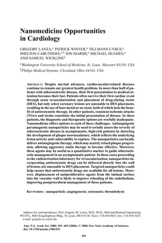
  • 6. 456 ANNALS NEW YORK ACADEMY OF SCIENCES FIGURE 1. MRI aortic wall signal enhancement with 3 -targeted paramagnetic nanoparticle (no drug) 1 week following treatment with 3 -targeted fumagillin or control (no drug) nanoparticles. signal enhancement 1 week after treatment with 3 -targeted nanoparti- cles lacking fumagillin was undiminished (18.1 ± 2.1%) (FIG. 1). Treatment with nontargeted fumagillin nanoparticles did not significantly diminish 3- integrin levels as determined by MRI signal enhancement 1 week after treat- ment, although a numerical decrease was observed (12.4 ± 0.9%). In this study, the total dose of fumagillin administered as a single injection in 3 -targeted nanoparticles was >10,000 times lower than the cumulative oral dose of TNP-470 reported by Moulton et al. Reduced 3 -integrin expression as determined by MRI molecular imaging and corroborated by decreased 3- integrin positive vessel density supported the potential reduction in dosage and increase in efficacy of chemotherapeutic agents using targeted nanomedicine approaches. Incorporation of fumagillin into paramagnetic nanoparticles al- lowed both aortic expression of 3 -integrin and local drug delivery to be assessed and quantified concomitantly and noninvasively. Moreover, the initial magnitude of the aortic signal response among hyperlipidemic rabbits receiv- ing 3 -targeted paramagnetic nanoparticles was correlated with the degree of change in MRI enhancement measured 7 days later with 3 -targeted paramagnetic nanoparticles (without drug). These data illustrate the concept of “rational drug dosing,” which provides noninvasive measures of treatment dosimetry and allows follow-up of response. For asymptomatic patients, nanomedicine approaches offer the ability to quantitatively interrogate the severity and distribution of disease more directly, to locally treat pathology with minimal doses, and to follow up response to
  • 7. LANZA et al.: NANOMEDICINE OPPORTUNITIES IN CARDIOLOGY 457 treatment noninvasively when there are no clinical symptoms or meaningful measures with which to titrate treatment efficacy. NANOMEDICINE APPROACH TO CORONARY REVASCULARIZATION Far too often the progression of atherosclerosis to acute coronary syndromes presents the need for acute revascularization. Fortunately, continuous advances from balloon angioplasty, bare-metal stents, and drug-covered stents such as heparin-coated stents, to the more recent, new class of drug-eluting stents (DES), have expanded our armamentarium for reopening stenotic vessels while preventing vascular reocclusion. Within the last few years, the use of conven- tional balloon angioplasty and bare-metal stent implantation, which were asso- ciated with clinical restenosis rates of 32–42% and 19–30%, respectively,35–37 have been improved with the local deposition of a pharmacologic agent to suppress neointimal proliferation. Current DES have reduced the rate of an- giographic restenosis to below 9% and diminished the frequency for repeat revascularization to below 5%.38,39 Unfortunately, DES cannot be routinely used for all lesions. In some situations, vessel tortuosity or the distal location of lesions prevents manipulation of the relatively inflexible DES. In other cases, the vessel diameter at the culprit lesion is too small for stent placement. As a result many lesions, in whole or part, do not receive the local antirestenotic therapy after revascularization. Moreover, despite the clear success of DES, the incidence of late instent thrombosis has arisen as an infrequent but serious complication of delayed endothelial healing.40 To avoid acute thrombosis, aggressive dual (and occa- sionally triple) antiplatelet therapy is employed for 6 months to a year. We now recognize that some patients are nonresponders to one or more of the drugs.41–43 In other instances, thrombosis presents when antithrombotic drugs are withheld secondary to bleeding complications or the need for emergent surgery. Late instent thrombosis has been linked to fatal outcomes,40 and the risk can persist up to 30 months after DES implantation.42,43 We anticipate that targeted local delivery of antirestenotic drugs such as paclitaxel or rapamycin into the stretch-injured arterial wall rather than the intimal surface will permit better healing and recovery of the endothelium. More rapid endothelial repair of the injured wall should substantially diminish the incidence of thrombosis and reduce the long-term requirement for aggressive antiplatelet therapy. Moreover, DES are now known to elicit unwanted effects on vessel healing and local endothelium-dependent vasomotor responses 6 months after implan- tation in the vessel distal to the intervened segment.44 Although still limited, data on the effect of sirolimus on vasomotor response are accumulating in animal models and patients alike. Swine coronary artery segments exposed to sirolimus for 48 h showed severe impairment of endothelial function.45 In patients, exercise-induced coronary vasoconstriction was noted in vessel
  • 8. 458 ANNALS NEW YORK ACADEMY OF SCIENCES segments adjacent to DES but not bare-metal stents.46 The mechanism for this effect is unclear. Higher rates of restenosis proximal to stent placement compared with the distal edge support an asymmetric downstream effect. We use a nanomedicine approach to address restenosis in lesions not amenable to current DES stent technology by intramural targeting and an- choring of rapamycin nanoparticles to the v 3 -integrin, present on smooth muscle and other plaque components (e.g., macrophages, T cells). In previous studies, we demonstrated that ligand-directed perfluorocarbon nanoparticles could penetrate balloon-injured vessel walls and target intramural biomark- ers, including tissue factor,47 collagen III, and integrins.48 We have recently reported that integrin-targeted PFC nanoparticles can provide effective intra- mural delivery of rapamycin and inhibit vascular stenosis following balloon overstretch injury. In these studies, femoral arteries of 12 rabbits on athero- genic diets for 3 weeks were subjected to balloon stretch injury via a catheter approach from the left common carotid artery. Using a double-balloon tech- nique, paramagnetic 3 -nanoparticles with rapamycin were administered to one artery while the contralateral vessel received targeted nanoparticles with- out drug or saline. Two weeks after nanoparticle treatment, plaque development was determined by MR angiography and by microscopic morphometric quan- tification. Routine MR angiograms were indistinguishable between control and targeted-vessel segments. Microscopic analysis of serial vascular sections 2 weeks after injury revealed that the intimal plaque to lumen area ratio of vessels treated with 3 -targeted rapamycin nanoparticles were significantly (P < 0.05) less (∼50%) than arteries receiving targeted nanoparticles without drug or saline (FIG. 2). Scanning electron microscopy of the intima performed 24 h after injury and treatment with 3 -targeted rapamycin nanoparticles demonstrated their binding to the underlying matrix and cells. Immunofluo- rescent imaging of the vessel wall ∼2 h after treatment demonstrated that the nanoparticles had penetrated into the media and adventitia, consistent with the MR images previously obtained. Although early, the results suggest that 3 -integrin-targeted nanoparticles can provide effective intramural therapy and may be a tool to extend the use of antirestenotic drugs to all revascularized sites with or without adjunctive stent placement. NANOMEDICINE APPROACH TO THROMBOLYSIS Stroke is the third leading cause of death in the United States and often results in functional impairment and long-term disability among survivors.49 Clinical trials have demonstrated that thrombolytic treatment such as tissue plasminogen activator (t-PA) can reduce or reverse ischemia in patients treated within the first 3 h of onset.50 However, intravenously administered throm- bolytic agents are associated with an increased incidence of intracerebral hem- orrhage and expanding stroke. This serious risk frequently delays the receipt of aggressive thrombolytic therapy until intracranial bleeding can be ruled out
  • 9. LANZA et al.: NANOMEDICINE OPPORTUNITIES IN CARDIOLOGY 459 FIGURE 2. Microscopic analysis of serial vascular sections 2 weeks after injury re- vealed that the intimal plaque to lumen area ratio of vessels treated with 3 -targeted rapamycin nanoparticles were significantly (P < 0.05) less than arteries receiving targeted nanoparticles without drug or saline. by CT study of the head. As a result of these delays, the window of opportunity to ameliorate neural damage is lost, and the personal and societal losses are magnified. The advent of perfluorocarbon nanoparticles to specifically deliver drug payloads to intravascular sites of interest presents a unique opportunity to tar- get clot-dissolving therapeutics to cerebral sites of embolism while decreas- ing the risk of hemorrhagic complications and increasing the effectiveness of thrombolytic therapy. We have previously demonstrated targeting of liquid perfluorocarbon nanoparticle emulsions to thrombi in vitro and in vivo,51 with concomitant enhancement of acoustic reflectivity from the targeted surfaces. Acoustic reflectivity enhancement of surfaces targeted with the nanoparticles arises because of the acoustic impedance mismatch between the adherent layer of nanoparticles and the surrounding media. We have recently demonstrated that nanoparticles modified with thrombolytic enzyme (streptokinase) can be targeted onto plasma clots and effect rapid dissolution in the presence of plas- minogen. In this series of experiments, acellular thrombi were produced from citrated human plasma combined with 500 mM calcium chloride and thrombin. Since this study was conducted in vitro, targeting of the nanoparticles to fibrin was accomplished using a three-step process in which biotinylated antifibrin antibody (NIB 1H10 52 ), avidin, and biotinylated nanoparticle emulsions (with or without streptokinase, depending on treatment group) were combined se- quentially with interval washings of unbound reagents. Acoustic microscopy was performed on targeted and control samples using a broadband, 25-MHz immersion transducer (Panametrics V324, Waltham, MA, USA) operated in
  • 10. 460 ANNALS NEW YORK ACADEMY OF SCIENCES pulse-echo mode. A computer-controlled pulser receiver was used to generate insonifying pulses and amplify the received echoes. The transducer was affixed to a three-axis, computer-controlled, motorized gantry. Radiofrequency (RF) data were acquired, digitized to 8 bits at 500 MHz for 2,048 point records, and stored to disk at every site as the transducer was scanned over each sample in a rectangular grid with 100- m resolution. In preparation for scanning, each sample was sealed within a chamber having a cellophane acoustic window, and which was filled with phosphate buffered saline (PBS). The sample chamber was submerged within a 37◦ C water bath, and the sample was scanned to yield a baseline measurement. The chamber was then emptied of PBS through an injection port and refilled with either plasminogen in PBS buffer (3 U/mL) or PBS alone. Scans were then performed at 15-min intervals for 3 h, and spatial registration was maintained at all times. RF data were analyzed to assess temporal changes in clot morphology and backscatter. A sliding Hamming window (0.2 sec duration) was applied and moved over the data in 2-nsec steps. The sum of the squared values within each segment, a quantity proportional to the reflected energy, was used as input to a peak-detection algorithm (implemented in LabVIEW, National Instruments Corp., Austin, TX, USA) and used to determine the arrival time of the echo from the thrombus surface. A similar technique was used to detect the echo from the nitrocellulose substrate in the same waveform. The difference between the echo arrival times of the clot surface and substrate determined the profile of the clot, and these values were used to generate surface plots for visualization of the sample volume. Backscatter was quantified by first applying a rectangular window to each waveform to isolate the reflection from the targeted surface, and then by calculating the log spectral difference with respect to the reflection from a steel plate. The average value within the usable bandwidth (10–30 MHz) was recorded in dB for each point in the scan, and this value was used to generate a C-scan image of the integrated backscatter from the sample surface. The detected clot volume was dramatically decreased (P < 0.05) for clots treated with fibrin-targeted streptokinase nanoparticles and exposed to plasminogen in buffer (FIG. 3). Treatment with fibrin-targeted streptokinase nanoparticles incubated in saline or fibrin-targeted nanoparticles without strep- tokinase incubated with plasminogen in buffer had no thrombolytic effect. The time to complete lysis varied with small changes in the synthesis process of the streptokinase nanoparticle formulations. Initial conjugates had to be left overnight for complete dissolution while more optimized agents formed later completely dissolved clots in vitro within an hour, often less than 15 min. Fibrin clot dissolution occurred from inside to outside. In some replicates, the clot measured at 1 h was a hollow fibrin shell, which immediately collapsed with slight motion. None of the control clots revealed morphologic or acoustic changes. The measurements presented here suggest that fibrin-targeted streptokinase nanoparticles could be used to promote local thrombolysis of plasma clots in vivo. We have previously shown that fibrin-targeted nanoparticles can
  • 11. LANZA et al.: NANOMEDICINE OPPORTUNITIES IN CARDIOLOGY 461 FIGURE 3. Mean normalized clot volume following 2-h treatment with fibrin-targeted nanoparticles streptokinase-modified or control perfluorocarbon nanoparticles incubated with plasminogen or saline. penetrate and acoustically enhance acute intravascular thromboses in dogs. Moreover, we have found that perfluorocarbon nanoparticles are constrained to the vasculature due to their nominal size, even in “leaky” vascular beds such as tumor neovasculature. Collectively, those results suggest that fibrin- targeted streptokinase nanoparticles could be used early in acute stroke or unstable angina with limited extravascular effects. SUMMARY Nanomedicine is a new evolving field referred to by many names, which promises to significantly enhance the tools available to clinicians to address some of the serious challenges responsible for profound mortality, morbidity, and numerous societal consequences. Unlike the simple pharmaceutics of the past, nanomedicine agents are typically three-dimensional, multicomponent systems, which require interdisciplinary expertise to produce and use. In this review, we have briefly introduced the opportunities associated with targeted perfluorocarbon nanoparticles in early atherosclerosis, in acute revasculariza- tion, and in thrombolytic therapy. The potential impact of these three concepts is enormous but pales in comparison with the advancements likely to evolve in this field over the coming decades. ACKNOWLEDGMENTS This research was supported by the NIH grants (HL-42950, HL-59865, HL-78631, NO1-CO-37007, and EB-01704), SCAI/Bracco Diagnostics, Inc.- ACIST Fellowship Program, and the American Heart Association. Philips
  • 12. 462 ANNALS NEW YORK ACADEMY OF SCIENCES Medical Systems (Cleveland, OH, USA) provided valuable equipment, soft- ware, and engineering support. REFERENCES 1. ANONYMOUS. 2005. American Heart Association, Vol. 2005. 2. LANZA, G., K. WALLACE, M. SCOTT, et al. 1996. A novel site-targeted ultra- sonic contrast agent with broad biomedical application. Circulation 94: 3334– 3340. 3. MATTREY, R.F. 1994. The potential role of perfluorochemicals (pfcs) in diagnostic imaging. Artif. Cells Blood Substit. Immobil. Biotechnol. 22: 295–313. 4. HALL, C.S., G.M. LANZA, J.H. ROSE, et al. 2000. Experimental determination of phase velocity of perfluorocarbons: applications to targeted contrast agents. IEEE Trans. Ultrason. Ferroelec. Freq. Contr. 47: 75–84. 5. HALL, C., J. MARSH, M. SCOTT, et al. 2001. Temperature dependence of ultrasonic enhancement with a site-targeted contrast agent. J. Acous. Soc. AM. 110: 1677– 1684. 6. LANZA, G., C. LORENZ, S. FISCHER, et al. 1998. Enhanced detection of thrombi with a novel fibrin-targeted magnetic resonance imaging agent. Acad. Radiol. 5(Suppl 1): s173–s176. 7. FLACKE, S., S. FISCHER, M. SCOTT, et al. 2001. A novel MRI contrast agent for molecular imaging of fibrin: implications for detecting vulnerable plaques. Cir- culation 104: 1280–1285. 8. WINTER, P., S. CARUTHERS, X. YU, et al. 2003. Improved molecular imaging contrast agent for detection of human thrombus. Mag. Reson. Med. 50: 411– 416. 9. YU, X., S.-K. SONG, J. CHEN, et al. 2000. High-resolution MRI characterization of human thrombus using a novel fibrin-targeted paramagnetic nanoparticle con- trast agent. Mag. Reson. Med. 44: 867–872. 10. LANZA, G.M., P.M. WINTER, A.M. NEUBAUER, et al. 2005. 1 H/19 Fmagnetic reso- nance molecular imaging with perfluorocarbon nanoparticles. Curr. Top. Dev. Biol. 70: 57–76. 11. CARUTHERS, S.D., A.M. NEUBAUER, F.D. HOCKETT, et al. 2006. In vitro demon- stration using 19f magnetic resonance to augment molecular imaging with para- magnetic perfluorocarbon nanoparticles at 1.5 tesla. Invest. Radiol. 41: 305– 312. 12. KOLODGIE, F.D., R. VIRMANI, A.P. BURKE, et al. 2004. Pathologic assessment of the vulnerable human coronary plaque. Heart 90: 1385–1391. 13. VIRMANI, R., F.D. KOLODGIE, A.P. BURKE, et al. 2005. Atherosclerotic plaque pro- gression and vulnerability to rupture: angiogenesis as a source of intraplaque hemorrhage. Arterioscler. Thromb. Vasc. Biol. 25: 2054–2061. 14. SCHAAR, J.A., J.E. MULLER, E. FALK, et al. 2004. Terminology for high-risk and vulnerable coronary artery plaques. Report of a meeting on the vulnera- ble plaque, June 17 and 18, 2003, Santorini. Greece. Eur. Heart J. 25: 1077– 1082. 15. OJIO, S., H. TAKATSU, T. TANAKA, et al. 2000. Considerable time from the onset of plaque rupture and/or thrombi until the onset of acute myocardial infarction in humans: coronary angiographic findings within 1 week before the onset of infarction. Circulation 102: 2063–2069.
  • 13. LANZA et al.: NANOMEDICINE OPPORTUNITIES IN CARDIOLOGY 463 16. BOTNAR, R.M., A. BUECKER, A.J. WIETHOFF, et al. 2004. In vivo magnetic reso- nance imaging of coronary thrombosis using a fibrin-binding molecular magnetic resonance contrast agent. Circulation 110: 1463–1466. 17. BOTNAR, R.M., A.S. PEREZ, S. WITTE, et al. 2004. In vivo molecular imaging of acute and subacute thrombosis using a fibrin-binding magnetic resonance imaging contrast agent. Circulation 109: 2023–2029. 18. KAHLON, R., J. SHAPERO & A.I. GOTLIEB. 1992. Angiogenesis in atherosclerosis. Can. J. Cardiol. 8: 60–64. 19. FAN, T.P., R. JAGGAR & R. BICKNELL. 1995. Controlling the vasculature: angiogen- esis, anti-angiogenesis and vascular targeting of gene therapy. Trends Pharmacol. Sci. 16: 57–66. 20. SUEISHI, K., Y. YONEMITSU, K. NAKAGAWA, et al. 1997. Atherosclerosis and angio- genesis. Its pathophysiological significance in humans as well as in an animal model induced by the gene transfer of vascular endothelial growth factor. Ann. N. Y. Acad. Sci. 811: 311–322; 322–324. 21. MOULTON, K. 2002. Plaque angiogenesis: its functions and regulation. Cold Spring Harb. Symp. Quant. Biol. 67: 471–482. 22. MOULTON, K.S. 2001. Plaque angiogenesis and atherosclerosis. Curr. Atheroscler. Rep. 3: 225–233. 23. MOULTON, K.S., E. HELLER, M.A. KONERDING, et al. 1999. Angiogenesis inhibitors endostatin or tnp-470 reduce intimal neovascularization and plaque growth in apolipoprotein e-deficient mice. Circulation 99: 1653–1655. 24. KLEIN, C.D. & G. FOLKERS. 2003. Understanding the selectivity of fumagillin for the methionine aminopeptidase type II. Oncol. Res. 13: 513–520. 25. RODRIGUEZ-NIETO, S., M.A. MEDINA & A.R. QUESADA. 2001. A re-evaluation of fumagillin selectivity towards endothelial cells. Anticancer Res. 21: 3457–3460. 26. LIU, S., J. WIDOM, C.W. KEMP, et al. 1998. Structure of human methionine aminopeptidase-2 complexed with fumagillin. Science 282: 1324–1327. 27. TRAN, H.T., G.R. BLUMENSCHEIN JR., C. LU, et al. 2004. Clinical and pharmacoki- netic study of TNP-470, an angiogenesis inhibitor, in combination with paclitaxel and carboplatin in patients with solid tumors. Cancer Chemother. Pharmacol. 54: 308–314. 28. HERBST, R.S., T.L. MADDEN, H.T. TRAN, et al. 2002. Safety and pharmacokinetic effects of TNP-470, an angiogenesis inhibitor, combined with paclitaxel in pa- tients with solid tumors: evidence for activity in non-small-cell lung cancer. J. Clin. Oncol. 20: 4440–4447. 29. LANZA, G.M., X. YU, P.M. WINTER, et al. 2002. Targeted antiproliferative drug delivery to vascular smooth muscle cells with a magnetic resonance imaging nanoparticle contrast agent: implications for rational therapy of restenosis. Cir- culation 106: 2842–2847. 30. CROWDER, K.C., M.S. HUGHES, J.N. MARSH, et al. 2005. Sonic activation of molecularly-targeted nanoparticles accelerates transmembrane lipid delivery to cancer cells through contact-mediated mechanisms: implications for enhanced local drug delivery. Ultrasound Med. Biol. 31: 1693–1700. 31. LANZA, G.M., D.R. ABENDSCHEIN, X. YU, et al. 2002. Molecular imaging and targeted drug delivery with a novel, ligand-directed paramagnetic nanoparticle technology. Acad. Radiol. 9(Suppl 2): S330–S331. 32. LANZA, G.M., P. WINTER, S. CARUTHERS, et al. 2004. Novel paramagnetic contrast agents for molecular imaging and targeted drug delivery. Curr. Pharm. Biotech- nol. 5: 495–507.
  • 14. 464 ANNALS NEW YORK ACADEMY OF SCIENCES 33. WINTER, P.M., A.M. MORAWSKI, S.D. CARUTHERS, et al. 2003. Molecular imag- ing of angiogenesis in early-stage atherosclerosis with alpha(v)beta3-integrin- targeted nanoparticles. Circulation 108: 2270–2274. 34. WILSON, M.W., J.M. LABERGE, R.K. KERLAN, et al. 2002. MR portal venography: preliminary results of fast acquisition without contrast material or breath holding. Acad. Radiol. 9: 1179–1184. 35. JAEGERE PD, P., R. DOMBURG RV, H. NATHOE, et al. 1999. Long-term clinical outcome after stent implantation in coronary arteries. Int. J. Cardiovasc. Interv. 2: 27–34. 36. KIMURA, T., T. TAMURA, H. YOKOI, et al. 1994. Long-term clinical and angiographic follow-up after placement of palmaz-schatz coronary stent: a single center ex- perience. J. Interv. Cardiol. 7: 129–139. 37. KEANE, D., A.J. AZAR, P. DE JAEGERE, et al. 1996. Clinical and angiographic out- come of elective stent implantation in small coronary vessels: an analysis of the benestent trial. Semin. Interv. Cardiol. 1: 255–262. 38. SERRUYS, P.W., P.A. LEMOS & B.A. VAN HOUT. 2004. Sirolimus eluting stent im- plantation for patients with multivessel disease: rationale for the arterial revas- cularisation therapies study part II (ARTS II). Heart 90: 995–998. 39. SERRUYS, P.W., E. REGAR & A.J. CARTER. 2002. Rapamycin eluting stent: the onset of a new era in interventional cardiology. Heart 87: 305–307. 40. ONG, A.T., E.P. MCFADDEN, E. REGAR, et al. 2005. Late angiographic stent throm- bosis (last) events with drug-eluting stents. J. Am. Coll. Cardiol. 45: 2088–2092. 41. WENAWESER, P. & O. HESS. 2005. Stent thrombosis is associated with an impaired response to antiplatelet therapy. J. Am. Coll. Cardiol. 46: CS5–CS6. 42. MCFADDEN, E.P., E. STABILE, E. REGAR, et al. 2004. Late thrombosis in drug- eluting coronary stents after discontinuation of antiplatelet therapy. Lancet 364: 1519–1521. 43. RODRIGUEZ, A.E., J. MIERES, C. FERNANDEZ-PEREIRA, et al. 2006. Coronary stent thrombosis in the current drug-eluting stent era: insights from the eraci iii trial. J. Am. Col. Cardiol. 47: 205–207. 44. HOFMA, S.H., W.J. VAN DER GIESSEN, B.M. VAN DALEN, et al. 2006. Indication of long-term endothelial dysfunction after sirolimus-eluting stent implantation. Eur. Heart J. 27: 166–170. 45. JEANMART, H., O. MALO, M. CARRIER, et al. 2002. Comparative study of cy- closporine and tacrolimus on coronary endothelial function. J. Heart Lung Trans- plant. 21: 990–998. 46. TOGNI, M., S. WINDECKER, R. COCCHIA, et al. 2005. Sirolimus-eluting stents as- sociated with paradoxic coronary vasoconstriction. J. Am. Coll. Cardiol. 46: 231–236. 47. LANZA, G., D. ABENDSCHEIN, C. HALL, et al. 2000. Molecular imaging of stretch- induced tissue factor expression in carotid arteries with intravascular ultrasound. Invest. Radiol. 35: 227–234. 48. CYRUS, T., D.R. ABENDSCHEIN, S.D. CARUTHERS, et al. 2006. MR three-dimensional molecular imaging of intramural biomarkers with targeted nanoparticles. J. Car- diovasc. Magn. Reson. 8: 535–541. 49. HINCHEY, J.A. & C. BENESCH. 2000. Thrombolytic therapy in patients with acute ischemic stroke. Arch. Neurol. 57: 1430–1436. 50. GROUP, T.N.I.O.N.D.A.S.R.-P.S.S. 1995. Tissue plasminogen activator for acute ischemic stroke. The National Institute of Neurological Disorders and Stroke rt-PA Stroke Study Group. N. Engl. J. Med. 333: 1581–1587.
  • 15. LANZA et al.: NANOMEDICINE OPPORTUNITIES IN CARDIOLOGY 465 51. LANZA, G.M., K.D. WALLACE, M.J. SCOTT, et al. 1996. A novel site-targeted ultra- sonic contrast agent with broad biomedical application. Circulation 95: 3334– 3340. 52. RAUT, S. & P. GAFFNEY. 1996. Evaluation of fibrin binding profile of two antifibrin monoclonal antibodies. Thromb. Haemost. 76: 56–64.