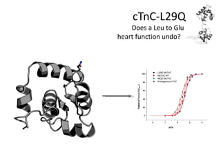
SlideShare ist ein Scribd-Unternehmen logo
1 von 40
cTnC-L29Q
    Does a Leu to Glu
heart function undo?
Topics
•   Introduce NMR spectroscopy
•   Review L29Q history and literature
•   Structure calculation by NMR
•   Dynamics measurement by NMR
•   Conclusions
What is NMR?
•   Nuclear Magnetic Resonance
    spectroscopy
•   Similar to other forms of
    spectroscopy
     – A photon of light causes a
       transition from a ground state to
       an excited state
•   In visible spectroscopy an
    electron absorbs the energy
•   In NMR, the absorbed photon
    promotes a transition of
    nuclear spin from ground to
    excited state
What is NMR?
•   Lifetime is ~109 times longer than conventional
    spectroscopies
•   Ground and excited states in NMR arise from
    the interaction of a nuclear magnetic dipole
    moment with an intense external magnetic
    field
•    The magnetic dipole arises from spin angular
    momentum
     – The spin angular momentum of a ½ nuclei can be
       either: +½ħ or -½ħ
     – The magnetic moment of s nuclear spin is
       proportional to its gyromagnetic ratio (γ)
What is NMR?
•   As the strength of the field
    increases so does the energy
    separation
     •   The net absorption depends on
         population difference
     •   Since NMR is insensitive need lots of
         material (i.e. mM concentrations)
     • Going from 14.1 T (600 MHz) to
       21.2 T (900 MHz) increases the             ∆E = (h / 2π )γ Bo
       signal to noise by ca. 84% and
       even from 18.8 T (800 MHz) to             Nβ      (h / 2π )γ Bo
       900 MHz increases it by 20%                  ≈ 1−
                                                 Nα           kT

                                                      S / N ∝ B0
                                                               3/2
What is NMR?
•   In a magnetic field nuclei precess
    about B0 at the resonance frequency
    (600 MHz = γ(1H atoms)*14.1 Tesla)
•   Pulse sample with a second magnetic
    field oscillating at the resonance
    frequency perpendicular to B0
•   Spins precess about B0 at their
    resonance frequency (bulk is in the
    transverse plane)
•   Measure the evolution speed of the
    spins (chemical shift; represented by
    p.p.m., but really defined as hz/mhz)
      – 10 ppm in a 600 MHz instrument
         represents 6000 Hz off from 600
         MHz
What is NMR?

•   Coupling: if nucleus A is near another
    nonequivalent nucleus B than when
    nucleus B is +½ and -½ nucleus A will
    experience different magnetic fields, and
    thus will have different chemical shifts
•   J-coupling: through bonds
•   Dipolar coupling: through space
The N-HSQC15


•   1
     H , 15N-HSQC correlates amide 1H with amide 15N
•   Spectra will change if magnetic environment changes
•   Can be used to obtain binding constants and predict binding sites
First FHC mutation in cTnC
•    In 2001, Hoffmann B, et al.
     identified in a 60 year old male
     patient
      – ECG revealed he had
         concentric hypertrophy of
         the left ventricle
•    Did not find it in 96 healthy
     volunteers, but authors were
     not willing to rule it out as
     “simple coincidence”
•    L29 serves to stabilize the A-
     helix


    Hoffmann B, et al. (2001) Human Mutation 17, 524
Function of L29


•   Differences in chemical shift of cTnC when cTnI1-80DD vs. cTnI1-80
•   L29 may be involved in binding to the cardiac specific N-terminal extension
    of cTnI


Finley et al., 1999
Function of L29
•   Deletion of 16-29 mimic phosphorylated state of
    contraction (Ward et al. 2002)
•   Cross-linking implicate cTnI1-64 interacts with I18C
    and R26C to cTnC (Ward et al. 2003)
•   Ward et al. (2004a) proved by looking at cTnI1-64
    NMR spectrum that when it is bisphosphorylated it
    does not bind to cNTnC, but does so when
    unphosphorylated
     –   Observed by monitoring broadening of 1D signals of cTnI1-64
         as cNTnC was titrated in
     –   Binds via Y25, Y28, and H33 of cTnI
•   Ward et al. (2004b) used 15N-HSQC data of
    cTnI1-64 to show that residues that flank the S22
    and S23 are less perturbed by cTnC when
    phosphorylated
Rosevear/Solaro Model
•   Rosevear and Solaro (Howarth
    et al., 2007) solved the NMR
    structure of cTnI1-32pp and
    proposed a mechanism
     –   Model suggested that R21 and
         R27 of cTnI interacts with E32 and
         D33 in site I and P11 forms a
         hydrophobic interaction with L29
•   Model also supported by
    cross-linking data(Warren et al.
    2009
     –   Also implicates cTnI147-163
         (bound to cNTnC) as a binding
         partner of the N-terminal
         extension of cTnI
Back to L29Q (Jaquet)

 •   Signal was reduced by ca. 14%
     at 208 and 222 nm.
 •   Results suggested that
     secondary structure contained
     ~2% less alpha helix for both
     apo and Ca2+ bound
 •   Found by peptide arrays that
     L29Q did not bind the N-
     terminal extension of cTnI,
     regardless of phosphorylation
     level (wt did, except for cTnIpp)


Schmidtmann A, et al. (2005) FEBS J. 6087-6097
Schmidtmann A, et al. Continued

•   ATPase assays and in vitro motility
    assays
•   pCa50 of L29Q was reduced when
    compared to WT (by ca. 0.1 units)
•   Found that phosphorylation had
    less of an impact on L29Q than WT
L29Q (Cheung)
•   FRET measurements in cTnC(L12W/N51C-
    IAEDANS) reconstituted thin filaments
•   No structural change in L29Q versus WT
•   Calcium sensitivity decreased for L29Q
    by 0.1 unit
•   No further decrease as a function of
    phosphorylation
      – Whereas wt decreased by
         approximately 0.2 units


Dong, W-J, et al. (2008) JBC 3424-3432
L29Q (Sykes)
•   No affect on Calcium binding
•   cTnI147-163 affinity was not
    altered by cTnI1-29 or cTnI-pp
      – Not true for WT-cTnC (as
         shown by OKB and
         Abbott et al)
•   And relaxation studies
    indicated that cTnI1-29 bound
    less efficiently to L29Q than
    WT


Baryshnikova, O, et al. (2008) JMB 735-751
L29Q (trout cardiac troponin C)
•   Trout troponin has an increased calcium
    affinity (2-3 fold)
     – Residues responsible are: N2, I28, Q29
          and D30 (Gillis et al., 2005)
     – Human cardiac cTnC: D2, V28, L29, G30
     – When cardiac contained these residues
          Ca sensitivty increased by 2-fold
•   Coordinate a second calcium weakly?
     – Not actually observed experimentally
     – Structure not much different than human
          cardiac (Blumenschein et al., 2004)
•   Trout cardiac troponin I lacks the N-terminal
    extension
     – Found that trout cTn is less sensitive to
          PKA than human cTn (Kirkpatrick et al.,
          2011)
L29Q (Davis and Tibbits)
•   Florescence Measurements:
     – Half maximal Ca2+ for cTnCF27W: 3.7 ± 0.2
         μM
     – L29Q: 2.8 ± 0.3 μM
     – NIQD: 2.0 ± 0.1 μM
•   Force pCa curves of skinned murine
    cardiomyocytes
     – WT: EC50 = 4.1 ± 0.5 μM
     – L29Q: EC50 = 3.0 ± 0.5 μM
     – NIQD: EC50 = 2.1 ± 0.5 μM
•   Stress that skinned cardiomyocytes are a better
    representation of reality than isolated thin
    filaments


Liang, B, et al. (2008) Physiol Genomics 257-266
L29Q (Potter)
•   Did not see a statistically
    significant increase in calcium
    sensitivity with skinned fibers,
    cardiac myofibrils, or regulated
    thin filaments (fluorescence)
     – although all had a “trend” towards a
       slight increase in calcium sensitivity
•   Porcine instead of murine muscle
•   Both Potter and Davis not
    controlling for phosphorylation
    levels, so may explain differences


Dweck, D, et al. (2008) JBC 33119-33128
L29Q (Pfitzer)

•   pCa50 unaffected by L29Q
•   Nor did PP1c treatment followed
    by PKA treatment yield any
    differences between wt and L29Q
•   Not just phosphorylating S22/S23
    anymore…
•   Unfortunately, they do not
    address differences between
    their results and Davis’s; actually
    they mention them as if they
    agree!


Neulen, A, et al. (2009) Basic Res Cardiol 751-760
Structure-function relationship
Assignment of Chemical shifts
• Easy for a single
  amino acid or even a
  small polypeptide…
Three-dimensional spectroscopy
•   Need to increase
    resolution for larger
    molecules
Assignment
of HSQC and
 Cα and Cβ
Structure prediction by ORBplus


                          AB interhelical angle (°)   CD interhelical angle (°)
      cNTnC(WT)                   134 ± 3                     118 ± 4
     cNTnC(Acys)                  142 ± 3                     109 ± 4
cNTnC(WT)-cTnI(147-163)           102 ± 4                      95 ± 6
    L29Q (ORBplus)                  143                        118.3
NMR experiment      Purpose                         Time
15
     N-HSQC         1
                        HN,15N pairs                30 min – 24 hours
13
     C-HSQC         1
                     H,13C pairs (aromatic          30 min – 24 hours
                    and/or aliphatic)
HNCACB              1
                     HN,13C (a and b; i,            48 – 64 hours
                    i-1)),15N
CBCA(CO)NNH         1
                        HN,13C (a and b; i-1),15N   48 – 64 hours
HC(CO)NH            1
                        HN,1H (i-1), 15N            72 hours
                                                                              •   Only represent a fraction
C(CO)NH             1
                        HN,13C (i-1),15N            72 hours
                                                                                  of the NMR experiments
15
     N-HSQC-NOESY   1
                        HN, 1H (j), 15N             72 – 144 hours                to help determine a
15
     N-HSQC-TOCSY   1
                        HN, 1H (i), 15N             72 hours                      protein structure
                                                                              •   Different for other
13
  C-HSQC-NOESY      1
                     H (aliphatic), 1H (j), 13C     100 – 144 hours
                                                                                  biological samples, such
(aliphatic)         (aliphatic)
                                                                                  as DNA
13
  C-HSQC-NOESY      1
                     H (aromatic), 1H (j), 13C      48 – 72 hours             •   Also need to add more
(aromatic)          (aromatic)                                                    experiments, if interested
HNHA                1
                     HN, 1H (a some b), 15N to      48 – 64 hours                 in solving a protein-ligand
                    obtain φ                                                      structure
HNHB                1
                        HN, 1H (b), 15N to obtain   48 – 64 hours
                    χ1
HN(CO)HB            1
                        HN, 1H (b), 15N to obtain   48 – 64 hours
                    χ1

Total                                               677 (28 days) – 944 (40
                                                    days)
Dihedral Restarints
NOESY
 •     The NOESY experiment measures the dipolar interaction between
       nuclei
 •     The intensity of an NOE is proportional to 1/r6 and can therefore
       provide distance measurements


Berg J.M., 2002
Structure calculation
•    Energy minimization: move atoms around
     to try and minimize energy
       – Define experimental restraints (and
          non-experimental, such as covalent
          bonds) as having energy
       – The higher the energy the greater the
          divergence a model is from the
          constraints
•    To avoid the structure from becoming
     trapped in a local minima simulated
     annealing is employed
       – Atoms are given a kinetic energy
          (associated with a high temperature
          and then cooled slowly
•    The ensemble represents a set of
     structures that satisfy the experimental
     restraints


Berg J.M., 2002
Structural Statistics for L29Q
R.m.s.d. from the average structure  Backbone atoms      Heavy Atoms
                  a
Ordered residues (Å)                    0.94 ± 0.18         1.40 ± 0.16
Total Distance Restraints                            1692
Intra Residual NOEs                                  1033
Short range (|i-j|=1) NOEs                            307
Medium range (1<|i-j|<5) NOEs                         191
Long range (|i-j|≥5) NOEs                             153
   2+
Ca distance restraints                                 8
Dihedral restraints                                   175
φ/ψ                                               154 (72/72)
χ1                                                     21
                          b
NOE violations/Structure
> 0.5 Å                                               0.0
> 0.3 Å                                               0.0
> 0.1 Å                                              3.35
Dihedral Violations/Structure (> 5º)                  0.0
Ramachadran plot statistics c
φ/ψ in most favored regions (%)                      96.6
φ/ψ in additionally allowed regions                   3.4
(%)
φ/ψ generously allowed regions (%)                    0.0
φ/ψ in disallowed regions (%)                         0.0
a
  Residues 3-49, 52-85; as calculated by psvs
b
  Violations are for the 20 NMR lowest energy
       structures
c
  Procheck for ordered residues listed above.
cNTnC(L29Q) structure


                                 AB interhelical angle (°)   CD interhelical angle (°)
             cNTnC(WT)                   134 ± 3                     118 ± 4
            cNTnC(Acys)                  142 ± 3                     109 ± 4
       cNTnC(WT)-cTnI(147-163)           102 ± 4                      95 ± 6
             sNTnC(WT)                    90 ± 3                      69 ± 5
           L29Q (ORBplus)                  143                        118.3
             L29Q (NMR)                  139 ± 5                     122 ± 7
cNTnC(L29Q) structure
Alignment with other ‘closed’
                  structures


L29Q (slate); cNTnC(WT), pdb code:1AP4 (magenta); cNTnC(Acys), pdb code: 2CTN (grey);
trout NTnC at 30°C, pdb code: 1R2U (orange); trout NTnC at 7°C, pdb code:1R6P (yellow);
sNTnC(E41A), pdb code: 1SMG (Green)
Alignment of loop 1


•   The structures were aligned between residues 15-27 and 40-48
    and the r.m.s.d. of the flexible loop in site 1 (residues 28-34)
    was determined to be (A) 3.5 Å, (B) 2.1 Å, and (C) 1.6 Å.

•   Loop 1 of cNTnC(L29Q) superimposes much better with cNTnC-
    cTnI(147-163) than cNTnC(Acys)
Dynamics of loop 1
•   Can determine the mobility of a backbone amide by determining its
    relaxation rates
     –   T1 is the relaxation time to return to thermal equilibrium
     –   T2 is the time it takes for transverse magnetization to be lost
     –   1
          H-15N NOE measures how altering the ground and excited state of one spin can affect the ground and
         excited state of another spin
•   Relaxation is caused by magnetic field fluctuations
     –   Can be caused by rapid internal (or external) motion
     –   direct interactions with nearby magnetic nuclei (DD), chemical shift effects (CSA), quadrupole-electric
         field gradient interaction (QR) and rapid modulation of J-coupling (SC)
S of L29Q compared with WT
2


              •   Since relaxation values are difficult
                  to interpret on their own, it is often
                  useful to use their values to calculate
                  the order parameter, S2
                   – Related to the amplitude of
                       internal motion
                   – If all orientations of the 1H-15N
                       vector are equally probable
                       than S2 = 0; if motion is rigid,
                       than S2 = 1.

              •   The data suggest that the order of
                  the loop is relatively unchanged
              •   There may be a slight increase in the
                  order at the end of the loop
Conclusions
•   L29Q did not alter the global structure of cNTnC
•   May slightly change the orientation of loop 1
     – New conformation may destabilize binding to cTnI1-29
     – Or may function just simply by destabilizing necessary hydrophobic interactions
       between cTnI1-29 and L29Q
     – Dynamics of loop were not significantly altered when compared to cNTnC
•   Maybe L29Q does nothing…
•   Limitations:
     – Only N-domain
     – Only Ca2+-bound (apo was not analyzed)
     – No cTnI147-163 or cTnI1-29 bound in structure
     – Low resolution of NMR so it’s difficult to be certain of change in the
        conformation of loop 1
Does a Leu to Glu
heart function undo?
…I still have no clue!
Acknowledgments

University of Alberta
Brian Sykes
Monica Li
Leo Spyracopoulos

Simon Fraser University
Glen Tibbits

King’s College London
Malcolm Irving
Yin-Biao Sun
(and everyone else)

Weitere ähnliche Inhalte

Was ist angesagt?

KEK PH 2017
KEK PH 2017KEK PH 2017
KEK PH 2017Son Cao
 
Strong Higgs Pair Production at the LHC with jet substructure
Strong Higgs Pair Production at the LHC with jet substructureStrong Higgs Pair Production at the LHC with jet substructure
Strong Higgs Pair Production at the LHC with jet substructurejuanrojochacon
 
Nitrogen Chemistry in Disffuse Interstellar Medium
Nitrogen Chemistry in Disffuse Interstellar MediumNitrogen Chemistry in Disffuse Interstellar Medium
Nitrogen Chemistry in Disffuse Interstellar MediumPrince Tiwari
 
Thermodynamic modeling of the platinum yttrium system
Thermodynamic modeling of the platinum yttrium systemThermodynamic modeling of the platinum yttrium system
Thermodynamic modeling of the platinum yttrium systemmbb5123
 
NMR studies of Complement complex and FkpA chaperone with substrate
NMR studies of Complement complex and FkpA chaperone with substrateNMR studies of Complement complex and FkpA chaperone with substrate
NMR studies of Complement complex and FkpA chaperone with substrateKonstantin Pervushin
 
Master Thesis Total Oxidation Over Cu Based Catalysts
Master Thesis  Total Oxidation Over Cu Based CatalystsMaster Thesis  Total Oxidation Over Cu Based Catalysts
Master Thesis Total Oxidation Over Cu Based Catalystsalbotamor
 
ICL - 2011, Ann Arbor, MI
ICL - 2011, Ann Arbor, MIICL - 2011, Ann Arbor, MI
ICL - 2011, Ann Arbor, MIelenamilliken
 

Was ist angesagt? (13)

MRI Physics RV
MRI Physics RVMRI Physics RV
MRI Physics RV
 
BioChar
BioCharBioChar
BioChar
 
KEK PH 2017
KEK PH 2017KEK PH 2017
KEK PH 2017
 
Strong Higgs Pair Production at the LHC with jet substructure
Strong Higgs Pair Production at the LHC with jet substructureStrong Higgs Pair Production at the LHC with jet substructure
Strong Higgs Pair Production at the LHC with jet substructure
 
Nitrogen Chemistry in Disffuse Interstellar Medium
Nitrogen Chemistry in Disffuse Interstellar MediumNitrogen Chemistry in Disffuse Interstellar Medium
Nitrogen Chemistry in Disffuse Interstellar Medium
 
Nmr 1
Nmr 1Nmr 1
Nmr 1
 
Radiogenic isotope
Radiogenic isotopeRadiogenic isotope
Radiogenic isotope
 
Thermodynamic modeling of the platinum yttrium system
Thermodynamic modeling of the platinum yttrium systemThermodynamic modeling of the platinum yttrium system
Thermodynamic modeling of the platinum yttrium system
 
NMR studies of Complement complex and FkpA chaperone with substrate
NMR studies of Complement complex and FkpA chaperone with substrateNMR studies of Complement complex and FkpA chaperone with substrate
NMR studies of Complement complex and FkpA chaperone with substrate
 
Presentation
PresentationPresentation
Presentation
 
NMRpresentationCombined
NMRpresentationCombinedNMRpresentationCombined
NMRpresentationCombined
 
Master Thesis Total Oxidation Over Cu Based Catalysts
Master Thesis  Total Oxidation Over Cu Based CatalystsMaster Thesis  Total Oxidation Over Cu Based Catalysts
Master Thesis Total Oxidation Over Cu Based Catalysts
 
ICL - 2011, Ann Arbor, MI
ICL - 2011, Ann Arbor, MIICL - 2011, Ann Arbor, MI
ICL - 2011, Ann Arbor, MI
 

Ähnlich wie cTnC-L29Q

UCSF Hyperpolarized MR #2: DNP Physics and Hardware (2019
UCSF Hyperpolarized MR #2: DNP Physics and Hardware (2019UCSF Hyperpolarized MR #2: DNP Physics and Hardware (2019
UCSF Hyperpolarized MR #2: DNP Physics and Hardware (2019Peder Larson
 
1H NUCLEAR MAGNETIC RESONANCE
1H NUCLEAR MAGNETIC RESONANCE1H NUCLEAR MAGNETIC RESONANCE
1H NUCLEAR MAGNETIC RESONANCEsantoshkumar3159
 
Introduction-NMR.pdf
Introduction-NMR.pdfIntroduction-NMR.pdf
Introduction-NMR.pdfAbid Zia
 
HETCOR,INADEQUATE.pptx
HETCOR,INADEQUATE.pptxHETCOR,INADEQUATE.pptx
HETCOR,INADEQUATE.pptxPratikKapse8
 
Measurement-induced long-distance entanglement of superconducting qubits usin...
Measurement-induced long-distance entanglement of superconducting qubits usin...Measurement-induced long-distance entanglement of superconducting qubits usin...
Measurement-induced long-distance entanglement of superconducting qubits usin...Ondrej Cernotik
 
use of nmr in structure ellucidation
 use of nmr in structure ellucidation use of nmr in structure ellucidation
use of nmr in structure ellucidationAnuradha Verma
 
Two dimensional nmr spectroscopy (practical application and spectral analysis
Two dimensional  nmr spectroscopy (practical application and spectral analysisTwo dimensional  nmr spectroscopy (practical application and spectral analysis
Two dimensional nmr spectroscopy (practical application and spectral analysisAwad Albalwi
 
PRINCIPLES of FT-NMR & 13C NMR
PRINCIPLES of FT-NMR & 13C NMRPRINCIPLES of FT-NMR & 13C NMR
PRINCIPLES of FT-NMR & 13C NMRAditya Sharma
 
Quantum networks with superconducting circuits and optomechanical transducers
Quantum networks with superconducting circuits and optomechanical transducersQuantum networks with superconducting circuits and optomechanical transducers
Quantum networks with superconducting circuits and optomechanical transducersOndrej Cernotik
 
NEPHAR 407 NMR_19.pdf
NEPHAR 407 NMR_19.pdfNEPHAR 407 NMR_19.pdf
NEPHAR 407 NMR_19.pdfAhmedKhlosy
 
T2KK/T2KO 実験での質量階層性、CP位相測定の可能性
T2KK/T2KO 実験での質量階層性、CP位相測定の可能性T2KK/T2KO 実験での質量階層性、CP位相測定の可能性
T2KK/T2KO 実験での質量階層性、CP位相測定の可能性Yoshitaro Takaesu
 

Ähnlich wie cTnC-L29Q (20)

NMR spectroscopy
NMR spectroscopyNMR spectroscopy
NMR spectroscopy
 
UCSF Hyperpolarized MR #2: DNP Physics and Hardware (2019
UCSF Hyperpolarized MR #2: DNP Physics and Hardware (2019UCSF Hyperpolarized MR #2: DNP Physics and Hardware (2019
UCSF Hyperpolarized MR #2: DNP Physics and Hardware (2019
 
1H NUCLEAR MAGNETIC RESONANCE
1H NUCLEAR MAGNETIC RESONANCE1H NUCLEAR MAGNETIC RESONANCE
1H NUCLEAR MAGNETIC RESONANCE
 
Introduction-NMR.pdf
Introduction-NMR.pdfIntroduction-NMR.pdf
Introduction-NMR.pdf
 
NMR
NMRNMR
NMR
 
Nmr spectroscopy
Nmr spectroscopyNmr spectroscopy
Nmr spectroscopy
 
HETCOR,INADEQUATE.pptx
HETCOR,INADEQUATE.pptxHETCOR,INADEQUATE.pptx
HETCOR,INADEQUATE.pptx
 
Measurement-induced long-distance entanglement of superconducting qubits usin...
Measurement-induced long-distance entanglement of superconducting qubits usin...Measurement-induced long-distance entanglement of superconducting qubits usin...
Measurement-induced long-distance entanglement of superconducting qubits usin...
 
Nmr
NmrNmr
Nmr
 
use of nmr in structure ellucidation
 use of nmr in structure ellucidation use of nmr in structure ellucidation
use of nmr in structure ellucidation
 
Two dimensional nmr spectroscopy (practical application and spectral analysis
Two dimensional  nmr spectroscopy (practical application and spectral analysisTwo dimensional  nmr spectroscopy (practical application and spectral analysis
Two dimensional nmr spectroscopy (practical application and spectral analysis
 
nmr
nmrnmr
nmr
 
NMR.pptx
NMR.pptxNMR.pptx
NMR.pptx
 
PRINCIPLES of FT-NMR & 13C NMR
PRINCIPLES of FT-NMR & 13C NMRPRINCIPLES of FT-NMR & 13C NMR
PRINCIPLES of FT-NMR & 13C NMR
 
Quantum networks with superconducting circuits and optomechanical transducers
Quantum networks with superconducting circuits and optomechanical transducersQuantum networks with superconducting circuits and optomechanical transducers
Quantum networks with superconducting circuits and optomechanical transducers
 
NEPHAR 407 NMR_19.pdf
NEPHAR 407 NMR_19.pdfNEPHAR 407 NMR_19.pdf
NEPHAR 407 NMR_19.pdf
 
T2KK/T2KO 実験での質量階層性、CP位相測定の可能性
T2KK/T2KO 実験での質量階層性、CP位相測定の可能性T2KK/T2KO 実験での質量階層性、CP位相測定の可能性
T2KK/T2KO 実験での質量階層性、CP位相測定の可能性
 
Ue-Li Pen - 21cm Cosmology
Ue-Li Pen - 21cm CosmologyUe-Li Pen - 21cm Cosmology
Ue-Li Pen - 21cm Cosmology
 
1 tc-renaissance2010
1 tc-renaissance20101 tc-renaissance2010
1 tc-renaissance2010
 
Introduction to mri
Introduction to mriIntroduction to mri
Introduction to mri
 

cTnC-L29Q

  • 1. cTnC-L29Q Does a Leu to Glu heart function undo?
  • 2. Topics • Introduce NMR spectroscopy • Review L29Q history and literature • Structure calculation by NMR • Dynamics measurement by NMR • Conclusions
  • 3. What is NMR? • Nuclear Magnetic Resonance spectroscopy • Similar to other forms of spectroscopy – A photon of light causes a transition from a ground state to an excited state • In visible spectroscopy an electron absorbs the energy • In NMR, the absorbed photon promotes a transition of nuclear spin from ground to excited state
  • 4. What is NMR? • Lifetime is ~109 times longer than conventional spectroscopies • Ground and excited states in NMR arise from the interaction of a nuclear magnetic dipole moment with an intense external magnetic field • The magnetic dipole arises from spin angular momentum – The spin angular momentum of a ½ nuclei can be either: +½ħ or -½ħ – The magnetic moment of s nuclear spin is proportional to its gyromagnetic ratio (γ)
  • 5. What is NMR? • As the strength of the field increases so does the energy separation • The net absorption depends on population difference • Since NMR is insensitive need lots of material (i.e. mM concentrations) • Going from 14.1 T (600 MHz) to 21.2 T (900 MHz) increases the ∆E = (h / 2π )γ Bo signal to noise by ca. 84% and even from 18.8 T (800 MHz) to Nβ (h / 2π )γ Bo 900 MHz increases it by 20% ≈ 1− Nα kT S / N ∝ B0 3/2
  • 6. What is NMR? • In a magnetic field nuclei precess about B0 at the resonance frequency (600 MHz = γ(1H atoms)*14.1 Tesla) • Pulse sample with a second magnetic field oscillating at the resonance frequency perpendicular to B0 • Spins precess about B0 at their resonance frequency (bulk is in the transverse plane) • Measure the evolution speed of the spins (chemical shift; represented by p.p.m., but really defined as hz/mhz) – 10 ppm in a 600 MHz instrument represents 6000 Hz off from 600 MHz
  • 7. What is NMR? • Coupling: if nucleus A is near another nonequivalent nucleus B than when nucleus B is +½ and -½ nucleus A will experience different magnetic fields, and thus will have different chemical shifts • J-coupling: through bonds • Dipolar coupling: through space
  • 8. The N-HSQC15 • 1 H , 15N-HSQC correlates amide 1H with amide 15N • Spectra will change if magnetic environment changes • Can be used to obtain binding constants and predict binding sites
  • 9. First FHC mutation in cTnC • In 2001, Hoffmann B, et al. identified in a 60 year old male patient – ECG revealed he had concentric hypertrophy of the left ventricle • Did not find it in 96 healthy volunteers, but authors were not willing to rule it out as “simple coincidence” • L29 serves to stabilize the A- helix Hoffmann B, et al. (2001) Human Mutation 17, 524
  • 10. Function of L29 • Differences in chemical shift of cTnC when cTnI1-80DD vs. cTnI1-80 • L29 may be involved in binding to the cardiac specific N-terminal extension of cTnI Finley et al., 1999
  • 11. Function of L29 • Deletion of 16-29 mimic phosphorylated state of contraction (Ward et al. 2002) • Cross-linking implicate cTnI1-64 interacts with I18C and R26C to cTnC (Ward et al. 2003) • Ward et al. (2004a) proved by looking at cTnI1-64 NMR spectrum that when it is bisphosphorylated it does not bind to cNTnC, but does so when unphosphorylated – Observed by monitoring broadening of 1D signals of cTnI1-64 as cNTnC was titrated in – Binds via Y25, Y28, and H33 of cTnI • Ward et al. (2004b) used 15N-HSQC data of cTnI1-64 to show that residues that flank the S22 and S23 are less perturbed by cTnC when phosphorylated
  • 12. Rosevear/Solaro Model • Rosevear and Solaro (Howarth et al., 2007) solved the NMR structure of cTnI1-32pp and proposed a mechanism – Model suggested that R21 and R27 of cTnI interacts with E32 and D33 in site I and P11 forms a hydrophobic interaction with L29 • Model also supported by cross-linking data(Warren et al. 2009 – Also implicates cTnI147-163 (bound to cNTnC) as a binding partner of the N-terminal extension of cTnI
  • 13. Back to L29Q (Jaquet) • Signal was reduced by ca. 14% at 208 and 222 nm. • Results suggested that secondary structure contained ~2% less alpha helix for both apo and Ca2+ bound • Found by peptide arrays that L29Q did not bind the N- terminal extension of cTnI, regardless of phosphorylation level (wt did, except for cTnIpp) Schmidtmann A, et al. (2005) FEBS J. 6087-6097
  • 14. Schmidtmann A, et al. Continued • ATPase assays and in vitro motility assays • pCa50 of L29Q was reduced when compared to WT (by ca. 0.1 units) • Found that phosphorylation had less of an impact on L29Q than WT
  • 15. L29Q (Cheung) • FRET measurements in cTnC(L12W/N51C- IAEDANS) reconstituted thin filaments • No structural change in L29Q versus WT • Calcium sensitivity decreased for L29Q by 0.1 unit • No further decrease as a function of phosphorylation – Whereas wt decreased by approximately 0.2 units Dong, W-J, et al. (2008) JBC 3424-3432
  • 16. L29Q (Sykes) • No affect on Calcium binding • cTnI147-163 affinity was not altered by cTnI1-29 or cTnI-pp – Not true for WT-cTnC (as shown by OKB and Abbott et al) • And relaxation studies indicated that cTnI1-29 bound less efficiently to L29Q than WT Baryshnikova, O, et al. (2008) JMB 735-751
  • 17. L29Q (trout cardiac troponin C) • Trout troponin has an increased calcium affinity (2-3 fold) – Residues responsible are: N2, I28, Q29 and D30 (Gillis et al., 2005) – Human cardiac cTnC: D2, V28, L29, G30 – When cardiac contained these residues Ca sensitivty increased by 2-fold • Coordinate a second calcium weakly? – Not actually observed experimentally – Structure not much different than human cardiac (Blumenschein et al., 2004) • Trout cardiac troponin I lacks the N-terminal extension – Found that trout cTn is less sensitive to PKA than human cTn (Kirkpatrick et al., 2011)
  • 18. L29Q (Davis and Tibbits) • Florescence Measurements: – Half maximal Ca2+ for cTnCF27W: 3.7 ± 0.2 μM – L29Q: 2.8 ± 0.3 μM – NIQD: 2.0 ± 0.1 μM • Force pCa curves of skinned murine cardiomyocytes – WT: EC50 = 4.1 ± 0.5 μM – L29Q: EC50 = 3.0 ± 0.5 μM – NIQD: EC50 = 2.1 ± 0.5 μM • Stress that skinned cardiomyocytes are a better representation of reality than isolated thin filaments Liang, B, et al. (2008) Physiol Genomics 257-266
  • 19. L29Q (Potter) • Did not see a statistically significant increase in calcium sensitivity with skinned fibers, cardiac myofibrils, or regulated thin filaments (fluorescence) – although all had a “trend” towards a slight increase in calcium sensitivity • Porcine instead of murine muscle • Both Potter and Davis not controlling for phosphorylation levels, so may explain differences Dweck, D, et al. (2008) JBC 33119-33128
  • 20. L29Q (Pfitzer) • pCa50 unaffected by L29Q • Nor did PP1c treatment followed by PKA treatment yield any differences between wt and L29Q • Not just phosphorylating S22/S23 anymore… • Unfortunately, they do not address differences between their results and Davis’s; actually they mention them as if they agree! Neulen, A, et al. (2009) Basic Res Cardiol 751-760
  • 22. Assignment of Chemical shifts • Easy for a single amino acid or even a small polypeptide…
  • 23. Three-dimensional spectroscopy • Need to increase resolution for larger molecules
  • 24. Assignment of HSQC and Cα and Cβ
  • 25. Structure prediction by ORBplus AB interhelical angle (°) CD interhelical angle (°) cNTnC(WT) 134 ± 3 118 ± 4 cNTnC(Acys) 142 ± 3 109 ± 4 cNTnC(WT)-cTnI(147-163) 102 ± 4 95 ± 6 L29Q (ORBplus) 143 118.3
  • 26. NMR experiment Purpose Time 15 N-HSQC 1 HN,15N pairs 30 min – 24 hours 13 C-HSQC 1 H,13C pairs (aromatic 30 min – 24 hours and/or aliphatic) HNCACB 1 HN,13C (a and b; i, 48 – 64 hours i-1)),15N CBCA(CO)NNH 1 HN,13C (a and b; i-1),15N 48 – 64 hours HC(CO)NH 1 HN,1H (i-1), 15N 72 hours • Only represent a fraction C(CO)NH 1 HN,13C (i-1),15N 72 hours of the NMR experiments 15 N-HSQC-NOESY 1 HN, 1H (j), 15N 72 – 144 hours to help determine a 15 N-HSQC-TOCSY 1 HN, 1H (i), 15N 72 hours protein structure • Different for other 13 C-HSQC-NOESY 1 H (aliphatic), 1H (j), 13C 100 – 144 hours biological samples, such (aliphatic) (aliphatic) as DNA 13 C-HSQC-NOESY 1 H (aromatic), 1H (j), 13C 48 – 72 hours • Also need to add more (aromatic) (aromatic) experiments, if interested HNHA 1 HN, 1H (a some b), 15N to 48 – 64 hours in solving a protein-ligand obtain φ structure HNHB 1 HN, 1H (b), 15N to obtain 48 – 64 hours χ1 HN(CO)HB 1 HN, 1H (b), 15N to obtain 48 – 64 hours χ1 Total 677 (28 days) – 944 (40 days)
  • 28. NOESY • The NOESY experiment measures the dipolar interaction between nuclei • The intensity of an NOE is proportional to 1/r6 and can therefore provide distance measurements Berg J.M., 2002
  • 29. Structure calculation • Energy minimization: move atoms around to try and minimize energy – Define experimental restraints (and non-experimental, such as covalent bonds) as having energy – The higher the energy the greater the divergence a model is from the constraints • To avoid the structure from becoming trapped in a local minima simulated annealing is employed – Atoms are given a kinetic energy (associated with a high temperature and then cooled slowly • The ensemble represents a set of structures that satisfy the experimental restraints Berg J.M., 2002
  • 30. Structural Statistics for L29Q R.m.s.d. from the average structure Backbone atoms Heavy Atoms a Ordered residues (Å) 0.94 ± 0.18 1.40 ± 0.16 Total Distance Restraints 1692 Intra Residual NOEs 1033 Short range (|i-j|=1) NOEs 307 Medium range (1<|i-j|<5) NOEs 191 Long range (|i-j|≥5) NOEs 153 2+ Ca distance restraints 8 Dihedral restraints 175 φ/ψ 154 (72/72) χ1 21 b NOE violations/Structure > 0.5 Å 0.0 > 0.3 Å 0.0 > 0.1 Å 3.35 Dihedral Violations/Structure (> 5º) 0.0 Ramachadran plot statistics c φ/ψ in most favored regions (%) 96.6 φ/ψ in additionally allowed regions 3.4 (%) φ/ψ generously allowed regions (%) 0.0 φ/ψ in disallowed regions (%) 0.0 a Residues 3-49, 52-85; as calculated by psvs b Violations are for the 20 NMR lowest energy structures c Procheck for ordered residues listed above.
  • 31. cNTnC(L29Q) structure AB interhelical angle (°) CD interhelical angle (°) cNTnC(WT) 134 ± 3 118 ± 4 cNTnC(Acys) 142 ± 3 109 ± 4 cNTnC(WT)-cTnI(147-163) 102 ± 4 95 ± 6 sNTnC(WT) 90 ± 3 69 ± 5 L29Q (ORBplus) 143 118.3 L29Q (NMR) 139 ± 5 122 ± 7
  • 33. Alignment with other ‘closed’ structures L29Q (slate); cNTnC(WT), pdb code:1AP4 (magenta); cNTnC(Acys), pdb code: 2CTN (grey); trout NTnC at 30°C, pdb code: 1R2U (orange); trout NTnC at 7°C, pdb code:1R6P (yellow); sNTnC(E41A), pdb code: 1SMG (Green)
  • 34. Alignment of loop 1 • The structures were aligned between residues 15-27 and 40-48 and the r.m.s.d. of the flexible loop in site 1 (residues 28-34) was determined to be (A) 3.5 Å, (B) 2.1 Å, and (C) 1.6 Å. • Loop 1 of cNTnC(L29Q) superimposes much better with cNTnC- cTnI(147-163) than cNTnC(Acys)
  • 35. Dynamics of loop 1 • Can determine the mobility of a backbone amide by determining its relaxation rates – T1 is the relaxation time to return to thermal equilibrium – T2 is the time it takes for transverse magnetization to be lost – 1 H-15N NOE measures how altering the ground and excited state of one spin can affect the ground and excited state of another spin • Relaxation is caused by magnetic field fluctuations – Can be caused by rapid internal (or external) motion – direct interactions with nearby magnetic nuclei (DD), chemical shift effects (CSA), quadrupole-electric field gradient interaction (QR) and rapid modulation of J-coupling (SC)
  • 36.
  • 37. S of L29Q compared with WT 2 • Since relaxation values are difficult to interpret on their own, it is often useful to use their values to calculate the order parameter, S2 – Related to the amplitude of internal motion – If all orientations of the 1H-15N vector are equally probable than S2 = 0; if motion is rigid, than S2 = 1. • The data suggest that the order of the loop is relatively unchanged • There may be a slight increase in the order at the end of the loop
  • 38. Conclusions • L29Q did not alter the global structure of cNTnC • May slightly change the orientation of loop 1 – New conformation may destabilize binding to cTnI1-29 – Or may function just simply by destabilizing necessary hydrophobic interactions between cTnI1-29 and L29Q – Dynamics of loop were not significantly altered when compared to cNTnC • Maybe L29Q does nothing… • Limitations: – Only N-domain – Only Ca2+-bound (apo was not analyzed) – No cTnI147-163 or cTnI1-29 bound in structure – Low resolution of NMR so it’s difficult to be certain of change in the conformation of loop 1
  • 39. Does a Leu to Glu heart function undo? …I still have no clue!
  • 40. Acknowledgments University of Alberta Brian Sykes Monica Li Leo Spyracopoulos Simon Fraser University Glen Tibbits King’s College London Malcolm Irving Yin-Biao Sun (and everyone else)

Hinweis der Redaktion

  1. Incidently, if you are trying to get a visa to go to the states, don’t say you’re a nuclear physicist…they don’t understand
  2. In part to develop a protocol for determing structures in the Sykes group
  3. The structures were all aligned by their secondary structural elements (residues 5-10,15-27, 35-37, 40-48, 54-64, 71-73, and 64-86). The structures overlaid are: