SlideShare ist ein Scribd-Unternehmen logo
1 von 71
Downloaden Sie, um offline zu lesen
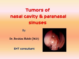
Tumors of
nasal cavity & paranasal
        sinuses
         By

Dr, Ibrahim Habib (M.D)

   ENT consultant
‫بسم هللا الرحمن الرحيم‬
‫{ أقم الصالة لدلوك الشمس إلى غسق الليل وقرآن الفجر إن قرآن الفجر كان مشهودا }‬
                                 ‫اإلسراء : 87‬
Introduction
Cancers of nose & PNS : 3% of Head & Neck cancers .
Age : 5th up to 7th decade .
Predominately of older males .
Exposure:
Wood, nickel-refining processes
Industrial fumes, leather tanning
Cigarette and Alcohol consumption:
No significant association has been shown
location

       3%

1%


            20%


            70%
• Floor : palatine process of maxilla
• Roof : cribriform plate .
Anatomy of maxillary antrum
Anterior : soft tissue of face .
Posterolateral : ITF , pterygopalatine F
Superior : Inferior orbital plate .
Inferiorly : hard palate ,
             superior alveolar ridge
Anatomy of ethmoid sinuses


Anterior : lacrimal bone .
Medialy : lateral nasal wall.
Superior : Fovea ethmoidalis .
Anatomy of sphenoid sinus
      Anteriorly : nasal cavity , ethmoid .
      Posteriorly : clivus , brainstem .
      Superiorly : pituitary fossa .
      Laterally : cavernous sinuses & optic N .
Anatomy of frontal sinus

Anteriorly :
soft tissue of forehead .
Inferiorly :
orbit .
Posteriorly :
anterior cranial fossa .
1- frontal sinus
2- ant. Ethmoid sinus
3- infundibulum
4- middle. Ethmoid
sinus
5- post. Ethmoid sinus
6- middle concha
7- sphenoid sinus
8- inf. concha
9- hard palate
Drainage of PNS
Maxillary sinus : middle meatus
Ethmoid sinuses “ anterior “ : middle meatus .
Ethmoid sinuses “ posterior “ : sphenoethmoid recess .
Sphenoid sinus : sphenoethmoid recess .
Frontal sinus : frontonasal duct .
Classification of sinonasal tumors
Benign ( epithelial )        Benign ( non             Malignant                  Malignant (non
sinonasal tumors             epithelial ) sinonasal   (epithelial )              epithelial ) sinonasal
                             tumours                  sinonasal tumours          tumours
- Schneiderian papilloma :   Leiomyoma                - squamous cell            - chondrosarcoma .
inverted .                   chondromyxoid fibroma    carcinoma :                - Rabdomyosarcoma
 Papillary ( septal ).                                Differentiated .           .
Cylinderical                                          Basaloid squamous .
 - Squamous papilloma (                               Adeosquamous
nasal vestibule )

- Adenoma .                                           Adenocarcinoma .             - lymphoproliferative
- Dermoid                                             Adenoid cystic .           Lymphoma
                                                      Mucoepidermoid             Midline malignant reticulosis
                                                                                 Plasmacytoma
                                                                                 - Terato carcinosarcoma
- Lobular capillary                                   Neuroendocrine             Hemangiopercytoma
hemangioma .                                          carcinoma .                Angiosarcoma
- Hemangiopericytoma .                                Hyallinizing clear cell    kaposi’sarcoma
- peripheral nerve sheath                             carcinoma
tumors
- Fibrous histocytoma .      myxoma , fibromyxoma.    - Melanoma .               Fibrosarcoma
- fibroma .                  ameloblastoma            - olfactory neuroblstoma   Osteogenic sarcoma
- osteoma .                                           .                          Malignant fibrous
- fibrosseus lesios .                                 - sinonasal                Histocytoma
                                                      undifferentiated
                                                      carcinoma (SNUC)


N.B. Secondary malignancy – Melanoma ,Thyroid , lung , kidney and G I T
Squamous Cell Carcinoma
• Most common sinonasal
malignancy
• 70% arise in antrum
• 30% arise in nasal cavity
• 15% with synchronus or
metachronus lesion
• Pre or co-existing papilloma is
risk factor
• 4-9%
• Look for necrosis on imaging
N.B. Squamous Cell Carcinoma in Inverted Papilloma
Adenocarcinoma
• 13-19% of SN
malignancies
• Arise from surface
epithelium and
seromucinous glands
• Intestinal, salivary,
neuroendocrine types
• Non-specific imaging
features
• Predilection for
ethmoid sinuses
Adenoid Cystic Ca
• <10% of SN malignancies
• 25% of adenocarcinomas
• Glandular origin
• Perineural growth pattern (60%)
• Neural cell adhesion molecule
(NCAM) in 93%
• Small lesions extend beyond
what is apparent
• Difficult to entirely remove
• Late recurrences and mets
Sinonasal Melanoma
• < 4% of SN neoplasms
• Melanocytes in mucosa
• Prefers nasal cavity
• Epistaxis
• Worse prognosis than
cutaneous types
• High recurrence and
mortality rates
Esthesioneuroblastoma
• Originate from olfactory
epithelium
• Two incidence peaks
• Adolescence
• 50 - 60 years
• Epistaxis
• High survival with
multimodality therapy
• Ca++ and peripheral cysts
Sinonasal Undifferentiated Ca (SNUC)
• Separate entity from SCCa,
ENB, and others
• Rare, high-grade malignancy
• 2-3:1 male predominance
• Broad age range from 3rd to
9th decades
• Characterized by aggressive
local growth, regional and
distant mets, and poor
survival
Sinonasal Lymphoma
• 44% of extranodal
lymphomas arise in SN
• Prefers nasal cavity
• Types
• T-cell (Asian)
• B-cell (US, Europe)
• T/NK-cell (LMG)
• Remodeling or erosion
• Homogeneous enhancement
Sarcomas and Other Malignancies
• Sarcomas
• Rhabdomyosarcoma
• Liposarcoma
• Leiomyosarcoma
• Fibrosarcoma
• Chondrosarcoma
• Osteosarcoma
• Plasmacytoma
• Metastases
symptoms
Early : asymptomatic .
Oral symptoms: 25-35%
, Toothache , trismus, alveolar ridge fullness, erosion ,
malocclosion .
Nasal findings: 50%
Obstruction, epistaxis, rhinorrhea , post nasal discharge , anosmia .
Ocular findings: 25%
Epiphora, diplopia, proptosis
Facial signs
Paresthesias, asymmetry
Physical examination
Nasal mass or polyposis .
Mass in the check or medical canthus .
Broadening of nasal dorsum .
Maxillary sinus involvement :
       Mass in palate or upper alveolus .
       Mass in upper gingivobuccal sulcus .
       Malocclusion or loose teeth .
Advanced : Trismus .
Orbital :
Periorbital swelling , proptosis .
Epiphora , impaired occular mobility
Uncommon : Neck mass
Nasal endoscopy that shows a tumor in the left nasal wall
Investigations


Aim : detect the disease & its extention .
Extention : orbit , skull base , dura , Intracranial , great
vessels .
Presence of regional or distant metastasis
Presentation of tumours of nose & PNS


  Nasal mass or polyposis   )mass in check )
Broadening of nasal dorsum , proptosis , restricted occular mobility
C T scan
- Ideal
- surrounding bone erosion or destruction .
Tum : -
     our
Calification .
Soft tissue denisty
Necrosis or hge
Vascular tum : enhancem
               ors          ent increase with contrast
Entrapped secretion : with low density
 Lym node : regional L.N. , ( retropharyngeal ) L.N.
       ph
. Staging
• Guide biopsy and surgery
• Treatm responseDistant m
         ent               etastasis .
Coronal section of nose & PNS shows soft tissue mass in region of Rt ethmoid air cell
pushing septum to other side with bony erosion of septum and fovea ethmoidalis )B)
CT Scan, of paranasal sinus, that shows the tumor( angiosarcoma ) in the left
                                  nasal cavity
MRI
Advantages :
- excellent delineation of tumour from
surrounding inflammatory soft tissue and
retained secretions.
- obtained in multiple planes .
- no exposure to ionizing radiation .
- no artifact in the presence of dental filling .
Figures 1 and 2: MR shows a 3.0 x 4.0-cm mass arising from the mucosa of the right
ethmoid region with some areas of necrosis; the surrounding bony structure is intact
             but its growth expands nasal septum and lamina papiracea -
Tumour                  secretion            inflammation


T1                      Intermediate signal     No enhancement          Low signal


T1 with contrast        Diffuse enhancement     No enhancement          Low signal


T2                      Intermediate signal     High signal             High signal


N.B. flow void --- vascular lesion .
With contrast -- perineural invasion, dural or intracranial involvements
L.N. -- Heterogenous on T2 , > 1 cm , peripheral enhancement with contrast using fat
suppresion
Angiography

Indications :
1- Evaluations of vascular tumours extention , vascular anatomy ,
selective embolization .
2- Skull base surgery with brain retraction , delineate intracranial
arterial and venous anatomy .
3- tumour encroaching on carotid a. , assess collaterals , may be
used with balloon occlusion testing .
P.E.T.
- Agent : 18 – F flurodeoxy glucose .
            C – 11 methionine .
- Principle : image metabolic activity of head & neck . Tumors including nose
& PNS
Assess : Local , regional or systemic metastasis . -
. Direct biopsy -
• Therapy response
• Recurrence vs.
treatment change
• Re-staging
- Result : inferior to C.T. & MRI .
Biopsy
Aim : confirm diagnosis & plan appropriate ttt.
Route : 1- transnasl .
           2- transoral .
           3- direct access to the sinus :
Maxillary sinus : Transnasal , medial wall of
maxillary sinus .
                     Caldwell – Luc . Procedure .
Ethmoid sinuses : Endoscopic ethmoidectomy -
                        External ethmoidectomy .
Sphenoid sinus : endoscopically
                        Trans – septally
Frontal sinus : its floor .
Staging of sinonasal tumours
Ohngern 1933 staged maxillary            Ohngern 1933 staged maxillary
                  sinus cancers (Infrastructure )         sinus cancers(Suprastructure)


Site          Infrastructure to Ohngern line            Suprastructure to Ohngern line


Symptoms      Early                                     Late


Spread        Oral , nasal , I.T.F                      Pterygomaxillary fossa , middle &
                                                        anterior cranial fossa


Treatment     More amenable to surgical resection       Less amenable to surgical resection


prognosis     Good                                      Bad


Ohngern line : an imaginary line drawn from maxillary tuberosity to inner canthus .
Ohngern 1933 staged maxillary sinus cancers
Staging of non maxillary sinonasal malignancies


Stage I : tumor confined to site of origin .
Stage II : spread to adjacent sinuses , skin , nasopharynx ,
ptergomaxillary fossa , and or orbit .
Stage III : involvement of skull base , pterygoid plate and
or intracranial extension .
Staging system for olfactory neuroblastoma


Stage I : confined to primary site .
Stage II : presence of nodal metastasis .
Stage III : presence of distant metastasis .
AJCC staging for PNS
       primary tumor ( T ) of maxillary sinus
- Tx primary T can’t be assessed .
- To : no evidence of primary T.
- Tis : carcinoma in situ .
- T1 : T limited to antral mucosa with no erosion nor
destruction of bone .
- T2 Tumour causing erosion or destruction except for
posterior antral wall , including extention into m.m. of
hard palate and / or middle nasal meatus .
AJCC staging for PNS
 primary tumor ( T ) of maxillary sinus
- T3 Tumour invade any of the following : bone of posterior wall of
maxillary sinus , subcutaneous tissue , skin of check , floor or
medial wall of orbit , I.T.F. , pterygoid plates , ethmoid sinuses .
- T4a (resectable): anterior orbit,
skin, infratemporal fossa, pterygoid
plates, cribriform plate, frontal or
sphenoid sinuses
- T4b (unresectable): orbital apex,
dura, brain, middle fossa, clivus,
nasopharynx, CNs (other than V2)-
Staging of ethmoid sinus
- T1 tumour confined to the ethmoid with or without bone
erosion .
- T2 Tumour extends into nasal cavity .
- T3 Tumour extends into ant. Orbit and / or maxillary
sinus .
- T4 Tumour with intracranial extension , orbital
extension including apex , involving sphenoid and / or
frontal sinus and / or skin of external nose .
Nodal involvement in sinonasal tumours
. Nodal involvement infrequent despite advanced stage
• Depends on primary site, extent, and histology
• 8-18% with nodes at presentaion
. Nodal stage based on:                 N1: Single ipsilat ≤ 3cm
                                        • N2:
• Number
                                        • a: Single ipsilat 3 – 6cm
• Uni- or bilateral
                                        • b: Multiple ipsilat ≤
• Size
                                        6cm
-Nodal drainage                         • c: Bilat or contralat ≤
• Facial, parotid, submandibular        6cm
• Retropharyngeal                       • N3: ≥ 6cm node
• Then L II
staging
- stage o         Tis         No         Mo
- stage I          T1         No         Mo
- stage II         T2         No         Mo
- stage III        T3          No       Mo
-                 T1-T3       N1        Mo
- stage IV A       T4         No         Mo
                  T4         N1         Mo
- stage IV B       any T       N2       Mo
                  any T       N2        Mo
- stage IV c       any T      any N     M1
( N ) lymph node . ( M ) distant metastasis .
TNM Staging of Maxillary Carcinomas

• Stage I: Limited to mucosa
• Stage II: Bone involvement
(NOT posterior wall)
• Stage III:
• T3 lesion
• TI or T2 lesions with N1
• Stage IV
• T4 lesion
• Any T with N2/N3 or M1
Management of sinonasal tumours
Surgical management         Indication        Surgical management           Indication
  of early primary                                      of
        lesion                                 Advanced primary
                                                      lesion


Infrastructure        lesions confined to      Radical maxillectomy   advanced lesions
maxillectomy          floor of maxillary sinus                        confined to maxillary
                      .                                               sinus advanced
                                                                      lesions confined to
                                                                      maxillary sinus


Medial maxillectomy   lesions confined to    Craniofacial resection   extension of disease
                      medial wall of                                  into the frontal
                      maxillary sinus                                 sinuses and / or
                                                                      cribriform plate


Partial or complete   lesions confined to    Palliative               disease is extended
septectomy            septum                 radiotherapy             into brain , sphenoid
                                                                      rostrum , cavernous
                                                                      sinus & internal
                                                                      carotid a
Midfacial degloving approach.. Surgical Treatment of Squamous Cell
                    Carcinoma of the Sinuses.
Combined bicoronal approach and Dieffenbach-Weber-Fergusson incision. Surgical
            Treatment of Squamous Cell Carcinoma of the Sinuses..
Management of orbit          Indication          Orbital complications             N.B.
in sinonasal tumors                                   where R.T.


Resection of a small   cases with minimal        epiphora , keratitis ,   complications with
portion of the         periorbital               diplopia , pain ,        pre-operative R.T. are
periorbita &           involvement without       exophthalmos , and       mostly minor and
reconstruct with       full penetration into     loss of vision .         transient .
fascial graft          the orbital fat .


Resection of orbit     with invasion of the                               complications are
                       periorbita , the                                   more frequent when
                       infraorbital nerve , or                            post operative R.T. is
                       the orbital apex                                   used
Reconstruction and Prosthetic Rehabilitation


- Aim : - prevent contracture of the check , to separate
oral & nasal cavities , and to provide support for the
globe .


- An obturator should be made preoperatively from an
impression of the hard palate .
. Algorithm to depict tissue options for midface reconstruction
Treatment of maxillary sinus carcinoma(A) 66-year-old woman with total maxillectomy defect
and orocutaneous fistula status after surgery and radiotherapy. (B) Cranial bone grafts used to
 reconstruct orbitozygomatic structure surrounded by rectus abdominus free flap. (C) 3-year
           postoperative result. (D) Intraoral view of 3-year postoperative result.
Management of tumours of nose &
                          PNS
                     (1) The Neck

No :
T1 – T2 :
electve ND is not generally performed.
T3 – T4 :
R.T. post. Operative . Upper neck & retro-ph. L.Ns .

N+ve with resectable 1ry :
MRND . Or dissect 1-V & retropharyngeal chain .
Management of tumours of nose &
                          PNS
          (1) The Neck )late node metastasis)
- 5 – 45% occure after 2-3 yrs .
- rarely occurs in absence of synchronous local or distant
recurrence you should search for .
- TTT aggressively : R.N.D.
- 5 yr survival rate was 39% after ttt of delayed metastasis
.
- N.B. None with nodes at presentation survived 3 years .
Radiotherapy as an adjuvant therapy in
       management of sinonasal tumours

- 1- combined with surgery in advanced resectable
lesions . Pre. Or post. Operative .


- 2- Single modality for :
- advanced unresectable lesions .
- patients unwilling or unable to undergo surgery .
- Average 5 yrs survival rates 10 – 15 % ( total doses up to 79 Gy ) .
chemotherapy as an adjuvant therapy in
      management of sinonasal tumours
- Combination chemotherapy with pre. Or post.
Operative R.T. in :
- Olfactory neuroblastoma & SN undifferentiated ca.
- Japanese researchers use combination of R.T. , intra-
arterial 5 – fluorouracil ( 5 FU ) and local debridement
or cryosurgery for maxillary sinus cancer .
- Knegt ‘s regimen in using topical chemotherapy as an
 adjuvant Therapy in management of sinonasal tumours
.The regimen
1-antrostomy and debulking of the tumour .
 2-The tumour bed is then packed with topical 5FU
emulsion .
3- The pack are removed and any residual necrotic
material is debrided as often as necessary .
 He reported 5-yr survival of 71% .
prognosis
The advancement of skull base surgery , cure rates for
patients with sinonasal tumours ,
form 39-76% have been achieved
Tumours have good chance of cure :
1- early maxillary tumours .
2- patients with nasal cavity tumours .
3- well differentiated adenocarcinoma 90% .
4- low grade minor salivary gland tumour .
5- olfactory neuroblastoma :
 100% stage A & 75% stage B & 60% stage C . Survival .
6- sq. cell ca. arising in inverted papilloma .
Tumours with bad prognosis
1- Advanced maxillary cancer .
2- lesions involving pterygoid plates or
pterygopalatine fossa .
3- lesions involving brain , dura , nasopharynx ,
sphenoid .
4- lesions involving orbital contents .
Tumours of nasal cavity & paranasal sinuses

Weitere ähnliche Inhalte

Was ist angesagt?

Neoplasms of nose and paranasal sinuses
Neoplasms of nose and paranasal  sinusesNeoplasms of nose and paranasal  sinuses
Neoplasms of nose and paranasal sinusesGurchand Behal
 
parapharyngeal space tumors
parapharyngeal space tumors parapharyngeal space tumors
parapharyngeal space tumors Mamoon Ameen
 
Juvenile nasopharyngeal angiofibroma
Juvenile nasopharyngeal angiofibromaJuvenile nasopharyngeal angiofibroma
Juvenile nasopharyngeal angiofibromapraneeth koduru
 
Spaces of middle ear and their surgical importance
Spaces of middle ear  and their surgical importanceSpaces of middle ear  and their surgical importance
Spaces of middle ear and their surgical importanceDr Soumya Singh
 
Maxillary sinus carcinoma
Maxillary sinus carcinomaMaxillary sinus carcinoma
Maxillary sinus carcinomaHarsha Yadav
 
Tumors of nose and paranasal sinus dr.sithanandhakumar -13.06.16
Tumors of nose and paranasal sinus  dr.sithanandhakumar -13.06.16Tumors of nose and paranasal sinus  dr.sithanandhakumar -13.06.16
Tumors of nose and paranasal sinus dr.sithanandhakumar -13.06.16ophthalmgmcri
 
Contact and Compact Endoscopy in ENT
Contact and Compact Endoscopy in ENTContact and Compact Endoscopy in ENT
Contact and Compact Endoscopy in ENTLakhan M S
 
Fisch approaches Dr Zeeshan Ahmad
Fisch approaches Dr Zeeshan AhmadFisch approaches Dr Zeeshan Ahmad
Fisch approaches Dr Zeeshan AhmadDr Zeeshan Ahmad
 
NASOPHARYNGEAL CARCINOMA
NASOPHARYNGEAL CARCINOMA NASOPHARYNGEAL CARCINOMA
NASOPHARYNGEAL CARCINOMA Mamoon Ameen
 
Tumours of oropharynx
Tumours of oropharynxTumours of oropharynx
Tumours of oropharynxVinay Bhat
 
surgical approaches to frontal sinus ppt
surgical approaches to frontal sinus pptsurgical approaches to frontal sinus ppt
surgical approaches to frontal sinus pptVaibhav Lahane
 

Was ist angesagt? (20)

Ca maxilla
Ca maxillaCa maxilla
Ca maxilla
 
Sinunasal malignacy
Sinunasal malignacySinunasal malignacy
Sinunasal malignacy
 
Neoplasms of nose and paranasal sinuses
Neoplasms of nose and paranasal  sinusesNeoplasms of nose and paranasal  sinuses
Neoplasms of nose and paranasal sinuses
 
Glomus Tumour
Glomus TumourGlomus Tumour
Glomus Tumour
 
parapharyngeal space tumors
parapharyngeal space tumors parapharyngeal space tumors
parapharyngeal space tumors
 
Juvenile nasopharyngeal angiofibroma
Juvenile nasopharyngeal angiofibromaJuvenile nasopharyngeal angiofibroma
Juvenile nasopharyngeal angiofibroma
 
Spaces of middle ear and their surgical importance
Spaces of middle ear  and their surgical importanceSpaces of middle ear  and their surgical importance
Spaces of middle ear and their surgical importance
 
Maxillary sinus carcinoma
Maxillary sinus carcinomaMaxillary sinus carcinoma
Maxillary sinus carcinoma
 
Glomus tumors
Glomus tumorsGlomus tumors
Glomus tumors
 
Vestibular schwannoma
Vestibular schwannomaVestibular schwannoma
Vestibular schwannoma
 
Tumors of nose and paranasal sinus dr.sithanandhakumar -13.06.16
Tumors of nose and paranasal sinus  dr.sithanandhakumar -13.06.16Tumors of nose and paranasal sinus  dr.sithanandhakumar -13.06.16
Tumors of nose and paranasal sinus dr.sithanandhakumar -13.06.16
 
Contact and Compact Endoscopy in ENT
Contact and Compact Endoscopy in ENTContact and Compact Endoscopy in ENT
Contact and Compact Endoscopy in ENT
 
JNA
JNAJNA
JNA
 
Fisch approaches Dr Zeeshan Ahmad
Fisch approaches Dr Zeeshan AhmadFisch approaches Dr Zeeshan Ahmad
Fisch approaches Dr Zeeshan Ahmad
 
NASOPHARYNGEAL CARCINOMA
NASOPHARYNGEAL CARCINOMA NASOPHARYNGEAL CARCINOMA
NASOPHARYNGEAL CARCINOMA
 
Tumours of oropharynx
Tumours of oropharynxTumours of oropharynx
Tumours of oropharynx
 
surgical approaches to frontal sinus ppt
surgical approaches to frontal sinus pptsurgical approaches to frontal sinus ppt
surgical approaches to frontal sinus ppt
 
Mucosal folds of the middle ear
Mucosal folds of the middle earMucosal folds of the middle ear
Mucosal folds of the middle ear
 
Esthesioneuroblastoma (ENB)
Esthesioneuroblastoma (ENB)Esthesioneuroblastoma (ENB)
Esthesioneuroblastoma (ENB)
 
Hypopharyngeal cancer
Hypopharyngeal cancer Hypopharyngeal cancer
Hypopharyngeal cancer
 

Ähnlich wie Tumours of nasal cavity & paranasal sinuses

Problem oriented approach in pediatric radiology
Problem oriented approach in pediatric radiologyProblem oriented approach in pediatric radiology
Problem oriented approach in pediatric radiologyAhmed Bahnassy
 
malignant sinonasal tumors+management.pptx
malignant sinonasal tumors+management.pptxmalignant sinonasal tumors+management.pptx
malignant sinonasal tumors+management.pptxEmanZayed17
 
Tumours of nasopharynx (2) itp class dr.davis - 03.06.16
Tumours of nasopharynx (2) itp class   dr.davis - 03.06.16Tumours of nasopharynx (2) itp class   dr.davis - 03.06.16
Tumours of nasopharynx (2) itp class dr.davis - 03.06.16ophthalmgmcri
 
Radiological approach to PNS (Paranasal sinus) Masses
Radiological approach to PNS (Paranasal sinus) MassesRadiological approach to PNS (Paranasal sinus) Masses
Radiological approach to PNS (Paranasal sinus) MassesDr Anuj Aggarwal
 
BENIGN NEOPLASMS OF THE NOSE AND PNS.pptx
BENIGN NEOPLASMS OF THE NOSE AND PNS.pptxBENIGN NEOPLASMS OF THE NOSE AND PNS.pptx
BENIGN NEOPLASMS OF THE NOSE AND PNS.pptxSayan Banerjee
 
Neoplasms of Salivary Glands
Neoplasms of Salivary GlandsNeoplasms of Salivary Glands
Neoplasms of Salivary GlandsKARTHIK K
 
Pre management salivary gland by dr pallavi jain
Pre management salivary gland by dr pallavi jainPre management salivary gland by dr pallavi jain
Pre management salivary gland by dr pallavi jainDr. Pallavi Jain
 
Management of sinonasal tract tumors 27082018
Management of sinonasal tract tumors 27082018Management of sinonasal tract tumors 27082018
Management of sinonasal tract tumors 27082018Varshu Goel
 
Neoplasms of nose and para nasal sinuses.ppt
Neoplasms of nose and para nasal sinuses.pptNeoplasms of nose and para nasal sinuses.ppt
Neoplasms of nose and para nasal sinuses.pptManu Babu
 
Sinonasal tumors dr rk
Sinonasal tumors dr rkSinonasal tumors dr rk
Sinonasal tumors dr rkraju kafle
 
Head and neck cancer Dr VIPIN V NAIR
Head and neck cancer Dr VIPIN V NAIRHead and neck cancer Dr VIPIN V NAIR
Head and neck cancer Dr VIPIN V NAIRPGIMER Chandigarh
 

Ähnlich wie Tumours of nasal cavity & paranasal sinuses (20)

Problem oriented approach in pediatric radiology
Problem oriented approach in pediatric radiologyProblem oriented approach in pediatric radiology
Problem oriented approach in pediatric radiology
 
malignant sinonasal tumors+management.pptx
malignant sinonasal tumors+management.pptxmalignant sinonasal tumors+management.pptx
malignant sinonasal tumors+management.pptx
 
Tumours of nasopharynx (2) itp class dr.davis - 03.06.16
Tumours of nasopharynx (2) itp class   dr.davis - 03.06.16Tumours of nasopharynx (2) itp class   dr.davis - 03.06.16
Tumours of nasopharynx (2) itp class dr.davis - 03.06.16
 
Radiological approach to PNS (Paranasal sinus) Masses
Radiological approach to PNS (Paranasal sinus) MassesRadiological approach to PNS (Paranasal sinus) Masses
Radiological approach to PNS (Paranasal sinus) Masses
 
Pleomorphic adenoma
Pleomorphic adenomaPleomorphic adenoma
Pleomorphic adenoma
 
Head and neck
Head and neckHead and neck
Head and neck
 
Parotid
ParotidParotid
Parotid
 
Salivary glands.pptx
Salivary glands.pptxSalivary glands.pptx
Salivary glands.pptx
 
Maxilla
MaxillaMaxilla
Maxilla
 
BENIGN NEOPLASMS OF THE NOSE AND PNS.pptx
BENIGN NEOPLASMS OF THE NOSE AND PNS.pptxBENIGN NEOPLASMS OF THE NOSE AND PNS.pptx
BENIGN NEOPLASMS OF THE NOSE AND PNS.pptx
 
Sino-nasal malignancy
Sino-nasal malignancySino-nasal malignancy
Sino-nasal malignancy
 
Neoplasms of Salivary Glands
Neoplasms of Salivary GlandsNeoplasms of Salivary Glands
Neoplasms of Salivary Glands
 
Pre management salivary gland by dr pallavi jain
Pre management salivary gland by dr pallavi jainPre management salivary gland by dr pallavi jain
Pre management salivary gland by dr pallavi jain
 
Management of sinonasal tract tumors 27082018
Management of sinonasal tract tumors 27082018Management of sinonasal tract tumors 27082018
Management of sinonasal tract tumors 27082018
 
Neoplasms of nose and para nasal sinuses.ppt
Neoplasms of nose and para nasal sinuses.pptNeoplasms of nose and para nasal sinuses.ppt
Neoplasms of nose and para nasal sinuses.ppt
 
Malignant tumor of neck
Malignant tumor of neckMalignant tumor of neck
Malignant tumor of neck
 
Ear carcinoma
Ear carcinomaEar carcinoma
Ear carcinoma
 
Sinonasal tumors dr rk
Sinonasal tumors dr rkSinonasal tumors dr rk
Sinonasal tumors dr rk
 
Head and Neck Imaging
Head and Neck ImagingHead and Neck Imaging
Head and Neck Imaging
 
Head and neck cancer Dr VIPIN V NAIR
Head and neck cancer Dr VIPIN V NAIRHead and neck cancer Dr VIPIN V NAIR
Head and neck cancer Dr VIPIN V NAIR
 

Mehr von Ibrahim Barakat

مفاتيح السعادة بين يديك د إبراهيم حبيب
مفاتيح السعادة بين يديك د إبراهيم حبيبمفاتيح السعادة بين يديك د إبراهيم حبيب
مفاتيح السعادة بين يديك د إبراهيم حبيبIbrahim Barakat
 
ترسيخ الحوار مع الآخرين د إبراهيم حبيب
ترسيخ الحوار مع الآخرين د إبراهيم حبيبترسيخ الحوار مع الآخرين د إبراهيم حبيب
ترسيخ الحوار مع الآخرين د إبراهيم حبيبIbrahim Barakat
 
الناشئة وتحديات العصر د إبراهيم حبيب بركات
الناشئة وتحديات العصر د إبراهيم حبيب بركاتالناشئة وتحديات العصر د إبراهيم حبيب بركات
الناشئة وتحديات العصر د إبراهيم حبيب بركاتIbrahim Barakat
 
Cochealer implant surgery
Cochealer implant surgeryCochealer implant surgery
Cochealer implant surgeryIbrahim Barakat
 
Pediatric airway obstruction
Pediatric airway obstructionPediatric airway obstruction
Pediatric airway obstructionIbrahim Barakat
 
Management of complications of acute otitis media
Management of complications of acute otitis media Management of complications of acute otitis media
Management of complications of acute otitis media Ibrahim Barakat
 
Management of complications of acute otitis media
Management of complications of acute otitis media Management of complications of acute otitis media
Management of complications of acute otitis media Ibrahim Barakat
 
Salivary gland diseases 1
Salivary gland diseases 1Salivary gland diseases 1
Salivary gland diseases 1Ibrahim Barakat
 
إلا من أـى الله بقلب سليم 1
إلا من أـى الله بقلب سليم 1إلا من أـى الله بقلب سليم 1
إلا من أـى الله بقلب سليم 1Ibrahim Barakat
 
إدارة الوقت و مواجهة التوتر
إدارة الوقت و مواجهة التوترإدارة الوقت و مواجهة التوتر
إدارة الوقت و مواجهة التوترIbrahim Barakat
 
Cancer of the nasopharynx
Cancer of the nasopharynxCancer of the nasopharynx
Cancer of the nasopharynxIbrahim Barakat
 
Cancer of the nasopharynx
Cancer of the nasopharynxCancer of the nasopharynx
Cancer of the nasopharynxIbrahim Barakat
 
Facial nerve paralysis ppt
Facial nerve paralysis pptFacial nerve paralysis ppt
Facial nerve paralysis pptIbrahim Barakat
 

Mehr von Ibrahim Barakat (15)

مفاتيح السعادة بين يديك د إبراهيم حبيب
مفاتيح السعادة بين يديك د إبراهيم حبيبمفاتيح السعادة بين يديك د إبراهيم حبيب
مفاتيح السعادة بين يديك د إبراهيم حبيب
 
ترسيخ الحوار مع الآخرين د إبراهيم حبيب
ترسيخ الحوار مع الآخرين د إبراهيم حبيبترسيخ الحوار مع الآخرين د إبراهيم حبيب
ترسيخ الحوار مع الآخرين د إبراهيم حبيب
 
الناشئة وتحديات العصر د إبراهيم حبيب بركات
الناشئة وتحديات العصر د إبراهيم حبيب بركاتالناشئة وتحديات العصر د إبراهيم حبيب بركات
الناشئة وتحديات العصر د إبراهيم حبيب بركات
 
Cochealer implant surgery
Cochealer implant surgeryCochealer implant surgery
Cochealer implant surgery
 
Pediatric airway obstruction
Pediatric airway obstructionPediatric airway obstruction
Pediatric airway obstruction
 
Management of complications of acute otitis media
Management of complications of acute otitis media Management of complications of acute otitis media
Management of complications of acute otitis media
 
Management of complications of acute otitis media
Management of complications of acute otitis media Management of complications of acute otitis media
Management of complications of acute otitis media
 
كن رحيما
كن رحيماكن رحيما
كن رحيما
 
العلم والعقل
العلم والعقلالعلم والعقل
العلم والعقل
 
Salivary gland diseases 1
Salivary gland diseases 1Salivary gland diseases 1
Salivary gland diseases 1
 
إلا من أـى الله بقلب سليم 1
إلا من أـى الله بقلب سليم 1إلا من أـى الله بقلب سليم 1
إلا من أـى الله بقلب سليم 1
 
إدارة الوقت و مواجهة التوتر
إدارة الوقت و مواجهة التوترإدارة الوقت و مواجهة التوتر
إدارة الوقت و مواجهة التوتر
 
Cancer of the nasopharynx
Cancer of the nasopharynxCancer of the nasopharynx
Cancer of the nasopharynx
 
Cancer of the nasopharynx
Cancer of the nasopharynxCancer of the nasopharynx
Cancer of the nasopharynx
 
Facial nerve paralysis ppt
Facial nerve paralysis pptFacial nerve paralysis ppt
Facial nerve paralysis ppt
 

Tumours of nasal cavity & paranasal sinuses

  • 1. Tumors of nasal cavity & paranasal sinuses By Dr, Ibrahim Habib (M.D) ENT consultant
  • 3.
  • 4. ‫{ أقم الصالة لدلوك الشمس إلى غسق الليل وقرآن الفجر إن قرآن الفجر كان مشهودا }‬ ‫اإلسراء : 87‬
  • 5. Introduction Cancers of nose & PNS : 3% of Head & Neck cancers . Age : 5th up to 7th decade . Predominately of older males . Exposure: Wood, nickel-refining processes Industrial fumes, leather tanning Cigarette and Alcohol consumption: No significant association has been shown
  • 6. location 3% 1% 20% 70%
  • 7. • Floor : palatine process of maxilla • Roof : cribriform plate .
  • 8. Anatomy of maxillary antrum Anterior : soft tissue of face . Posterolateral : ITF , pterygopalatine F Superior : Inferior orbital plate . Inferiorly : hard palate , superior alveolar ridge
  • 9. Anatomy of ethmoid sinuses Anterior : lacrimal bone . Medialy : lateral nasal wall. Superior : Fovea ethmoidalis .
  • 10. Anatomy of sphenoid sinus Anteriorly : nasal cavity , ethmoid . Posteriorly : clivus , brainstem . Superiorly : pituitary fossa . Laterally : cavernous sinuses & optic N .
  • 11. Anatomy of frontal sinus Anteriorly : soft tissue of forehead . Inferiorly : orbit . Posteriorly : anterior cranial fossa .
  • 12. 1- frontal sinus 2- ant. Ethmoid sinus 3- infundibulum 4- middle. Ethmoid sinus 5- post. Ethmoid sinus 6- middle concha 7- sphenoid sinus 8- inf. concha 9- hard palate
  • 13. Drainage of PNS Maxillary sinus : middle meatus Ethmoid sinuses “ anterior “ : middle meatus . Ethmoid sinuses “ posterior “ : sphenoethmoid recess . Sphenoid sinus : sphenoethmoid recess . Frontal sinus : frontonasal duct .
  • 14.
  • 16. Benign ( epithelial ) Benign ( non Malignant Malignant (non sinonasal tumors epithelial ) sinonasal (epithelial ) epithelial ) sinonasal tumours sinonasal tumours tumours - Schneiderian papilloma : Leiomyoma - squamous cell - chondrosarcoma . inverted . chondromyxoid fibroma carcinoma : - Rabdomyosarcoma Papillary ( septal ). Differentiated . . Cylinderical Basaloid squamous . - Squamous papilloma ( Adeosquamous nasal vestibule ) - Adenoma . Adenocarcinoma . - lymphoproliferative - Dermoid Adenoid cystic . Lymphoma Mucoepidermoid Midline malignant reticulosis Plasmacytoma - Terato carcinosarcoma - Lobular capillary Neuroendocrine Hemangiopercytoma hemangioma . carcinoma . Angiosarcoma - Hemangiopericytoma . Hyallinizing clear cell kaposi’sarcoma - peripheral nerve sheath carcinoma tumors - Fibrous histocytoma . myxoma , fibromyxoma. - Melanoma . Fibrosarcoma - fibroma . ameloblastoma - olfactory neuroblstoma Osteogenic sarcoma - osteoma . . Malignant fibrous - fibrosseus lesios . - sinonasal Histocytoma undifferentiated carcinoma (SNUC) N.B. Secondary malignancy – Melanoma ,Thyroid , lung , kidney and G I T
  • 17. Squamous Cell Carcinoma • Most common sinonasal malignancy • 70% arise in antrum • 30% arise in nasal cavity • 15% with synchronus or metachronus lesion • Pre or co-existing papilloma is risk factor • 4-9% • Look for necrosis on imaging N.B. Squamous Cell Carcinoma in Inverted Papilloma
  • 18. Adenocarcinoma • 13-19% of SN malignancies • Arise from surface epithelium and seromucinous glands • Intestinal, salivary, neuroendocrine types • Non-specific imaging features • Predilection for ethmoid sinuses
  • 19. Adenoid Cystic Ca • <10% of SN malignancies • 25% of adenocarcinomas • Glandular origin • Perineural growth pattern (60%) • Neural cell adhesion molecule (NCAM) in 93% • Small lesions extend beyond what is apparent • Difficult to entirely remove • Late recurrences and mets
  • 20. Sinonasal Melanoma • < 4% of SN neoplasms • Melanocytes in mucosa • Prefers nasal cavity • Epistaxis • Worse prognosis than cutaneous types • High recurrence and mortality rates
  • 21. Esthesioneuroblastoma • Originate from olfactory epithelium • Two incidence peaks • Adolescence • 50 - 60 years • Epistaxis • High survival with multimodality therapy • Ca++ and peripheral cysts
  • 22. Sinonasal Undifferentiated Ca (SNUC) • Separate entity from SCCa, ENB, and others • Rare, high-grade malignancy • 2-3:1 male predominance • Broad age range from 3rd to 9th decades • Characterized by aggressive local growth, regional and distant mets, and poor survival
  • 23. Sinonasal Lymphoma • 44% of extranodal lymphomas arise in SN • Prefers nasal cavity • Types • T-cell (Asian) • B-cell (US, Europe) • T/NK-cell (LMG) • Remodeling or erosion • Homogeneous enhancement
  • 24. Sarcomas and Other Malignancies • Sarcomas • Rhabdomyosarcoma • Liposarcoma • Leiomyosarcoma • Fibrosarcoma • Chondrosarcoma • Osteosarcoma • Plasmacytoma • Metastases
  • 25.
  • 26. symptoms Early : asymptomatic . Oral symptoms: 25-35% , Toothache , trismus, alveolar ridge fullness, erosion , malocclosion . Nasal findings: 50% Obstruction, epistaxis, rhinorrhea , post nasal discharge , anosmia . Ocular findings: 25% Epiphora, diplopia, proptosis Facial signs Paresthesias, asymmetry
  • 27. Physical examination Nasal mass or polyposis . Mass in the check or medical canthus . Broadening of nasal dorsum . Maxillary sinus involvement : Mass in palate or upper alveolus . Mass in upper gingivobuccal sulcus . Malocclusion or loose teeth . Advanced : Trismus . Orbital : Periorbital swelling , proptosis . Epiphora , impaired occular mobility Uncommon : Neck mass
  • 28. Nasal endoscopy that shows a tumor in the left nasal wall
  • 29. Investigations Aim : detect the disease & its extention . Extention : orbit , skull base , dura , Intracranial , great vessels . Presence of regional or distant metastasis
  • 30. Presentation of tumours of nose & PNS Nasal mass or polyposis )mass in check )
  • 31. Broadening of nasal dorsum , proptosis , restricted occular mobility
  • 32. C T scan - Ideal - surrounding bone erosion or destruction . Tum : - our Calification . Soft tissue denisty Necrosis or hge Vascular tum : enhancem ors ent increase with contrast Entrapped secretion : with low density Lym node : regional L.N. , ( retropharyngeal ) L.N. ph . Staging • Guide biopsy and surgery • Treatm responseDistant m ent etastasis .
  • 33. Coronal section of nose & PNS shows soft tissue mass in region of Rt ethmoid air cell pushing septum to other side with bony erosion of septum and fovea ethmoidalis )B)
  • 34. CT Scan, of paranasal sinus, that shows the tumor( angiosarcoma ) in the left nasal cavity
  • 35. MRI Advantages : - excellent delineation of tumour from surrounding inflammatory soft tissue and retained secretions. - obtained in multiple planes . - no exposure to ionizing radiation . - no artifact in the presence of dental filling .
  • 36. Figures 1 and 2: MR shows a 3.0 x 4.0-cm mass arising from the mucosa of the right ethmoid region with some areas of necrosis; the surrounding bony structure is intact but its growth expands nasal septum and lamina papiracea -
  • 37. Tumour secretion inflammation T1 Intermediate signal No enhancement Low signal T1 with contrast Diffuse enhancement No enhancement Low signal T2 Intermediate signal High signal High signal N.B. flow void --- vascular lesion . With contrast -- perineural invasion, dural or intracranial involvements L.N. -- Heterogenous on T2 , > 1 cm , peripheral enhancement with contrast using fat suppresion
  • 38. Angiography Indications : 1- Evaluations of vascular tumours extention , vascular anatomy , selective embolization . 2- Skull base surgery with brain retraction , delineate intracranial arterial and venous anatomy . 3- tumour encroaching on carotid a. , assess collaterals , may be used with balloon occlusion testing .
  • 39. P.E.T. - Agent : 18 – F flurodeoxy glucose . C – 11 methionine . - Principle : image metabolic activity of head & neck . Tumors including nose & PNS Assess : Local , regional or systemic metastasis . - . Direct biopsy - • Therapy response • Recurrence vs. treatment change • Re-staging - Result : inferior to C.T. & MRI .
  • 40. Biopsy Aim : confirm diagnosis & plan appropriate ttt. Route : 1- transnasl . 2- transoral . 3- direct access to the sinus : Maxillary sinus : Transnasal , medial wall of maxillary sinus . Caldwell – Luc . Procedure . Ethmoid sinuses : Endoscopic ethmoidectomy - External ethmoidectomy . Sphenoid sinus : endoscopically Trans – septally Frontal sinus : its floor .
  • 41.
  • 43. Ohngern 1933 staged maxillary Ohngern 1933 staged maxillary sinus cancers (Infrastructure ) sinus cancers(Suprastructure) Site Infrastructure to Ohngern line Suprastructure to Ohngern line Symptoms Early Late Spread Oral , nasal , I.T.F Pterygomaxillary fossa , middle & anterior cranial fossa Treatment More amenable to surgical resection Less amenable to surgical resection prognosis Good Bad Ohngern line : an imaginary line drawn from maxillary tuberosity to inner canthus . Ohngern 1933 staged maxillary sinus cancers
  • 44. Staging of non maxillary sinonasal malignancies Stage I : tumor confined to site of origin . Stage II : spread to adjacent sinuses , skin , nasopharynx , ptergomaxillary fossa , and or orbit . Stage III : involvement of skull base , pterygoid plate and or intracranial extension .
  • 45. Staging system for olfactory neuroblastoma Stage I : confined to primary site . Stage II : presence of nodal metastasis . Stage III : presence of distant metastasis .
  • 46. AJCC staging for PNS primary tumor ( T ) of maxillary sinus - Tx primary T can’t be assessed . - To : no evidence of primary T. - Tis : carcinoma in situ . - T1 : T limited to antral mucosa with no erosion nor destruction of bone . - T2 Tumour causing erosion or destruction except for posterior antral wall , including extention into m.m. of hard palate and / or middle nasal meatus .
  • 47. AJCC staging for PNS primary tumor ( T ) of maxillary sinus - T3 Tumour invade any of the following : bone of posterior wall of maxillary sinus , subcutaneous tissue , skin of check , floor or medial wall of orbit , I.T.F. , pterygoid plates , ethmoid sinuses . - T4a (resectable): anterior orbit, skin, infratemporal fossa, pterygoid plates, cribriform plate, frontal or sphenoid sinuses - T4b (unresectable): orbital apex, dura, brain, middle fossa, clivus, nasopharynx, CNs (other than V2)-
  • 48. Staging of ethmoid sinus - T1 tumour confined to the ethmoid with or without bone erosion . - T2 Tumour extends into nasal cavity . - T3 Tumour extends into ant. Orbit and / or maxillary sinus . - T4 Tumour with intracranial extension , orbital extension including apex , involving sphenoid and / or frontal sinus and / or skin of external nose .
  • 49. Nodal involvement in sinonasal tumours . Nodal involvement infrequent despite advanced stage • Depends on primary site, extent, and histology • 8-18% with nodes at presentaion . Nodal stage based on: N1: Single ipsilat ≤ 3cm • N2: • Number • a: Single ipsilat 3 – 6cm • Uni- or bilateral • b: Multiple ipsilat ≤ • Size 6cm -Nodal drainage • c: Bilat or contralat ≤ • Facial, parotid, submandibular 6cm • Retropharyngeal • N3: ≥ 6cm node • Then L II
  • 50. staging - stage o Tis No Mo - stage I T1 No Mo - stage II T2 No Mo - stage III T3 No Mo - T1-T3 N1 Mo - stage IV A T4 No Mo T4 N1 Mo - stage IV B any T N2 Mo any T N2 Mo - stage IV c any T any N M1 ( N ) lymph node . ( M ) distant metastasis .
  • 51. TNM Staging of Maxillary Carcinomas • Stage I: Limited to mucosa • Stage II: Bone involvement (NOT posterior wall) • Stage III: • T3 lesion • TI or T2 lesions with N1 • Stage IV • T4 lesion • Any T with N2/N3 or M1
  • 52.
  • 54. Surgical management Indication Surgical management Indication of early primary of lesion Advanced primary lesion Infrastructure lesions confined to Radical maxillectomy advanced lesions maxillectomy floor of maxillary sinus confined to maxillary . sinus advanced lesions confined to maxillary sinus Medial maxillectomy lesions confined to Craniofacial resection extension of disease medial wall of into the frontal maxillary sinus sinuses and / or cribriform plate Partial or complete lesions confined to Palliative disease is extended septectomy septum radiotherapy into brain , sphenoid rostrum , cavernous sinus & internal carotid a
  • 55. Midfacial degloving approach.. Surgical Treatment of Squamous Cell Carcinoma of the Sinuses.
  • 56. Combined bicoronal approach and Dieffenbach-Weber-Fergusson incision. Surgical Treatment of Squamous Cell Carcinoma of the Sinuses..
  • 57. Management of orbit Indication Orbital complications N.B. in sinonasal tumors where R.T. Resection of a small cases with minimal epiphora , keratitis , complications with portion of the periorbital diplopia , pain , pre-operative R.T. are periorbita & involvement without exophthalmos , and mostly minor and reconstruct with full penetration into loss of vision . transient . fascial graft the orbital fat . Resection of orbit with invasion of the complications are periorbita , the more frequent when infraorbital nerve , or post operative R.T. is the orbital apex used
  • 58. Reconstruction and Prosthetic Rehabilitation - Aim : - prevent contracture of the check , to separate oral & nasal cavities , and to provide support for the globe . - An obturator should be made preoperatively from an impression of the hard palate .
  • 59. . Algorithm to depict tissue options for midface reconstruction
  • 60. Treatment of maxillary sinus carcinoma(A) 66-year-old woman with total maxillectomy defect and orocutaneous fistula status after surgery and radiotherapy. (B) Cranial bone grafts used to reconstruct orbitozygomatic structure surrounded by rectus abdominus free flap. (C) 3-year postoperative result. (D) Intraoral view of 3-year postoperative result.
  • 61. Management of tumours of nose & PNS (1) The Neck No : T1 – T2 : electve ND is not generally performed. T3 – T4 : R.T. post. Operative . Upper neck & retro-ph. L.Ns . N+ve with resectable 1ry : MRND . Or dissect 1-V & retropharyngeal chain .
  • 62. Management of tumours of nose & PNS (1) The Neck )late node metastasis) - 5 – 45% occure after 2-3 yrs . - rarely occurs in absence of synchronous local or distant recurrence you should search for . - TTT aggressively : R.N.D. - 5 yr survival rate was 39% after ttt of delayed metastasis . - N.B. None with nodes at presentation survived 3 years .
  • 63. Radiotherapy as an adjuvant therapy in management of sinonasal tumours - 1- combined with surgery in advanced resectable lesions . Pre. Or post. Operative . - 2- Single modality for : - advanced unresectable lesions . - patients unwilling or unable to undergo surgery . - Average 5 yrs survival rates 10 – 15 % ( total doses up to 79 Gy ) .
  • 64. chemotherapy as an adjuvant therapy in management of sinonasal tumours - Combination chemotherapy with pre. Or post. Operative R.T. in : - Olfactory neuroblastoma & SN undifferentiated ca. - Japanese researchers use combination of R.T. , intra- arterial 5 – fluorouracil ( 5 FU ) and local debridement or cryosurgery for maxillary sinus cancer .
  • 65. - Knegt ‘s regimen in using topical chemotherapy as an adjuvant Therapy in management of sinonasal tumours .The regimen 1-antrostomy and debulking of the tumour . 2-The tumour bed is then packed with topical 5FU emulsion . 3- The pack are removed and any residual necrotic material is debrided as often as necessary . He reported 5-yr survival of 71% .
  • 66. prognosis The advancement of skull base surgery , cure rates for patients with sinonasal tumours , form 39-76% have been achieved
  • 67. Tumours have good chance of cure : 1- early maxillary tumours . 2- patients with nasal cavity tumours . 3- well differentiated adenocarcinoma 90% . 4- low grade minor salivary gland tumour . 5- olfactory neuroblastoma : 100% stage A & 75% stage B & 60% stage C . Survival . 6- sq. cell ca. arising in inverted papilloma .
  • 68. Tumours with bad prognosis 1- Advanced maxillary cancer . 2- lesions involving pterygoid plates or pterygopalatine fossa . 3- lesions involving brain , dura , nasopharynx , sphenoid . 4- lesions involving orbital contents .