
Recommended
More Related Content
What's hot
What's hot (20)
Similar to Skull masters
Similar to Skull masters (20)
Recently uploaded
Recently uploaded (20)
Skull masters
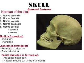
- 1. Skull is formed of: - Cranium - Mandible Cranium is formed of: - Brain box (calvaria) - Facial skeleton Facial skeleton is formed of: - An upper fixed part - A lower mobile part (the mandbile) General features SKULL Normae of the skull 1. Norma verticalis 2. Norma frontalis 3. Norma lateralis 4. Norma occipitalis 5. Norma basalis: - externa - interna
- 2. Norma verticalis (Superior view) Bones to be identified: • Frontal bone • Parietal bones • Occiptial bone Sutures: • Sagittal suture, between the 2 parietal bones • Coronal suture, between the frontal & the two parietal bones • Lambdoid suture, between the occipital and the two parietal bones Features: • Bregma, point of meeting of sagittal & coronal sutures • Lambda, point of meeting of sagittal & lambdoid sutures • Frontal tuber, most prominent area on the frontal bone • Parietal tuber, most prominent area on the parietal bone • Parietal foramen, transmits the parietal emissary vein Frontal bone Frontal tuber Bregma Coronal suture Sagittal suture Parietal tuber Parietal foramen Lambda Lambdoid suture Occipital bone Parietalbone
- 3. Anterior fontanelle: •A rhomboidal membrane that closes the gap present at the site of the bregma in the fetal skull. •Obliterated in the middle of the second year after birth. Frontal bone Parietal bone Lambdoid suture Occipital bone Posterior fontanelle Sagittal suture Coronal suture Anterior fontanelle Posterior fontanelle: •A triangular membrane that closes the gap present at the site of the lambda in the fetal skull. •Obliterated in the first 3 months after birth.
- 4. Norma frontalis (Anterior view) Bones to be identified: •Frontal bone •Nasal bones •Zygomatic bones •Maxillary bones •Mandible Sutures: •Fronto-nasal •Fronto-zygomatic •Inte-rnasal •Inter-maxillary •Maxillo-zygomatic Frontal bone Frontal tuber Glabella Nasal bone Zygomatic bone Maxilla Mental foramen Body of the mandible Ramus of the mandible Infra-orbital foramen Zygomatico-facial foramen Supra-orbital foramen
- 5. • Three large openings: -2 orbital openings -anterior nasal aperture; bound by the nasal bones and maxillae • Anterior nasal spine; a median projection on the lower margin of the anterior nasal aperture • Nasion, point of meeting of fronto- nasal & internasal sutures • Glabella, elevation above the nasion • Supercilliary arches, transverse ridges extending laterally from the glabella • Frontal tuber, the most prominent area of the frontal bone • Foramina: 1. Supra-orbital foramen (notch) 2. Infra-orbital foramen 3. Zygomatico-facial foramen 4. Mental foramen Frontal bone Frontal tuber Glabella Mental foramen Supra-orbital foramen Features of norma frontalis: Nasion Zygomatico-facial foramen Infra-orbital foramen Anterior nasal spine
- 6. Norma lateralis (Lateral view) Bones to be identified: Above the level of zygomatic arch: • Frontal bone • Parietal bone • Occipital bone • Greater wing of sphenoid bone • Squamous part of temporal bone Zygomatic process of temporal bone (zygoma) Temporal process of zygomatic bone Zygomatic arch Frontal bone Parietalbone O ccipital bone Greater wing of sphenoid bone Squamous part of temporal bone Zygomatic arch, Formed by the temporal process of zygomatic bone and zygomatic process of temporal bone (zygoma)
- 7. Bones to be identified: Below the level of zygomatic arch: • Mastoid part of temporal bone, projects downwards forming the mastoid process • Tympanic part of temporal bone • Pterygoid process of sphenoid bone • Maxilla Squamous part of temporal bone Mastoid part of temporal bone Tympanic part of temporal bone Greater wing of sphenoid bone Pterygoid process of sphenoid bone Maxilla Tympanic part of temporal bone Mastoid process Ramus of the mandible Squamous part of temporal bone
- 8. Features of norma lateralis Styloid process • Pterion, region of meeting of frontal, parietal, squamous part of temporal and greater wing of sphenoid bones. It lies 35mm behind and 12mm above the fronto-zygomatic suture. • Asterion, region of meeting of lambdoid, parietomastoid and occipitomastoid sutures. Pterion Asterion
- 9. • Temporal line, begins behind the lateral orbital margin, follows a curved course on the frontal, parietal and temporal bones, forms a ridge at the base of mastoid bone (supra- mastoid crest) and continues with the posterior root of zygoma. • External acoustic meatus, below the posterior root of the zygoma • Pterygoid process, formed of a lateral and medial pterygoid plates separated by the pterygoid fossa Superior & inferior Temporal lines Supramastoid crest Mastoid part of temporal bone Mastoid process External acoustic meatus Lateral & medial pterygoid platesPterygoid fossa
- 10. • Temporal fossa, the area bound by the temporal line • Infratemporal fossa, the space below the greater wing of sphenoid and deep to the ramus of the mandible. • Pterygo-maxillary fissure, between the maxilla (anteriorly) and the pterygoid process (posteriorly), it connects the infratemporal fossa with the pterygopalatine fossa. • Inferior orbital fissure, connects the infratemporal fossa with the orbital cavity. Temporal lines Temporal fossa Greater wing of sphenoid bone Pterygoid process of sphenoid bone Maxilla Squamous part of temporal bone Pterygo-maxillary fissure • Styloid process, a long slender process directed downwards and forwards Styloid process
- 11. Norma occipitalis (Posterior view) Bones to be identified: • Parietal bones • Squamous part of occiptial bone • Mastoid part of temporal bone Features to be identified: • External occipital protuberance • External occipital crest • Superior nuchal lines • Highest nuchal lines • Lambda • Asterion • Mastoid foramen • Parietal foramen Sutures to be identified: • Sagittal suture • Lambdoid suture • Parietomastoid suture • Occipitomastoid suture Parietal bone Squamous part of occipital bone Mastoid bone External occipital protuberance Parietal foramen Sagittal sutureLambda Asterion Sup nuchal line Occipitomastoid suture Lambdoid suture Parietomastoid suture External occipital crest Inferior nuchal line
- 13. Anterior part • Formed by the Bony Palate • The anterior ¾ of the palate is formed by the palatine processes of both maxillae (separated by the intermaxillary suture) • The posterior ¼ is formed by the horizontal plates of both palatine bones (separated by the interpalatine suture) Palatine processes of maxillary bones Horizontal plates of palatine bones Interpalatine suture Intermaxillary suture Incisive fossa
- 14. Features of the anterior part • Alveolar arch, formed by the alveolar processes of both maxillae • Cruciform suture, separates the parts forming the hard palate. • Incisive fossa, an anterior median depression. • Greater palatine foramen (one on each side), on the posterior and lateral part of the bony palate. It leads to the greater palatine canal • Lesser palatine foramina (two small foramina) behind the greater palatine foramen. • Vascular groove, runs forward close to the alveolar arch • Posterior nasal spine, a median projection from the posterior border of the bony palate Incisive fossa Alveolar arch Greater palatine foramen Lesser palatine foraminaPosterior nasal spine Interpalatine suture Palato-maxillary suture Intermaxillary suture Cruciform suture
- 15. • Body of sphenoid bone • Pterygoid process of sphenois • Greater wing of sphenoid • Petrous part of temporal bone • Tympanic part of temporal bone • Mastoid part of temporal bone • Mandibular fossa Body of sphenoid bone Petrous part of temporal bone Groove for cartilagenous part of auditory tube Tympanic part of temporal bone Styloid process Mandibular fossa Mastoid part of temporal bone Greater wing of sphenoid Middle part Extends from the posterior margin of hard palate to the anterior margin of foramen magnum
- 16. Features: •Vomer, a median bone forming the posterior part of the nasal septum •Posterior nasal apertures •Pterygoid process, formed of lateral & medial plates with the pterygoid fossa in between. •Pterygoid hamulus •Pterygomaxillary fissure •Maxillary tuberosity, a rough prominence behind the last molar tooth •Infratemporal surface of greater wing of sphenoid Pterygoid hamulus Posterior nasal apertures Vomer Medial pterygoid plate Lateral pterygoid plate Greater wing of sphenoid
- 17. Foramina to be identified: • Foramen ovale, on the infratemporal surface of greater wing of sphenoid • Foramen spinosum, behind the foramen ovale • Foramen lacerum, at the apex of petrous temporal bone • Carotid canal • Stylomastoid foramen, between the styloid and mastoid processes. Foramen ovale Foramen spinosum Foramen lacerum Carotid canal Stylomastoid foramen Groove for cartilagenous part of auditory tube
- 18. Posterior part Formed mainly by the different parts of the occipital bone, surrounding the foramen magnum Bones to be identified: • Basilar part of occipital bone, in front of foramen magnum • Condylar parts, one on each side of foramen magnum • Squamous part, behind the foramen magnum Features: •Pharyngeal tubercle, a small elevation on the basilar part •Occipital condyles, on each side of foramen magnum •Condylar fossa, behind the occipital condyles •External occipital protuberance •External occipital crest •Inferior nuchal lines •Superior nuchal lines Basilar part Squamous part Foramen magnum Pharyngeal tubercle Occipital condyles (condylar part) External occipital protuberance Condylar fossa Condylar canal External occipital crest Inferior nuchal lines Superior nuchal lines
- 19. Foramina: • Foramen magnum, the largest in the skull • Jugular foramen, between the occipital and petrous temporal bones • Hypoglossal canal, above the occipital condyle • Condylar canal Foramen magnum Jugular foramen Jugular fossa Condylar canal Mastoid foramen
- 20. Norma Basalis Interna Divided into; anterior, middle and posterior cranial fossae Features of the anterior cranial fossa • Frontal crest, a median ridge on the inner aspect of frontal bone • Cribriform plate of ethmoid bone, in the median region of the fossa, forming the roof of the nasal cavity. It shows a median projection called the crista galli • Orbital plate of frontal bone, on each side of the cribriform plate of ethmoid. • Body of sphenoid bone, behind the cribriform plate • Lesser wing of sphenoid, on each side of the sphenoid bone. • Anterior clinoid process The medial end of the lesser wing Frontal crest Cribriform plate of ethmoid Orbital plate of frontal bone Body of sphenoid bone Lesser wing of sphenoid Crista galli Anterior clinoid process Anterior cranial fossa Bound posteriorly by the lesser wings of sphenoid
- 21. Foramina: • Foramen cecum, between the crista galli and frontal crest • Antherior ethmoidal canal, on the lateral margin of the cribriform plate of ethmoid • Posterior ethmoidal canal, at the posterolateral angle of the cribriform plate of ethmoid. Foramen cecum Antherior ethmoidal canal Posterior ethmoidal canal
- 22. Middle cranial fossa • Bound anteriorly by the lesser wings of sphenoid and posteriorly by the upper border of petrous temporal bone • Narrow in the middle and expands greatly laterally Bones to be identified; • Body of sphenoid, in the median region • Greater wing of sphenoid • Squamous part of temporal bone • Anterior surface of petrous temporal bone Body of sphenoid Greater wing of sphenoid Squamous part of temporal bone Petrous part of temporal bone F. magnum
- 23. Features of the middle cranial fossa Tegmen tympani Acruate eminence Trigeminal impression Groove for ICA Hypophyseal fossa Posterior clinoid process Foramen lacerum Sulcus for optic chiasma Anterior clinoid process • Hypophyseal fossa (sella turcica), a median depression on the body of sphenoid. Bound anteriorly by the tuberculum sellae and posteriorly by the dorsum sellae. The upper end of the dorsum sellae forms two projections (the posterior clinoid processes) • Groove for Internal Carotid Artery, on the lateral aspect of the body of sphenoid • Trigeminal impression, a depression at the apex of petrous temporal bone • Acruate eminence, an elevation on the anterior surface of petrous temporal bone • Tegmen tympani (roof of the middle ear) lies lateral to the arcuate eminence.
- 24. Foramina and openings in the middle cranial fossa; • Superior orbital fissure, a fissure between the greater and lesser wings of sphenoid • Foramen rotundum, behind the medial end of superior orbital fissure, leads to the pterygopalatine fossa • Foramen ovale, behind the foramen rotundum. • Foramen spinosum, behind the foramen ovale • Foramen lacerum, at the apex of petrous temporal bone Superior orbital fissure Foramen rotundum Foramen ovale Foramen spinosum Foramen lacerum Groove for greater petrosal nerve Groove for lesser petrosal nerve
- 25. Posterior cranial fossa Boundaries: posterior clinoid process, upper border of petrous temporal bone, groove for transvers sinus, internal occipital protuberance Bones to be identified; • Basilar part of occipital bone, in front of F magnum. Together with the basilar part of sphenoid they form the clivus. • Condylar part of occipital bone, on each side of F magnum. • Squamous part of occipital bone, behind F magnum. • Posterior surface of petrous temporal bone, lateral to the occipital bone • Mastoid part of temporal bone, behind and lateral to the petrous part. Basilar part of occipital bone Basilar part of sphenoid Squamous part of occipital bone Petrous temporal bone Clivus Condylar part of occipital bone Mastoid part of temporal bone
- 26. Features of the posterior cranial fossa • Internal occipital protuberance, a median elevation on the squamous part of occipital bone • Internal occipital crest, a median ridge extending from the internal occipital protuberance to the F. magnum • Groove for transverse venous sinus, extends from the internal occipital protuberance to the mastoid part of temporal bone • Sigmoid sulcus, an S-shaped sulcus extending from the end of the groove for transverse sinus to the jugular foramen • Groove for superior petrosal sinus, running along the upper border of petrous temporal bone • Groove for inferior petrosal sinus, running along the ptrosphenoidal fissure (between petrous bone and occipital bone) Internal occipital protuberance Internal occipital crest Groove for transverse venous sinus Sigmoid sulcus Groove for superior petrosal sinus Groove for inferior petrosal sinus
- 27. Foramina to be identified: • Foramen magnum • Jugular foramen, between the occipital and petrous bones • Hypoglossal canal, above the occipital condyle • Condylar canal, connects the codylar fossa (seen on the norma basalis externa) with the sigmoid sulcus • Mastoid foramen, on the line of the occipito-mastoid suture (opens into the sigmoid sulcus) • Internal acoustic meatus, on the posterior surface of petrous bone Foramen magnum Jugular foramen Hypoglossal canal Condylar canal Mastoid foramen Internal acoustic meatus Foramen spinosum Foramen ovale Foramen rotundum Foramen lacerum
- 28. Mandible • Formed of a body and 2 rami • The body is horse shoe-shaped and formed of 2 halves fused together at the symphysis menti • The ramus forms 2 upward projections; coronoid process anteriorly and condylar process posteriorly • The condylar process constitutes the head and neck of the mandible. The small depression on the front of the neck is called pterygoid fovea • Angle of the mandible, the junction between the base of the mandible (lower border) and the posterior border of the ramus • Mandibular notch, the concave upper border of the ramus (the depression between the 2 processes) Features of the outer surface of mandible • Mental protuberance, a median elevation close to the lower border. It forms a projection on each side called mental tubercle • Mental foramen, lateral to the mental protuberance • Oblique line, a ridge extending from the anterior border of the ramus onto the outer surface of the body • Alveolar border, the upper border of the body Head of mandible Neck of mandible Condylar process Mandibular notch Oblique line Angle of the mandible Mental foramen Mental tubercle Coronoid process Pterygoid fovea Ramus B o d y Base of the mandible
- 29. Features of the inner surface of mandible • Mylohyoid line, an oblique line on the inner surface of the body • Submandibular fossa, a depression below the mylohoid line • Sublingual fossa, a small depression above the mylohoid line • Superior and inferior genial tubercles, 2 small median projections on the back of smyphysis menti • Digastric fossa, a small depression on the base on each side of the symphysis menti • Mandibular foramen, on the inner aspect of the ramus. It leads to the mandibular canal which ends below the 2nd premolar tooth and bifurcates into incisive canal (continues below the incisor teeth) and the mental canal which opens at the mental foramen • Lingula, a small projection of bone at the mandibular foramen • Mylohyoid groove, a narrow groove on the inner surface of the ramus, from the mandibular foramen and below the mylohyoid line. Mylohyoid line Submandibular fossa Sublingual fossa Genial tubercles Digastric fossa Mandibular foramen Lingula Mylohyoid groove