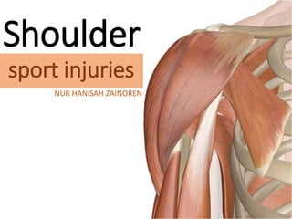
SHOULDER SPORT INJURIES
•Download as PPTX, PDF•
24 likes•10,548 views

This is only a part of Shoulder Sport Injuries. Content: shoulder instability

Recommended
More Related Content
What's hot
What's hot (20)
Viewers also liked
Viewers also liked (20)
Similar to SHOULDER SPORT INJURIES
Similar to SHOULDER SPORT INJURIES (20)
More from hanisahwarrior
More from hanisahwarrior (20)
Recently uploaded
Recently uploaded (20)
SHOULDER SPORT INJURIES
- 1. Shoulder sport injuries NUR HANISAH ZAINOREN
- 2. • Sternoclavicular injury • Acromioclavicular injury • Biceps tendon injury •• Superior labral injury •• Shoulder instability
- 8. SHOULDER INS
- 9. SHOULDER INS A B R OA D term used for shoulder problems, where the head of humerus is not stable in glenoid
- 10. • The shoulder by virtue of its anatomy and biomechanics is one of most unstable and frequently dislocated joints in body • Its account nearly 50% of all dislocations
- 12. Why shoulders become unstable? 1) Structural changes due to major traumaor recurrent micro-trauma 2) Unbalanced muscle recruitment resulting in humeral head being displaced upon the glenoid
- 13. TYPES • Traumatic structural instability Type I • Atraumatic (or minimally traumatic) structural instability Type II • Atraumatic non- structural instability Type III
- 14. PATHOGENETIC CLASSIFICATION TYPE 1 TYPE 2 TYPE 3 Type of disorder Traumatic structural instability Atraumatic (or minimally traumatic) structural instability Atraumatic non- structural instability Trauma Yes No No Articular surface damage Yes Yes No Capsular problem Bankart lesion Dysfunctional Dysfunctional Laxity Unilateral Uni/bilateral Bilateral Muscle patterning Normal Normal Abnormal
- 15. TRAUMATIC ANTERIOR INSTABILITY • Commonest type of instability ( >95% cases) • Recurrent dislocation may results in: • Bankart’s lesion • Hill-Sachs lesion • Recurrent subluxation alternately occur with dislocation • Patient age >50, often associated with tears of rotator cuff
- 16. Bankart lesion(stripping of glenoid labrum & periosteum from the antero-inferior surface of the glenoid)
- 18. Hill-sachs lesion (depression on the humeral head in its posterolateral quadrant, caused by impingement by the anterior edge of the glenoid on the head as it dislocates)
- 19. Clinical features: • History of the shoulder “coming out”, perhaps during a sporting event • The 1st episode of acute dislocation: patient may be able to describe the mechanism precisely (ie: an applied force with the shoulder in abduction, external rotation and extension
- 20. Recurrent subluxation: symptoms and signs are less obvious A catching sensation numbness/weakness (“dead arm syndrome”) Whenever shoulder is used with the arm in the overhead position
- 22. Clinical examination: • Apprehension test Patient senses that the humeral head is about to slip out anteriorly and his or her body tautens in apprehension
- 23. • Relocation test Opposite to apprehension test
- 25. Investigations: • Most cases ca be diagnosed from the history and examination alone • Hill-Sach lesion (when it is best present) is best shown by an AP X-ray with shoulder internally rotated, or in axillary view • MRI or MR arthrography: demonstration of bone lesions and labral tears • Arthroscopy: to define labral tear • Examination under anesthesia: to determine the direction of instability
- 26. Treatment: • Indication for surgery: – Frequent dislocation – Recurrent subluxation – Fear of dislocation • Two type operation are employed : – Anatomical repairs – Bankart repairs – Non Anatomical repairs – Laterjet-Bristow procedure
- 27. Bankart repair
- 28. Bankart repair
- 30. ATRAUMATIC OR MINIMALLY TRAUMATIC INSTABILITY STRUCTURAL NON-STRUCTURAL
- 31. ATRAUMATIC STRUCTURAL INSTABILITY • Acquired multidirectional instability due to – Repetitive microtrauma – Forceful movement that lead to overall laxity • Recognized problem in athletes, particularly swimmers and throwers • They develop symptoms of instability due to overload and fatigue in the stabilizing muscles of the shoulder
- 32. • Dislocation may occur in several different directions • Important to rule out the presence of any pathological conditions
- 33. Treatments : • Rehabilitative measures (Physiotherapy) – Focus on strengthening the muscles that involved in stabilizing the shoulder – Aim at restoring muscular coordination and control • Surgical treatment – Capsular shift
- 34. Capsular shift
- 35. ATRAUMATIC NON-STRUCTURAL INSTABILITY • Instability of muscle pattern • In younger patients who can voluntarily slip the shoulder out of joint as a trick, then it go on to dislocate repeatedly (habitual dislocation)
- 36. Treatment : –Aim to regain normal neuromuscular control and patterning –Rehabilitation programs –Surgery need to be avoided if possible
- 37. POSTERIOR INSTABILITY • Other name: luxatio erecta • Humeral head riding back on the posterior lip of the glenoids • Due to a violent jerk in an unusual position or following an epileptic fit or a severe electric shock
- 38. • Dislocation may be associated with: • Fractures of proximal humerus • Reverse Bankart’s lesion • Reverse Hill-Sach lesion
- 39. A P
- 40. Humeral head looks globular the so-called light bulb appearance
- 41. Humeral head looks globular the so-called light bulb appearance
- 42. Examination: – Posterior drawer test – Jerk test
- 43. Posterior drawer test Scapular spine and coracoid process in one hand, Humeral head pushed backwards with the other
- 44. Jerk test Stabilize the scapula with one hand, while the other hand holds the elbow with the arm in 90° abduction and internal rotation. Firm axial compression force is applied on the glenohumeral joint The arm is horizontally adducted while maintaining the firm axial load
- 45. Treatment : – For posterior instability, the initial treatment should be non-operative physiotherapy – Surgery may be indicated • If at least 4-6month of an appropriate rehabilitation program has failed • dislocation has been ruled out • patient is emotionally stable and fit for surgery – ‘Reverse’ Bankart procedure is done
- 47. INFERIOR INSTABILITY •Instability which occurs particularly when carrying something heavy with the arm •Occur some weeks after injury to shoulder girdle •Due to temporary weakness of the shoulder muscles, usually because of prolonged splintage of the arm and lack of exercise
- 48. •X-ray: • Head of humerus has subluxated inferiorly • Further views with patient carrying a 10kg weight, shows the head of humerus lying below the glenoid socket on the affected side •Usually corrects by itself after a period of normal muscular activity, but physiotherapy will help to speed up the process
- 51. MULTIDIRECTIONAL INSTABILITY • The primary abnormality in multidirectional instability is a loose, redundant inferior pouch • It is important to distinguish multidirectional instability from routine undirectional dislocation • MDI can be define as global (anterior, posterior and inferior) or at least two direction
- 52. • Patient with MDI may have variety of symptoms including pain, instability, weakness, paresthesia, fatigue and difficulty in throwing or lifting • The treatment for MDI is rehabilitation program that emphasizes the strengthening of rotator cuff and scapular rotator • If rehabilitation fails to resolve patient symptoms most commonly surgical procedure remains the inferior capsular shift
- 53. CONCLUSION Once the sling or splint is removed. This will help to improve range of motion as well as the stability and strength to shoulder. 5. Rehabilitation Most surgical procedures are performed right after the dislocation if it has been determined there has been any damage to nerves, muscles, tendons or blood vessels. If shoulder continues to become dislocated, need to have surgery to keep it stabilized. 4. Surgery To expand the range of motion. Strengthen the muscles in rotator cuff so they are better able to support the shoulder and help to prevent another dislocation. 3. Exercises It should remain in a sling until the tissues are healed. Rehabilitation is needed to get back the strength and mobility. 5 to 7 days or more. NSAIDS (ibuprofen) can help to remove the inflammation and pain. 2. Immobilization The shoulder should only be reduced, or put back into the socket by a trained professional in the medical field. For most young adults who are less than 30 years old, reduction is the best course of action. 1. Reduction
- 54. REFERENCES • Apley’s System of Orthopaedics and Fractures, 9th edition • Campbell Operative Orthopaedics 10th edition, Volume Three • Google images
- 55. Thank you.