
Recomendados
Más contenido relacionado
La actualidad más candente
La actualidad más candente (20)
Destacado
Destacado (16)
Similar a Clase de endocrino thalia
Similar a Clase de endocrino thalia (20)
Clase de endocrino thalia
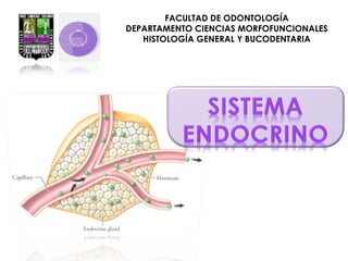
- 1. UNIVERSIDAD DE CARABOBO FACULTAD DE ODONTOLOGÍA DEPARTAMENTO CIENCIAS MORFOFUNCIONALES HISTOLOGÍA GENERAL Y BUCODENTARIA
- 3. DESCRIBIR MACRO Y MICROSCÓPICAMENTE LOS ÓRGANOS PERTENECIENTES AL SISTEMA ENDOCRINO Y RELACIONAR CON SU FUNCIÓN.
- 4. • El sistema endocrino esta formado por un conjunto de glándulas que no poseen conductos y que secretan hormonas al torrente sanguíneo que se encargan de coordinar las actividades metabólicas de ciertos órganos y tejidos
- 6. Son mensajeros químicos producidos por las glándulas endocrinas y se desplazan por el torrente sanguíneo a células u órganos blancos
- 10. La Neurohipófisis se divide en: •Pars nervosa •Infundíbulo •La Adenohipófisis se divide en: a)Pars distalis b)Pars tuberalis c)Pars intermedia
- 11. Se compone de células cromófobas y cromófilas Las cromófilas se dividen en : Acidófilas: •SOMATOTROPAS: secretan somatotropina. •MAMOTROPAS: secretan prolactina Basófilos : •CORTICOTROPAS:Secretan la hormona adenocorticotropica. •TIROTROPAS: secretan tirotrpina •GONADOTROPAS: secretan la hormona foliculoestimulante y la estimulante de las celula intersticial
- 12. No tiene un aspecto muy organizado Histológicamente • PITUICITOS • AXONES AMIELINICOS CUERPOS HERRING • DE almacenan las hormonas oxitocina hormona antidiurética
- 19. metabolismo celular crecimiento Facilita procesos mentales Estimulo de metabolismo de carbohidratos y grasas Frecuencia cardiaca Acción muscular Peso corporal
- 24. •Las células Principales son pequeñas ,de núcleo redondo y central y se tiñen escasamente. Secretan la Paratohormona (PTH) antagónico a la Calcitonina. que posee un efecto •Las celulas Oxífilas, son menos abundantes que las Principales, de mayor tamaño y son fuertemente acidófilas. Su función se desconoce.
- 25. aumenta la concentración de Ca2+ en los líquidos La Paratohormona corporales : Calcitonina Tiroides Inhibe la reabsorción ósea Ca 2+ en sa ngre Ca2+ normal Regula Vitamina D Ca2+ en sangre Intestinos reabsorben Ca2+ Riñones reabsorben Ca2+ Ca 2+ en sa ngre Paratiroides Paratohormona Estimula la reabsorción ósea
- 29. Sus células se acomodan en grupos redondeados, envueltos por capilares. Las células son cilíndricas, con núcleo esférico, citoplasma Células que forman cordones paralelos entre sí y con capilares que los rodean. Sus células son poliédricas, con citoplasma acidófilo, con gran número de inclusiones lipídicas
- 30. Cercana a la médula; sus células se acomodan en forma de red, son pequeñas, con citoplasma acidófilo, pequeñas inclusiones lipídicas y gránulos de pigmento pardo. Las células de la médula son poliédricas y se acomodan en cordones que forman una densa red en cuyas mallas hay capilares y vénulas.
- 31. Aldosterona y Desoxicorticosterona Cortisol y corticosterona Dehidropiandosterona y androstediona Adrenalina y noradrenalina
- 35. Hipotálamo Catecolaminas Catecolaminas Adrenalina Adrenalina Incrementa frecuencia Incrementa frecuencia Aumenta flujo Aumenta flujo sanguíneo, liberación sanguíneo, liberación de glucosa de glucosa Catecolamina Catecolamina Noradrenalina Noradrenalina Eleva la presión Eleva la presión arterial por arterial por vasoconstricción vasoconstricción Nervios simpáticos Preganglionares Responsable de respuesta rápida Hipófisis Mineralocorticoides Mineralocorticoides Controlan líquido Controlan líquido corporal : : corporal Excreción KKyy Excreción Reabsorción Na Reabsorción Na ACTH Responsable de respuesta lenta Glucocorticoides Glucocorticoides Regulan metabolismo Regulan metabolismo de: de: Carbohidratos grasas Carbohidratos grasas yy proteínas, proteínas, ++ Gluconeogénesis, Gluconeogénesis, libera Ac. Grasos, libera Ac. Grasos, Antiinflamtorios, Antiinflamtorios, reduce reduce permeabilidad capilar permeabilidad capilar
- 37. “ Un profesor trabaja para la eternidad: nadie puede predecir dónde acabará su influencia” H.B. Adams
Notas del editor
- LM, human pituitary, at low power. From Japanese slide set (Humio Mizoguti, Kobe Univ Sch Med), slide 515 (= 57-1). Japan515-L.jpg. 72(550)6.
- LM, human pituitary, showing anterior (at left) and posterior (at right) lobes. The pars intermedia ("intermediate lobe") is very poorly developed in humans, and consists merely of a modest layer of darker cells lying against the posterior lobe, to the right of the white cleft between the anterior and posterior lobes. From Japanese slide set (Humio Mizoguti, Kobe Univ Sch Med), slide 516 (= 57-2). Japan516.jpg. 72(600)6.
- LM, human pituitary, stained with H&E. From Stan Erlandsen Medical Histology slide set, slide MH-9B3. MH-9B3-L.jpg. 72(600)6.
- LM, posterior pituitary (= pars nervosa). Note the promient nuclei of pituicytes, which are glial cells. In this histological preparation you can't make out axon terminals (containing oxytocin or vasopressin) derived from neurosecretory neurons in the hypothalamus. There are no neuron cell bodies in the posterior pituitary. Taken by AKC from a Burton L. Baker LM slide (hypoxed female rat). 1980C12(154)CL.jpg. 72(700)6.
- (Left) The hypothalamus produces two hormones, ADH and oxytocin, which are stored and secreted by the posterior pituitary. (Right) The hypothalamus controls the secretions of the anterior pituitary, and the anterior pituitary controls the secretions of the thyroid, adrenal cortex, and gonads, which are also endocrine glands.
- LM, human parathyroid, H&E stain. In the left half of the field are the usual chief cells. At right is a large cluster of oxyphil cells. In the oxyphil cells, note the small, dense nuclei, and the clear, eosinophilic cytoplasm. The large white circles are extracted fat cells. Photograph taken of slide from U-M medical histology class set by AKC. 1992G23(50)CL.jpg. 72(650)6.
- LM, human parathyroid, H&E stain. In the left half of the field are the usual chief cells. At right is a large cluster of oxyphil cells. In the oxyphil cells, note the small, dense nuclei, and the clear, eosinophilic cytoplasm. The large white circles are extracted fat cells. Photograph taken of slide from U-M medical histology class set by AKC. 1992G23(50)CL.jpg. 72(650)6.
- LM, human adrenal cortex, stained with H&E. From left to right, capsule , zona glomerulosa, zona fasciculata, zona reticularis, medulla. From Japanese slide collection(Humio Mizoguti, Kobe Univ Sch Med), slide 547 (= 57-8). Japan547-L.jpg. 72(700)6.
- LM, human adrenal medulla, source of epinephrine and nor-epinephrine (= adrenaline and nor-adrenaline). There is some zona reticularis at the top of the picture (cells more reddish). The somewhat brownish appearance of the medulla is due to staining with potassium dichromate -- the cells that make norepinephrine and epinephrine are chromaffin cells. From Japanese slide collection (Humio Mizoguti, Kobe Univ Sch Med), slide 565 (= 57-13). Japan565-L.jpg. 72(600)6.
- LM, human adrenal medulla, source of epinephrine and nor-epinephrine (= adrenaline and nor-adrenaline). There is some zona reticularis at the top of the picture (cells more reddish). The somewhat brownish appearance of the medulla is due to staining with potassium dichromate -- the cells that make norepinephrine and epinephrine are chromaffin cells. From Japanese slide collection (Humio Mizoguti, Kobe Univ Sch Med), slide 565 (= 57-13). Japan565-L.jpg. 72(600)6.