Evaluation of accelerated collagen cross-linking for the treatment of melting keratitis in 8 dogs
•
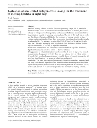
3 gefällt mir•968 views

Accelerated collagen cross-linking was used to treat melting keratitis in 8 dogs. The following results were observed: 1) Pain improvement was seen in all cases within 3 days after treatment. 2) Epithelial healing was observed within 15 days for all cases. 3) Disappearance of cellular infiltration was observed in all cases by day 7. 4) Corneal vascularization disappeared in 4 cases and was reduced in 4 cases by 1 month. 5) At 1 month, all eyes had visual function despite variable corneal scarring. No recurrent infections occurred. The study suggests that accelerated collagen cross-linking is a valuable treatment for melting keratitis in dogs.

Empfohlen
Empfohlen
Weitere ähnliche Inhalte
Was ist angesagt?
Was ist angesagt? (20)
Ähnlich wie Evaluation of accelerated collagen cross-linking for the treatment of melting keratitis in 8 dogs
Ähnlich wie Evaluation of accelerated collagen cross-linking for the treatment of melting keratitis in 8 dogs (20)
Mehr von Frank FAMOSE
Mehr von Frank FAMOSE (20)
Kürzlich hochgeladen
Kürzlich hochgeladen (20)
Evaluation of accelerated collagen cross-linking for the treatment of melting keratitis in 8 dogs
- 1. Evaluation of accelerated collagen cross-linking for the treatment of melting keratitis in eight dogs Frank Famose Service d’Ophtalmologie, Clinique Veterinaire des Acacias, 42 avenue Lucien Servanty, 31700 Blagnac, France Address communications to: F. Famose Tel.: +33 5 61 71 24 02 Fax: +33 5 61 71 65 52 e-mail: frankfamose@gmail.com Abstract Objective Melting keratitis is serious condition presenting a high risk of permanent blindness and is caused by infectious or noninfectious factors. In humans, the clinical efficacy of collagen cross-linking (CXL) has been described in the treatment of refrac- tory infectious keratitis by arresting keratomalacia. The aim of this study was to evalu- ate the efficacy of accelerated CXL for the treatment of melting keratitis in dogs. Animal studied and Procedure Eight dogs were treated for unilateral melting keratitis by accelerated CXL. Corneas were irradiated by UVA (370 nm) at 30 mW/cm² irradi- ance for 3 min after soaking by 0.1% riboflavin in 20% dextran for 30 min. Follow- up was conducted 3, 7, 14, and 30 days after treatment. Results Pain improvement was observed for all cases within 3 days after treatment. Epithelial healing was observed within 15 days for all cases. Disappearance of cellular infiltration was observed for all cases at day 7. The corneal vascularization disappeared for 4 of 8 dogs and was reduced for 4 of 8 dogs within 1 month. At 1 month, all cases presented a variable degree of corneal scarring, but all eyes had visual function. No recurrent infection was observed. Conclusions The main observation of this study is that all the cases have presented with the same clinical result regardless of the presence and the sensitivity of the infectious agents and regardless of the duration of the condition prior to CXL treatment. Accel- erated CXL appears to be a valuable option for the treatment of melting keratitis. Key Words: accelerated CXL, cross-linking, dogs, melting keratitis, optical coherence tomography, riboflavin INTRODUCTION In dogs, melting keratitis is serious condition presenting a high risk of permanent blindness.1–3 In melting kerati- tis, stromal damage is initiated by various mechanisms including bacterial proliferation, toxin secretion, micro- bial or corneal proteases activation leading to keratomala- cia or corneal melting. An imbalance between the endogenous and exogenous matrix metalloproteinases (MMP) and the proteinases present in the cornea and the precorneal tear film leads to the destruction of the colla- gen of the cornea.4–7 Microbial infection is usually sus- pected to drive the inflammatory state responsible for corneal melting but cannot always be demonstrated.1 There are few corneal pathogens that are associated with primary corneal infections,8,9 and infectious corneal melt- ing is typically due to secondary bacterial or fungal infec- tions.1–3 Risk factors for the development of secondary infectious keratitis include ocular trauma and corneal exposition because of lagophthalmos, particularly in brachycephalic breeds, with reduced corneal sensitivity, and ocular surface conditions such as keratoconjunctivitis sicca or other quantitative or qualitative tear film defi- ciencies. Melting can also occur in the absence of infec- tion, and the imbalance between proteolytic enzymes and protease inhibitors is thought to be due to the produc- tion of collagenolytic agents by resident and inflamma- tory cells.4–6 Medical treatment is based on the administration of topical antibiotics either commercially available prepara- tions or reinforced compounded preparations10 and on the topical use of protease inhibitors.7,11 Failure of the treat- ment can lead to loss of vision because of the evolution of keratomalacia and corneal perforation. These failures are usually managed by tectonic surgery. Traditional surgical procedures are conjunctival grafts, biomaterial grafts, or amniotic membrane transplantation.12–15 These techniques lead to variable degrees of corneal opacity. © 2013 American College of Veterinary Ophthalmologists Veterinary Ophthalmology (2013) 1–10 DOI:10.1111/vop.12085
- 2. Collagen cross-linking (CXL) is a technique which cre- ates intrafibrillar covalent bonds in the collagen fibers of the corneal stroma via the photo-activation of riboflavin by ultraviolet-A (UVA) light. It has been used since 1998 in human ophthalmology for the treatment of ectatic conditions of the cornea, such as progressive keratoconus, pellucid marginal degeneration, and ectatic complications of refractive surgeries.16–19 For these indications, safety and efficacy of this procedure have been widely estab- lished.16–25 The antimicrobial activity of CXL against numerous bacteria and fungi26 has been demonstrated under experimental conditions. Efficacy against fungal species is very variable: no effect on Candida and Fusari- um cultures;26,27 minimal/moderate effect on Fusarium in vivo in rabbits;28 and an amphotericin B pretreatment– dependent effect on Candida albicans, Fusarium spp, and Aspergillus fumigatus.29 An increased collagen resistance against enzymatic digestion30 has been demonstrated under experimental conditions in vitro. More recently, the clinical efficacy of CXL has been described in the treatment of presumed infectious keratitis and corneal melting.31–38 Because of the promising results obtained in human ophthalmology and the recently described application of CXL in dogs and cats in a pilot study,39 the aim of this study was to evaluate the efficacy of accelerated CXL for the treatment of melting keratitis in dogs. MATERIALS AND METHODS Inclusion criteria For this prospective uncontrolled study, case recruitment was based on a clinical diagnosis of a present or impend- ing melting keratitis characterized by epithelial and ante- rior stromal loss, cellular infiltration, corneal vascularization, and loss of corneal integrity. The cases were recruited after referral by the initial veterinarian after worsening of ocular signs despite the initial medical treat- ment. Cases with impending or confirmed corneal perfo- ration were excluded from this study. All animals were included after obtaining their owner’s consent. All procedures were performed in accordance with the French guidelines for animal care. Ophthalmologic examination Keratitis evaluation was made under the following criteria: The specific signs of keratitis were evaluated by slit- lamp examination (HawkeyeTM , Dioptrix, Toulouse, France). A clinical score modified from Tajima et al.40 (0–3; 0 = absent, 1 = mild, 2 = moderate, 3 = severe) was used to grade the severity of eye symptoms and signs, which included mucopurulent discharge, corneal edema, corneal vascularization, conjunctivitis, blepharitis, and uve- itis. The highest possible total score was 18. Cellular infil- tration was noted as ‘present’ or ‘absent’ and was a component of the diagnosis of the corneal melting, includ- ing corneal edema and stromal dissolution.1,39 A pain score modified from the Hackett and McDonald scoring41 (0–1; 0 = absent, 1 = present) was used to grade pain signs that included prostration, aggressive behavior, blepharo- spasm, enophthalmos, photophobia, ocular pruritus, and defense reaction to examination. The highest possible total score was 7. Tear production was evaluated by Schirmer test I with the absorbent strip of paper (Test de Schirmer, Virbac, Carros, France) placed in the temporal third of the infe- rior conjunctival fornix and reading after 1 min (normal values 10–15 mm). A sample for bacteriological diagnosis (culture and antibiotic sensitivity) was performed on each animal by corneal scraping with a Kimura spatula (Moria Surgical, Antony, France) and submitted for bacteriologic analysis at a local laboratory dedicated to veterinary bacte- riology (Laboratoire Meynaud, Toulouse, France). Measurements of the corneal thickness and the ulceration size Dogs were anesthetized with 300 lg/m² medetomidine (DomitorTM , Pfizer, New York, USA) and 5 mg/kg keta- mine (Imalgene 1000TM , Merial, Lyon, France) delivered intravenously followed by 0.5–2% isoflurane (Isoflurane BelamontTM , Nicholas Piramal Ltd., London, UK) in oxygen after endotracheal intubation. Dogs were maintained in dorsal recumbency with a vac- uum cushion. Pachymetry was performed with an Optical Coherence Tomography (OCT) device (iVueTM , Optovue, Fremont, CA, USA) as described elsewhere42 . Measure- ments were performed with the focus at the center of the corneal lesion. Minimal and maximal corneal thicknesses were evaluated with the caliper tool on the OCT pictures obtained (Fig. 1). Ulceration size was measured with a manual caliper. As the corneal ulcers have sometimes an elliptic or irregular Figure 1. OCT pachymetry. Corneal thickness is measured at its maximum and its minimum. Case no 3. © 2013 American College of Veterinary Ophthalmologists, Veterinary Ophthalmology, 1–10 2 f a m o s e
- 3. shape, the greater axis length was noted as the ulcer size (in mm). Because it can interfere with UVA absorption,43 fluorescein staining was not used for initial measurements of epithelial defects. Treatment by accelerated cross-linking The eyelids were kept open with the use of a lid specu- lum. After the instillation of a topical anesthetic (oxybu- procaine 0.4%, Cebesine 0.4%TM , Bausch Lomb- Chauvin, Montpellier, France), the ulcer margins were cleaned and the debris removed from the corneal surface with a microsurgery sponge. A solution of isotonic ribofla- vin (riboflavin 0,1%, dextran 20%, VibexTM , Avedro, Wal- tham MA, USA) was instilled on the cornea for 30 min (one drop every 2 minutes), and the corneal surface was rinsed with balanced salt solution (BSS pour irrigation oc- ulaireTM , Alcon, Fort Worth, TX, USA) at the end of riboflavin instillation. Penetration of riboflavin through the cornea was confirmed by visualizing the fluorescence of the riboflavin in the anterior chamber with slit-lamp bi- omicroscopy using the cobalt blue light. Corneas were irradiated with UVA (wave length = 370 nm) for 3 min with an irradiance of 30 mW/cm² (5.4 J/cm² per dose) using the KXLTM System (Avedro, Waltham MA, USA). The beam of 9 mm in diameter was centered and focused on the center of the corneal lesion. Care was taken not to irradiate the corneal lim- bus. One drop of riboflavin was instilled after 2 min of irradiation. All animals have received a single CXL treat- ment. Postoperative treatment With patient’s safety in mind, each eye was treated twice daily with one drop of a tobramycin solution (Tobrex 0.3%TM , Alcon, Rueil-Malmaison, France) after the CXL treatment until complete epithelial healing. All anticolla- genase treatments have been stopped. Follow-up The aim of the study was to evaluate corneal healing and symptom reduction over a 30-day follow-up period. Eyes were examined 3, 7, 14, and 30 days after treatment using the same pretreatment protocol minus the Schirmer test. Epithelial integrity was evaluated after instillation of a drop of fluorescein solution (fluorescein 0.5% collyre uni- dose TVM, Laboratoires TVM, Lempdes, France) and rinsing (Ocryl nettoyant oculaire, Laboratoires TVM, Lempdes, France) before cobalt-blue-light illumination (Hawkeye, Dioptrix, France). Fluorescein dye staining of the cornea was interpreted as a positive result. In cases of epithelial healing, topical treatment was stopped. In cases of epithelial defect, topical antibiotic treatment (Tobrex 0.3%TM two drops/day) was maintained until the next examination. Photographs were taken for each case at each follow-up appointment. RESULTS Preoperative features Eight dogs were treated between April and December 2012 at a single site (Clinique veterinaire des Acacias, Toulouse-Blagnac, France) by the same clinician (Frank Famose). Table 1 presents individual preoperative fea- tures. Five of eight dogs were from brachycephalic breeds, two were Cavalier King Charles Spaniels with macro- palpebral fissures, and the last one was a Great Dane. Keratitis duration before CXL treatment ranged from 7 days to 1 month. None of the cases had a bacteriologic analysis before initiation of the first treatment, and 5 of 8 received a surgical treatment (one epithelial debridement and four nictitant membrane flaps). Among eight samples submitted for bacteriologic analy- sis, three were positive. Pseudomonas aeruginosa was isolated from case no. 2 and case no. °4 and was resistant to am- inosides (gentamicin and tobramycin) and sensitive to qui- nolones (marbofloxacin and enrofloxacin). In case no. 7, a Pseudomonas aeruginosa was isolated and was sensitive to aminosides (gentamicin, tobramycin, kanamycin, framyce- tin) and to quinolones (marbofloxacin, enrofloxacin). Maximal corneal thickness ranged from 543 to 1370 lm, and minimal corneal thickness ranged from 292 to 810 lm. The relative depth of the ulceration ranged from 29 to 56% of the corneal thickness. The size of epithelial defect ranged from 2 to 8 mm in maximal diameter. Postoperative features Individual scores are summarized on the Table 2. Epithelial healing was observed at the 7th day post- treatment for 6 of 8 cases and at 15 days for the two last cases (Fig. 2). Mean clinical and pain score showed a marked decrease from the 3rd to the 7th day after treatment (Fig. 2). Cel- lular infiltration of the cornea was present for all cases at the time of treatment and had disappeared 7 days after CXL treatment (Fig. 3). Corneal vascularization was present in all cases from day 8, even in the cases in which it was not observed at D1. At 30 days post-treatment, it had disappeared in 4 of 8 cases and was mild in 4 of 8 cases (Fig. 4). At 1 month, all cases presented a variable degree of corneal fibrosis ranging from mild (Fig. 5) to marked (Figs 6 and 7) and melanosis (Figs 5 and 7), but all eyes had visual function. No recurrent infection was observed during the study. DISCUSSION We have used accelerated collagen cross-linking for the treatment of melting keratitis in eight cases. All treated animals showed reduced pain 3 days after treatment. This observation is similar to results in humans in which © 2013 American College of Veterinary Ophthalmologists, Veterinary Ophthalmology, 1–10 t r e a t m e n t o f m e l t i n g k e r a t i t i s 3
- 4. ocular pain improvement was achieved over the same time duration.31–33 Complete epithelial healing was achieved in 7 days for 6 of 8 dogs and within 14 days after treatment for 2 of 8. In the description of Spiess et al., epithelial healing was achieved in 13 days for one dog and 30 and 40 days for the two others. The difference in healing rates between these two studies could be explained by the difference in the epithelial removal before riboflavin application. Epi- thelium was removed on a surface of 9–11 mm in diame- ter in Spiess et al. study and was limited to the ulcer margins in the present study as described by Rosetta et al.34 Resolution of corneal melting was observed in all trea- ted eyes within 7 days after treatment. This effect was observed by biomicroscopic examination as a reduction in corneal thickness and cellular infiltration with recovery of corneal transparency. These results are similar to those in the literature31–38,44 and could be attributable to direct effects of CXL. Clinical observations and experimental data have shown that CXL may have three distinct effects on cornea: (1) A bactericidal effect because of free radical libera- tion by photo-activation of riboflavin. This effect is manifested by bacterial DNA and membrane altera- tions.45 Although this effect has been well estab- lished in experimental conditions, immediate reduction in the bacterial load has not been demon- strated in the different clinical reports.31–39 (2) An increase in the corneal resistance because of mechanical and biochemical modifications of cor- neal structure46,47 and the creation of intralamellar covalent collagen bonds.48 This mechanism is thought to be limited to the first anterior 200 lm of the treated cornea49 and to contribute to increased resistance to proteases.30 Experimentally, an increase in mechanical rigidity and in resistance to proteolytic enzymes has been shown for human and swine corneas.30,50–52 Similar data are not avail- able for dogs, but as long as architecture and colla- gen structure of canine and human corneas are very similar, it can be expected that the corneal effects of CXL should be similar. (3) A reduction in the corneal inflammatory response by induction of apoptosis of the keratocytes located in the anterior part of the stroma.53,54 This may con- tribute to modification of the local immune response mediated by Langerhans and dendritic cells and may reduce corneal melting and vascularization as suggested by Makdoumi et al.44 In our cases, we observed a dra- matic reduction in cellular infiltration and in corneal melting. Corneal vascularization increased in the first 7 days and regressed over the next 3 weeks but persisted in a reduced state in 4 of 8 eyes. As far as we know, limited data have been published about the corneal effects of CXL in dogs under clinical conditions. The first publication is very recent39 and presents an adap- Table 1. Individual preoperative features Case number Case no. 1 Case no. 2 Case no. 3 Case no. 4 Case no. 5 Case no. 6 Case no. 7 Case no. 8 Breed Cavalier King Charles Carlin Shih Tzu Shih Tzu Pekingese Cavalier King Charles French Bulldog Great Dane Age 1 year 2 year 8 year 3 year 10 year 6 year 8 year 1 year Affected eye OS OS OD OS OD OS OD OD Duration prior to CXL (from first symptoms) 3 weeks 1 week 1 month 2 weeks 2 weeks 2 weeks 2 weeks 1 week Initial bacteriology NP* NP NP NP NP NP NP NP Previous medical treatment (local) Framycetin Ciprofloxacin NAC† Gentamicin 14 mg/mL NAC Framycetin Na- Hyaluronate Ciprofloxacin Atropine Gentamicin 14 mg/mL NAC Framycetin Tobramycin NAC Tobramycin NAC Ciprofloxacin Previous surgical treatment Epithelial scraping None 3rd eyelid flap 3rd eyelid flap None 3rd eyelid flap 3rd eyelid flap None Schirmer test (mm/min) 12 14 12 8 17 12 12 13 Presurgical bacteriology Sterile Pseudomonas aeruginosa Sterile Pseudomonas aeruginosa Sterile Sterile Pseudomonas aeruginosa Sterile Min. CT‡ (lm) 557 580 475 570 292 606 340 810 Max. CT (lm) 1002 1300 703 800 660 980 543 1370 Ulcer depth (% corneal Thickness) 44% 55% 32% 29% 56% 38% 37% 41% Ulcer size (mm) 3 8 8 3 2 4 8 2 *NP: Not performed. † NAC: N-acetylcysteine. ‡ CT: Corneal thickness. © 2013 American College of Veterinary Ophthalmologists, Veterinary Ophthalmology, 1–10 4 f a m o s e
- 5. tation of the treatment protocol used for human keratoc- onus described by Wollensack16 with the use of a com- pounded riboflavin solution. Accelerated cross-linking (with the KXLTM ) is a recent adaptation of this traditional tech- nique based on the use of a higher irradiance UV light source. The energy delivered by both protocols (30 mW/ cm² for 3 min for the accelerated protocol or 3 mW/cm² for 30 min for the traditional one) is the same (5.4 J/cm²) achieving the same biological effects.55,56 However, in experimental conditions, the biochemical stiffness of the cornea seems to decrease in higher influences due to rapid oxygen depletion, because CXL is an oxygen-dependant process.57 The influence on the treatment of melting kerati- tis is unknown. The use of accelerated cross-linking reduces operating time and thus the duration of anesthesia. In human patients, no additional adverse effects have been observed with irradiances 3 mW/cm².55,56 The isotonic riboflavin solution used in the present study (VibexTM ) is commercially available in Europe. Its concentration is the same as the compounded solution used by Spiess et al. and is ready to use. The absorption spectra of fluorescein and riboflavin for UV-A are very close. The presence of fluores- cein in the anterior stroma limits UV-A absorption and could explain some differences observed in healing rates in human patients43 by reducing the photochemical reaction of UV-A and riboflavin. In the present study, to avoid the risk of interference, fluorescein staining was used only in the follow-up period. Case inclusion was based on clinical diagnosis of melt- ing keratitis by the observation of epithelial loss, corneal edema, cellular infiltration, and stromal dissolution. As stated earlier, microbial infection is usually suspected to drive the inflammatory state responsible for corneal melt- ing by the contamination of a previous epithelial defect.3 However, a microbial infection cannot always be identi- fied. Although bacterial contamination was suspected in all cases, only 3 of 8 cases had a positive culture in our study, Table 2. Summary of individual scores Case number Score D1 D4 D8 D15 D31 Case no. 1 Clinical score 10 6 4 1 0 Pain score 5 2 0 0 0 Epithelial defect + + À À À Cellular infiltration + + À À À Case no. 2 Clinical score 15 8 4 2 1 Pain score 6 3 0 0 0 Epithelial defect + + À À À Cellular infiltration + + À À À Case no. 3 Clinical score 10 7 3 1 0 Pain score 4 3 0 0 0 Epithelial defect + + + À À Cellular infiltration + À À À À Case no. 4 Clinical score 7 5 2 1 0 Pain score 4 3 1 0 0 Epithelial defect + + À À À Cellular infiltration + À À À À Case no. 5 Clinical score 9 5 2 1 1 Pain score 6 5 1 0 0 Epithelial defect + + À À À Cellular infiltration + + À À À Case no. 6 Clinical score 11 7 2 1 1 Pain score 4 3 0 0 0 Epithelial defect + + À À À Cellular infiltration + + À À À Case no. 7 Clinical score 11 8 3 1 1 Pain score 6 5 1 0 0 Epithelial defect + + + À À Cellular infiltration + À À À À Case no. 8 Clinical score 10 6 1 1 0 Pain score 6 2 0 0 0 Epithelial defect + + À À À Cellular infiltration + + À À À Mean clinical score 10.375 6.5 2.625 1.125 0.5 Mean pain score 5.125 3.25 0.375 0 0 With a cellular infiltration (nb/8) 8 5 0 0 0 With an epithelial defect (nb/8) 8 8 2 0 0 Figure 2. Evolution of the mean clinical score (blue), of the mean pain score (red), and of the number of dogs with epithelial defect (green) during the 30-day follow-up period. © 2013 American College of Veterinary Ophthalmologists, Veterinary Ophthalmology, 1–10 t r e a t m e n t o f m e l t i n g k e r a t i t i s 5
- 6. which correlates with literature data.58 This fact can be explained by the inhibition of bacteria by the medical treatments prescribed before the presentation and also by the hypothesis of a sterile keratitis due to the activation of the proteases caused by mechanisms other than bacterial proliferation.1,3,4 This observation joins the results pre- sented by Spiess et al.39 in which none of the six cases (three dogs and three cats) had a positive bacteriologic culture. In the present study, the failure of the antibiotic treatments performed before referral could be explained by the presence of resistant bacteria, but bacteriologic analysis isolated them only in one case. Other causes, such as the advanced stage of the disease when presented to the veterinarian, poor patient, and/or owner compliance could influence the course of the disease. The use of tobramycin in the postoperative phase may be a bias for case no. 7 for which the germ identified was sensitive. In this case, the postoperative evolution was exactly the same as the other cases, and the use of tobra- mycin did not show any added value in comparison. The main observation of this study is that all the cases have presented with the same outcome regardless of the presence and the sensitivity of the isolated bacteria and regardless of the duration of the condition prior to CXL treatment. Similar observations have been reported by Makdoumi in a series of 25 human cases.44 His conclusion is that CXL should be considered as the initial treatment for keratitis without the use of antibiotics, arguing that this could be a way of reducing the risk of antibiotic resis- tance. In the present study, topical antibiotic therapy was maintained until complete epithelial healing, for the pre- vention of a secondary bacterial contamination, and according to the lack of data about efficacy of CXL treat- ment in dogs. The next step of the definitive evaluation of the CXL treatment would be the follow-up without any postoperative treatment. Although justified on a scientific basis to compare CXL with a traditional treatment, no control group was included in our series. Animals were presented after a pre- vious medical treatment (topical antibiotics and, in some cases, antiproteases) with no improvement in or worsening of the clinical signs at the time of referral. Treatment with antiproteases could have yielded similar results as described in this study, and such therapy would have been a possible alternative. Because no control group was included, a comparison of treatment effects was not possi- ble. However, all antiprotease treatments were stopped at the time of CXL, and the arrest of corneal melting is therefore probably attributable to the CXL procedure. In human patients, three kinds of adverse effects of CXL have been described: postoperative infection, herpes- virus exacerbation, and corneal endothelial lesions. In the treatment of human keratoconus by CXL, three cases of postoperative infections have been described in the days or weeks following the procedure.59–61 In all cases, stromal infection was present 3 to 5 days after the procedure and was attributed to the surgical removal of corneal epithe- (a) (b) Figure 4. Case no. 4 Evolution of vascularization from pretreatment presentation (a) to 1 month post-treatment presentation (b). Residual vascularization is scored ‘mild’. (a) (b) Figure 3. Case no. 1 Initial presentation (a) and 7 days after the treatment (b). Cellular infiltration and corneal edema are reduced, and transparency of the corneal peripheral to the lesion has greatly increased. Epithelial healing was complete. © 2013 American College of Veterinary Ophthalmologists, Veterinary Ophthalmology, 1–10 6 f a m o s e
- 7. lium. These observations are not transposable to this study in which epithelial loss was an inclusion criterium, and previous bacterial infection was suspected. In the pres- ent study, no epithelial debridement was performed, but only a careful cleaning of cellular debris before the instil- lation of riboflavin, as described by Rosetta et al.34 No postoperative infection was observed during the follow-up period, as described in the human studies.23–25,34 A case of herpetic keratitis has been described in a human patient 5 days after his treatment for keratoconus by CXL.62 Viral reactivation was described as a superficial keratitis and materialized by dendritic ulceration and as an anterior uveitis. In dogs, canine herpesvirus has a worldwide distri- bution and a high-exposure prevalence,63 and ocular symptoms in adult dogs have been occasionally described.64,65 The risk of viral activation by CXL treat- ment is unknown and should be investigated further. Endothelial lesions may be directly attributable to the CXL treatment by the cytolytic effect of riboflavin photo- activation on endothelial cells.66,67 Experimentally, the maximal absorption depth for UVA in a riboflavin-satu- rated cornea is thought to be approximately 300 lm for this protocol. UVA absorption does not stop at 300 lm, but absorption levels drop below the toxic threshold in the deeper parts of the cornea and eye as a whole. Stiffen- ing effects of CXL seem to be limited to the more superfi- cial 200 lm of the stroma,47,49 and apoptotic effects of the procedure appear to be rare in the deeper parts of the cor- nea (deeper than 300 lm).49 This concept, often referred to as ‘riboflavin shielding’, correlates directly with the minimal corneal thickness necessary for safe CXL treat- ment. In this study, corneal thickness was significantly 300 lm in 7 of 8 cases and was 292 lm in case no. 5. With the owner’s consent, we took the risk to treat such a thin cornea. No endothelial effects were observed in any treated cases. Before the treatment, many eyes presented a thick cellular infiltration, which can interfere with UV penetration and produce a heterogeneous photo-activation of riboflavin. No difference in the results was observed between these different cases regardless of the initial cor- neal thickness or infiltration. In cases of melting keratitis, corneal loss is sometimes much more pronounced than the cases we have treated, and the residual stroma can be less than 300 lm. In human patients, infected corneas with a thickness 300 lm have been successfully treated by CXL after soaking with hypotonic riboflavin34 . Very thin corneas or corneas with impending perforation have not been included in this study. An extension of our study to a larger group should allow us to optimize treatment parameters according to the size and the depth of the cor- neal loss. In the study of Spiess et al.39 two brachycephalic dogs were treated, and one of these demonstrated the develop- ment of corneal melanosis during the second month follow- ing the CXL treatment. In the present study, at 30 days after the treatment, corneal melanosis was observed in 3 of 5 brachycephalic dogs, but not in the two Cavalier King Charles Spaniels that presented with macropalpebral fis- (a) (b) Figure 5. Case no. 2. Pretreatment presentation (a) and 1 month after treatment (b) with persistence of central superficial corneal vascularization scored as ‘mild’ and central corneal melanosis. (a) (b) Figure 6. Case no. 3. Pretreatment presentation (a) and 1 month after CXL, with central corneal fibrosis and ‘mild’ vascularization. No corneal melanosis is visible (b). © 2013 American College of Veterinary Ophthalmologists, Veterinary Ophthalmology, 1–10 t r e a t m e n t o f m e l t i n g k e r a t i t i s 7
- 8. sures nor in the Great Dane with euryblepharon. As stated by Spiess et al., it is not clear whether the keratitis or the CXL treatment or both were significant factors in the devel- opment of melanosis in these three patients. In this series, a variable degree of corneal fibrosis has been observed in all cases. Corneal haze has been described in human patients after CXL treatment for keratoconus.18–22 In this study, it is not clear whether the corneal fibrosis can be attributable to the initial keratitis, the treatment procedure, or both. Fol- low-up for a longer period could be useful to evaluate the long-term effects of the treatment. All the dogs treated in this study have presented a com- plete healing or at least limited sequelae of their keratitis, regardless of the presence of bacterial agents, the exten- sion of the initial corneal lesions, the duration of the dis- ease before treatment, and the previous treatments. The excellent results achieved in this small series with no observable adverse effects in the short-term suggest that CXL could be a valuable therapeutic option for melting keratitis. Previously published observations were con- firmed. The CXL procedure needs a precise focalization of the UV beam and thus needs general anesthesia. Because the duration of the anesthesia is reduced in com- parison with the traditional CXL protocol, accelerated cross-linking could present a practical advantage while providing the same biological effects. However, acceler- ated CXL is performed with a commercially available riboflavin (VibexTM ) whose price is significantly higher than compounded riboflavin. These disadvantages (anes- thesia, price) have to be taken into account in the treat- ment decision. Based on the results of this study, CXL could also be considered as primary treatment for keratitis with or without the use of antibiotics. Further studies in this direction will likely contribute to a better knowledge of melting keratitis treatment and may also be useful in comparative ophthalmology. REFERENCES 1. Gilger BC, Ollivier FJ, Bentley E. Diseases and surgery of the canine cornea and sclera. In: Veterinary Ophthalmology, 4thedn. (ed. Gelatt KN) Blackwell Publishing, Ames, 2007; 690–752. 2. Ollivier FJ. Bacterial corneal diseases in dogs and cats. Clinical Techniques in Small Animal Practice 2003; 18: 193–198. 3. Miller WW. Evaluation and management of corneal ulcerations: a systematic approach. Clinical Techniques in Small Animal Practice 2001; 16: 51–57. 4. Ollivier FJ, Gilger BC, Barrie KP et al. Proteinases of the cornea and preocular tear film. Veterinary Ophthalmology 2007; 10: 199– 206. 5. Wang L, Pan Q, Xue Q et al. Evaluation of matrix metalloproteinase concentration in precorneal tear film from dogs with Pseudomonas aeruginosa-associated keratitis. American Journal of Veterinary Research 2008; 69: 1341–1345. 6. Brejchova K, Liskova P, Cejkova J et al. Role of matrix metalloproteinase in recurrent corneal melting. Experimental Eye Research 2010; 90: 583–590. 7. Ollivier FJ, Brooks DE, Kallberg ME et al. Evaluation of various compounds to inhibit activity of matrix metalloproteinases in the tear film of horses with ulcerative keratitis. American Journal of Veterinary Research 2003; 63: 1081–1087. 8. Murphy JM, Lavach JD, Severin GA. Survey of conjunctival flora in dogs with clinical signs of external eye disease. Journal of the American Veterinary Medical Association 1978; 172: 66–68. 9. Ledbetter EC, Dubovi EJ, Kim SG et al. Experimental primary ocular canine herpesvirus-1 infection in adult dogs. American Journal of Veterinary Research 2009; 70: 513–521. 10. Regnier A. Clinical pharmacology and therapeutics part 2: antimicrobial, antiinflammatory agents and antiglaucoma drugs. In: Veterinary Ophthalmology. 4th edn. (ed. Gelatt KN) Blackwell Publishing, Ames, 2007, 288–331. 11. Brooks DE, Ollivier FJ. Matrix metalloproteinase inhibition in corneal ulceration. Veterinary Clinics of North America: Small Animal Practice 2004; 34: 611–622. 12. Hollingsworth SR. Corneal surgical techniques. Clinical Techniques in Small Animal Practice 2003; 16: 161–167. 13. Bussieres M, Krohne SG, Stiles J et al. The use of porcine small intestinal submucosa for the repair of full-thickness corneal defects in dogs, cats and horses. Veterinary Ophthalmology 2004; 7: 352–359. 14. Goulle F. Use of porcine small intestinal submucosa for corneal reconstruction in dogs and cats: 106 cases. Journal of Small Animal Practice 2012; 53: 34–43. 15. Barachetti L, Giudice C, Mortellaro C. Amniotic membrane transplantation for the treatment of feline corneal sequestrum: a pilot study. Veterinary Ophthalmology 2010; 13: 326–330. 16. Wollensack G, Spoerl E, Seiler T. Riboflavin/ Ultraviolet-A-induced collagen crosslinking for the treatment of Keratoconus. American Journal of Ophthalmology 2003; 135: 620– 627. (a) (b) Figure 7. Case no. 7. Pretreatment presentation (a) and 1 month after CXL, with central corneal fibrosis, mild vascularization, and nasal corneal melanosis (b). © 2013 American College of Veterinary Ophthalmologists, Veterinary Ophthalmology, 1–10 8 f a m o s e
- 9. 17. Hafezi F, Kanellopoulous J, Wiltfrang R et al. Corneal collagen cross-linking with riboflavin and ultraviolet A to treat induced keratectasia after laser in situ keratomileusis ectasia and keratoconus. Journal of Cataract and Refractive Surgery 2007; 33: 2035–2040. 18. Kymionis GD, Diakonis VF, Kalyvianaki M et al. One-year follow-up of corneal confocal microscopy after corneal cross-linking in patients with post laser in situ keratomileusis and keratoconus. American Journal of Ophthalmology 2009; 147: 774–778. 19. Kymionis GD, Karavitaki AE, Kounis GA et al. Management of pellucid marginal corneal degeneration with simultaneous customized photorefractive keratectomy and collagen cross-linking. Journal of Cataract and Refractive Surgery 2009; 35: 1298–1301. 20. Caporossi A, Baiocchi S, Mazzotta C et al. Parasurgical therapy for keratoconus by riboflavin-ultraviolet type A rays induced cross-linking of corneal collagen: preliminary refractive results in an Italian study. Journal of Cataract and Refractive Surgery 2006; 32: 837–845. 21. Raiskup-Wolf F, Hoyer A, Spoerl E et al. Collagen crosslinking with riboflavin and ultraviolet-A light in keratoconus: long-term results. Journal of Cataract and Refractive Surgery 2008; 34: 796–801. 22. Spoerl E, Mrochen M, Sliney D et al. Safety of UVA-Riboflavin cross-linking of the cornea. Cornea 2007; 26: 385–389. 23. Dhawan S, Rao K, Natrajan S. Complications of Corneal Collagen Cross-Linking. Journal of Ophthalmology 2011. DOI: 10.1155/2011/899015. [Epub ahead of print]. 24. Koller T, Mrochen M, Seiler T. Complication and failure rates after corneal crosslinking. Journal of Cataract and Refractive Surgery 2009; 35: 1358–1362. 25. Spoerl E, Hoyer A, Pillunat LE et al. Corneal cross-linking and safety issues. The Open Ophthalmology Journal 2011; 5: 14–16. 26. Martins SA, Combs JC, Noguera G, et al. Antimicrobial efficacy of riboflavin/UVA combination (365 nm) in vitro for bacterial and fungal isolates: a potential new treatment for infectious keratitis. Investigative Ophthalmology Visual Science 2008; 49: 3402–3408. 27. Kashiwabushi RT, Carvalho FRS, Khan YA et al. Assessment of fungal viability after long-wave ultraviolet light irradiation combined with riboflavin administration. Graefe’s Archives for Clinical and Experimental Ophthalmology 2013; 251: 521–527. 28. Galperin G, Berra M, Tau J et al. Treatment of fungal keratitis from Fusarium infection by corneal cross-linking. Cornea 2012; 31: 176–180. 29. Sauer A, Letscher-Bru V, Speeg-Schatz C et al. In vitro efficacy of antifungal treatment using riboflavin/UV-A (365 nm) combination and amphotericin B. Investigative Ophthalmology and Visual Science 2010; 51: 3950–3953. 30. Spoerl E, Wollensack G, Seiler T. Increased resistance of cross-linked cornea against enzymatic digestion. Current Eye Research 2004; 29: 35–40. 31. Makdoumi K, Mortensen J, Crafoord S. Infectious keratitis treated with corneal crosslinking. Cornea 2010; 19: 1353–1358. 32. Moren H, Malmsj€o M, Mortensen J et al. Riboflavin and ultraviolet a collagen crosslinking for the treatment of keratitis. Cornea 2010; 29: 102–104. 33. Panda A, Krishna SN, Kumar S. Photo-activated riboflavin therapy of refractory corneal ulcers. Cornea 2012; 31: 1210–1213. 34. Rosetta P, Vinciguerra R, Romano MR et al. corneal collagen cross-linking window absorption. Cornea 2013; 32: 550–554. 35. Al-Sabai N, Koppen C, Tassignon MJ. UVA/Riboflavin crosslinking as treatment for corneal melting. Bulletin de la Societe Belge d’Ophtalmologie 2010; 315: 13–17. 36. Anwar HM, El-Danasoury AM, Hashem AN. Corneal collagen crosslinking in the treatment of infectious keratitis. Clinical Ophthalmology 2011; 5: 1277–1280. 37. Iseli HP, Thiel MA, Hafezi F et al. Ultraviolet A/riboflavin corneal cross-linking for infectious keratitis associated with corneal melts. Cornea 2008; 27: 590–594. 38. Price MO, Tenkman LR, Schrier M et al. Photoactivated riboflavin treatment of infectious keratitis using collagen cross-linking technology. Journal of Refractive Surgery 2012; 28: 706–713. 39. Spiess BM, Pot SA, Florin M et al. Corneal collagen cross-linking (CXL) for the treatment of melting keratitis in cats and dogs: a pilot study. Veterinary Ophthalmology 2013; DOI: 10.1111/vop.12027. [Epub ahead of print]. 40. Tajima K, Sinjyo A, Ito T et al. Methicillin-resistant Staphylococcus aureus keratitis in a dog. Veterinary Ophthalmology 2013; 16: 240– 243. 41. Munger RJ. Veterinary ophthalmology in laboratory animal studies. Veterinary Ophthalmology 2002; 5: 167–175. 42. Famose F. Assessment of the use of spectral domain optical coherence tomography (SD-OCT) for evaluation of the healthy and pathological cornea in dogs and cats. Veterinary Ophthalmology 2013; DOI: 10.1111/vop.12028. [Epub ahead of print]. 43. Richoz O. UV-A absorption rate of fluorescein and its impact on the efficacy of collagen cross-linking (CXL) for infectious corneal melting. Abstract from the 8th International Congress of Corneal Cross-Linking December 7-8, 2012, Geneva, Switzerland. 44. Makdoumi K, Mortensen J, Sorkhabi O et al. UVA-Riboflavin photochemical therapy of bacterial keratitis: a pilot study. Graefe’s Archives of Clinical and Experimental Ophthalmology 2012; 250: 95–102. 45. Ruane PH, Edrich H, Gampp D et al. Photochemical inactivation of selected viruses and bacteria in platelet concentrates using riboflavin and light. Transfusion 2004; 44: 877–885. 46. Messmer EM, Meyer P, Herwig MC et al. Morphological and immunohistochemical changes after corneal cross-linking. Cornea 2013; 32: 111–117. 47. Wollensack G, Spoerl E, Seiler T. Stress-strain measurements of human and porcine corneas after riboflavin-ultraviolet-A-induced cross-linking. Journal of Cataract and Refractive Surgery 2003; 29: 1780–1785. 48. McCall AS, Kraft S, Edelhauser HF et al. Mechanisms of corneal tissue cross-linking in response to treatment with topical riboflavin and long-wavelength ultraviolet radiation (UVA). Investigative Ophthalmology Visual Science 2010; 51: 129–138. 49. Kohlhaas M, Spoerl E, Schilde T et al. Biochemical evidence of the distribution of cross-links in corneas treated with riboflavin and ultraviolet A light. Journal of Cataract and Refractive Surgery 2006; 32: 279–283. 50. Spoerl E, Huhle M, Kasper M et al. Artificial stiffening of the cornea by induction of intrastromal cross-links. Ophthalmologe 1997; 94: 902–906. 51. Spoerl E, Huhle M, Seiler T. Induction of cross-links in corneal tissue. Experimental Eye Research 1998; 66: 97–103. 52. Kontadakis GA, Ginis H, Karyotakis N et al. In vitro effect of corneal collagen cross-linking on corneal hydration properties and stiffness. Graefe’s Archives of Clinical and Experimental Ophthalmology 2013; 251: 543–547. 53. Wollensack G, Iomdina E, Didert DD et al. Wound healing in the rabbit cornea after corneal collagen cross-linking with riboflavin and UVA. Cornea 2007; 26: 600–605. 54. Wang F. UVA/Riboflavin-induced apoptosis in mouse cornea. Ophthalmologica 2008; 222: 369–372. © 2013 American College of Veterinary Ophthalmologists, Veterinary Ophthalmology, 1–10 t r e a t m e n t o f m e l t i n g k e r a t i t i s 9
- 10. 55. Kanellopoulos AJ. Long term results of a prospective randomized bilateral eye comparison trial of higher fluence, shorter duration ultraviolet A radiation, and riboflavin collagen cross linking for progressive keratoconus. Clinical Ophthalmology 2012; 6: 97–101. 56. Celik HU, Alag€oz N, Yildirim Y et al. Accelerated corneal crosslinking concurrent with laser in situ keratomileusis. Journal of Cataract and Refractive Surgery 2012; 38: 1424–1431. 57. Richoz O. Corneal Biomedical properties at different Corneal Cross-Linking fluences. Abstract from the 8th International Congress of Corneal Cross-Linking. December 7-8, 2012, Geneva, Switzerland. 58. Massa KL, Murphy CJ, Hartmann FA et al. Usefulness of aerobic microbial culture and cytologic evaluation of corneal specimens in the diagnosis of infectious ulcerative keratitis in animals. Journal of the American Veterinary Medical Association 1999; 215: 1671–1674. 59. Sharma N, Maharana P, Singh G et al. Pseudomonas keratitis after collagen crosslinking for keratoconus: case report and review of literature. Journal of Cataract and Refractive Surgery 2010; 36: 517–520. 60. Pollhammer M, Cursiefen C. Bacterial keratitis early after corneal crosslinking with riboflavin and ultraviolet-A. Journal of Cataract and Refractive Surgery 2009; 35: 588–589. 61. Perez-Santonja JJ, Artola A, Javaloy J et al. Microbial keratitis after corneal collagen crosslinking. Journal of Cataract and Refractive Surgery 2009; 35: 1138–1140. 62. Kymionis GD, Portaliou DM, Bouzoukis DI et al. Herpetic keratitis with iritis after corneal crosslinking with riboflavin and ultraviolet A for keratoconus. Journal of Cataract and Refractive Surgery 2007; 33: 1282–1284. 63. Ledbetter EC, Kim SG, Dubovi EJ. Outbreak of ocular disease associated with naturally-acquired canine herpesvirus-1 infection in a closed domestic dog colony. Veterinary Ophthalmology 2009; 12: 242–247. 64. Gervais KJ, Pirie CG, Ledbetter EC et al. Acute primary canine herpesvirus-1 dendritic ulcerative keratitis in an adult dog. Veterinary Ophthalmology 2012; 15: 133–138. 65. Ledbetter EC, Marfurt CF, Dubielzig RR. Metaherpetic corneal disease in a dog associated with partial limbic stem cell deficiency and neurotrophic keratitis. Veterinary Ophthalmology. 2012; DOI: 10.1111/j.1463-5224.2012.01064.x. [Epub ahead of print]. 66. Gokhale N. Corneal endothelial damage after collagen cross- linking treatment. Cornea 2011; 30: 1495–1498. 67. Bagga B, Pahuja S, Murthy S et al. Endothelial failure after collagen cross-linking with riboflavin an UV-A: case report with literature review. Cornea 2012; 31: 1197–1200. © 2013 American College of Veterinary Ophthalmologists, Veterinary Ophthalmology, 1–10 10 f a m o s e