Is Platelet-rich Plasma the Perfect Enhancement Factor
•
7 likes•4,324 views

Review paper on Growth Factors in Dentistry

Recommended
More Related Content
What's hot
What's hot (20)
Similar to Is Platelet-rich Plasma the Perfect Enhancement Factor
Similar to Is Platelet-rich Plasma the Perfect Enhancement Factor (20)
More from Andres Sanchez DDS, MS, Dip ABP
More from Andres Sanchez DDS, MS, Dip ABP (13)
Is Platelet-rich Plasma the Perfect Enhancement Factor
- 1. Is Platelet-rich Plasma the Perfect Enhancement Factor? A Current Review Andrés R. Sánchez, DDS1/ Phillip J. Sheridan, DDS, MS2/Leo I. Kupp, DDS, PhD3 Guided bone regeneration is an accepted surgical method employed in implant dentistry to increase the quantity and quality of the host bone in areas of localized alveolar defects. The lack of predictabil- ity in osseous regenerative procedures with various grafting materials suggests that improvement in the osteoinductive properties of these materials is highly desirable. Platelet-rich plasma (PRP), a modi- fication of fibrin glue made from autologous blood, is being used to deliver growth factors in high con- centration to sites requiring osseous grafting. Growth factors released from the platelets include platelet-derived growth factor, transforming growth factor , platelet-derived epidermal growth factor, platelet-derived angiogenesis factor, insulin-like growth factor 1, and platelet factor 4. These factors signal the local mesenchymal and epithelial cells to migrate, divide, and increase collagen and matrix synthesis. PRP has been suggested for use to increase the rate of bone deposition and quality of bone regeneration when augmenting sites prior to or in conjunction with dental implant placement. Only 6 human studies using PRP have been found in the dental implant literature and 5 were case series or reports. Thus, there is clearly a lack of scientific evidence to support the use of PRP in combination with bone grafts during augmentation procedures. This novel and potentially promising technique requires well-designed, controlled studies to provide evidence of efficacy. (INT J ORAL MAXILLOFAC IMPLANTS 2003;18:93–103) Key words: guided bone regeneration, growth factors, plasmapheresis, platelet gel, platelet-rich plasma G uided bone regeneration (GBR) is an accepted surgical procedure intended to increase the quantity and quality of host bone in localized vest the bone increase the time and cost of the surgery and require a second surgical site, thus pos- sibly increasing postoperative discomfort. Another defects of the alveolar ridge.1 Methods described to drawback to obtaining autogenous bone is the lim- increase the rate of bone formation and to augment ited amount that can be obtained from intraoral the bone quantity include the utilization of auto- sites such as the maxillary tuberosity, extraction grafts, allografts, xenografts, and alloplastic bone sites, edentulous alveolar areas, mandibular sym- substitutes. Autogenous bone is the ideal material physis, ramus, and retromolar areas. for increasing bone volume, but procedures to har- The allografts most commonly used are deminer- alized freeze-dried allograft (DFDBA) and freeze- dried bone allograft (FDBA), and controversy exists with respect to the osteoinductive potential of these 1Resident, Section of Periodontics, Department of Dental Special- materials. It has been shown that the inductive ties, Mayo Clinic, Rochester, Minnesota. capacity varies for DFDBA processed from different 2Consultant, Section of Periodontics, Department of Dental Spe- bone banks, and even different batches from the cialties, Mayo Clinic, Rochester, Minnesota. same bank respond differently. Also, the bioactivity 3 Senior Associate Consultant, Section of Periodontics, Depart- of DFDBA seems to be dependent on the age of the ment of Dental Specialties, Mayo Clinic, Rochester, Minnesota. donor, since the younger the donor, the more osteoinductive properties in the graft material. 2 Reprint requests: Dr Andrés R. Sánchez, Section of Periodontics, Department of Dental Specialties W-4, Mayo Clinic, 200 First Controversial results and patient concerns about Street SW, Rochester, MN 55905. E-mail: disease transmission have encouraged the develop- sanchez.andres@mayo.edu ment of xenografts and alloplastic alternatives. Both The International Journal of Oral & Maxillofacial Implants 93 COPYRIGHT © 2002 BY QUINTESSENCE PUBLISHING CO, INC. PRINTING OF THIS DOCUMENT IS RESTRICTED TO PERSONAL USE ONLY. NO PART OF THIS ARTICLE MAY BE REPRODUCED OR TRANSMITTED IN ANY FORM WITHOUT WRITTEN PERMISSION FROM THE PUBLISHER.
- 2. SÁNCHEZ ET AL have shown good biocompatibility and osteocon- Factors released from the platelets are summa- ductive potential, but the clinical outcomes with rized in Table 1 and include platelet-derived growth these bone substitutes are unpredictable. The lack factor (PDGF), transforming growth factor of predictability in osseous regenerative procedures (TGF- ), platelet-derived epidermal growth factor using bone grafts suggests that improvement in the (PDEGF), platelet-derived angiogenesis factor osteoinductive/osteoconductive properties of these (PDAF), insulin-like growth factor 1 (IGF-1), and materials is highly desirable. platelet factor 4 (PF-4).8 In 1990 Gibble and Ness introduced fibrin glue, alternatively referred to as fibrin sealant or fibrin Platelet-derived Growth Factor gel, a biomaterial that was developed in response to PDGF is a family of polypeptide growth factors con- the necessity for improved hemostatic agents with sisting of 2-chain polypeptides linked by disulfide adhesive properties.3 Platelet-rich plasma gel (PRP bonds with a molecular mass ranging from 27,000 to gel) is an autologous modification of fibrin glue that 30,000 daltons. Exposure to PDGF is known to has been described and used in various applications stimulate DNA and protein synthesis in bone, as well with apparent clinical success.4 PRP obtained from as bone resorption. PDGF is stored in the alpha autologous blood is used to deliver growth factors granules of platelets and released during the clotting in high concentrations to the site of the bone defect cascade and is also found in monocytes, macro- or a region requiring augmentation.5 phages, smooth muscle cells, and endothelial cells. The purpose of this review of PRP was to PDGF acts as a potent mitogen in serum for mes- describe the constituents participating in the heal- enchymal cells, including fibroblasts, and smooth ing process, discuss the different techniques and muscle cells. The effect of PDGF is dependent upon available technology for procurement and prepara- the presence of other growth factors, and it also tion, discuss risks and possible applications in serves as a powerful chemoattractant for smooth implant dentistry, review human studies published muscle cells, fibroblasts, macrophages, and leuko- to date, and provide guidance for future research. cytes. In addition to its angiogenic properties, it stimulates collagen and matrix formation in vivo.9–11 METHODS Transforming Growth Factor TGF- is a 2-chain polypeptide that is linked The literature search was conducted using the together by disulfide bonds with a molecular mass MEDLINE database, from 1960 to June 2002 in of 25,000 daltons. It exists as 3 different gene prod- the English language. The articles were successively ucts: TGF- 1, TGF- 2, and TGF- 3. TGF- 1 is searched by keyword or title using the following found in high concentrations in bone and platelets.9 words: PRP, platelet-rich plasma, autologous gel, TGF- is a fundamental regulatory molecule that and platelet gel. Articles concerned with the subject acts by both autocrine and paracrine mechanisms. It of review were included if directly related to the use is now appreciated that TGF- action is not limited of PRP in combination with bone grafts in sites to the local environment of the cells that produce it intended for future dental implant placement. and that endocrine circulation of TGF- con- tributes prominently to the pathogenesis of chronic fibrotic and autoimmune diseases and serves as a PLATELET-RICH PLASMA AND prognostic marker in several diseases, including car- ITS CONSTITUENTS cinogenesis and atherosclerosis.12 When released by platelets or secreted by PRP gel is formed by mixing PRP, derived from macrophages, TGF- exerts its effects on adjacent centrifugation of autologous whole blood, with cells, including fibroblasts, marrow stem cells, thrombin and calcium chloride. PRP gel includes a endothelial cells, and preosteoblasts. TGF- stimu- high concentration of platelets and a native concen- lates angiogenesis and the production of fibronectin, tration of fibrinogen. The platelet concentrate is glycosaminoglycans, and collagen in connective tis- activated by the addition of thrombin and calcium sue.10 One of the most important functions of TGF- chloride, and this results in the release of a cascade seems to be the chemotaxis and mitogenesis of of growth factors from the platelet alpha ( ) gran- osteoblast precursors. In addition, this polypeptide ules.6 The growth factors are a diverse group of inhibits osteoclast formation and resorption, favor- polypeptides that have important roles in the regu- ing bone formation.13 This local connective tissue lation of growth and development of a variety of response to TGF- in vivo is strongly anabolic and tissues.7 leads to fibrosis and angiogenesis.14 94 Volume 18, Number 1, 2003 COPYRIGHT © 2002 BY QUINTESSENCE PUBLISHING CO, INC. PRINTING OF THIS DOCUMENT IS RESTRICTED TO PERSONAL USE ONLY. NO PART OF THIS ARTICLE MAY BE REPRODUCED OR TRANSMITTED IN ANY FORM WITHOUT WRITTEN PERMISSION FROM THE PUBLISHER.
- 3. SÁNCHEZ ET AL Table 1 Summary of Growth Factors Released from Platelets Growth Molecular factor properties Source cells Target Action PDGF Cationic polypeptide Platelets, macrophages, Fibroblasts, smooth Stimulates chemotaxis/mitogenesis in fibro- (Mr = 30 kda) monocytes, endothelial muscle cells, glial blast/glial/smooth muscle cells; regulates cells, smooth muscle cells cells, macrophages/ collagenase secretion/collagen synthesis; neutrophils stimulates macrophage/neutrophil chemotaxis TGF- 2-chain polypeptide Platelets, T-lymphocytes, Fibroblasts, Stimulates/inhibits endothelial, fibroblastic, (Mr = 25 kda); 3 diff- macrophages/monocytes, marrow stem cells, and osteoblastic mitogenesis; regulates colla- erent gene products neutrophils endothelial cells, gen synthesis/collagenase secretion; regu- in humans: TGF- 1, epithelial cells, lates mitogenic effects of other growth TGF- 2, TGF- 3 preosteoblasts factors; stimulates endothelial chemotaxis and angiogenesis PDEGF 53-amino acid poly- Platelets, macrophages, Fibroblasts, Stimulates endothelial chemotaxis/angiogen- peptide (Mr = 6 kda) monocytes endothelial cells, esis; regulates collagenase secretion; stimu- epithelial cells lates epithelial/mesenchymal mitogenesis PDAF Acidic polypeptide Platelets, endothelial cells Endothelial cells Increases angiogenesis and vessel permea- (Mr = 45 kda) bility; stimulates mitogenesis for endothelial cells by direct or indirect actions; several cy- tokines and growth factors up-regulate PDAF, including IGF-1, TGF alpha and beta, PDGF, bFGF, PDEGF, and IL-1 beta IGF-1 Single-chain polypep- Osteoblasts, macrophages, Fibroblasts, osteo- Stimulates cartilage growth, bone matrix form- tide (Mr = 47 kda) monocytes, chondrocytes blasts, chondro- ation, and replication of preosteoblasts and 47% homology with cytes osteoblasts; acts as an autocrine and para- insulin crine factor; in combination with PDGF can enhance the rate and quality of wound healing PF-4 Homotetramer Platelets Fibroblasts, Chemoattractant for neutrophils and fibro- (Mr = 29 kda) neutrophils blasts; potent antiheparin agent PDGF = platelet-derived growth factor; TGF- = transforming growth factor beta; PDEGF = platelet-derived epidermal growth factor; PDAF = platelet-derived angiogenesis factor; IGF-1 = insulin-like growth factor 1; PF-4 = platelet factor 4; bFGF = basic fibroblast growth factor. A fundamental mechanism of the antiprolifera- Platelet-derived Angiogenesis Factor tive (catabolic) action of TGF- is its ability to PDAF has the capacity to induce vascularization in antagonize the mitogenic effects of other peptide vivo. It stimulates vascular endothelial cells by growth factors such as PDEGF and PDGF.14 Even direct or indirect actions, and it is involved in the in a single cell type, the nature of growth factor process by which new blood vessels invade devascu- action may depend on the context set by other sub- larized tissue.18 Several cytokines and growth factors stances present. For example, TGF- stimulates up-regulate PDAF, including IGF-1, TGF- and , growth of certain fibroblasts in vitro in the presence PDGF, basic fibroblast growth factor (bFGF), of PDGF but inhibits their growth if epidermal PDEGF, and interleukin 1 (IL-1 ). This factor is growth factor is present.15 In vivo, TGF- enhances highly expressed by the induction of hypoxia. the proliferation and migration of macrophages, fibroblasts, and endothelial cells while inhibiting the Insulin-like Growth Factor-1 proliferation of vascular endothelial cells in vitro.16 IGF-1 is a single-chain polypeptide hormone weighing 7,500 daltons. IGF-1 has 47% homology Platelet-derived Epidermal Growth Factor with insulin and it stimulates cartilage growth, bone PDEGF was discovered by Cohen in 196217 and matrix formation, and replication of preosteoblasts was the first growth factor described. It stimulates and osteoblasts. IGF-1 may directly stimulate the epidermal regeneration, promotes wound healing cells it activates (autocrine factor) and increase the by stimulating the proliferation of keratinocytes and alkaline phosphatase activity in osteoblastic cells. dermal fibroblasts, and enhances the effects and IGF-1 transcripts have been isolated from production of other growth factors. macrophages in wounds, suggesting that this growth factor may also act as a local messenger (paracrine factor). 9 IGF-1 in combination with PDGF can enhance the rate and quality of wound healing. The International Journal of Oral & Maxillofacial Implants 95 COPYRIGHT © 2002 BY QUINTESSENCE PUBLISHING CO, INC. PRINTING OF THIS DOCUMENT IS RESTRICTED TO PERSONAL USE ONLY. NO PART OF THIS ARTICLE MAY BE REPRODUCED OR TRANSMITTED IN ANY FORM WITHOUT WRITTEN PERMISSION FROM THE PUBLISHER.
- 4. SÁNCHEZ ET AL Platelet Factor 4 duce many of the growth factors, which direct PF-4 is a chemoattractant for neutrophils also repair until the wound is healed. released from alpha granules, which may be par- tially responsible for the initial influx of neutrophils into wounds.19 It also acts as a chemoattractant for THE BIOLOGIC RESPONSE TO PRP GEL fibroblasts and is a potent antiheparin agent. The plasmapheresis technique concentrates the platelet count in the PRP by 338% in comparison to ROLE OF PLATELET-RICH PLASMA IN the total blood platelet count. PRP is an autologous THE PROCESS OF WOUND HEALING preparation; thus any concerns of disease transmis- sion or immunogenic reactions that exist with allo- The process of wound healing can be divided into 3 graft or xenograft preparations are eliminated.5 different stages: biochemical activation, cellular The properties of PRP are based on the produc- activation, and cellular response.19 tion and release of multiple growth and differentia- Biochemical activation involves the translation of tion factors upon platelet activation. These factors mechanical injury into biochemical signals that can are critical in the regulation and stimulation of the be understood by the body. The trigger that starts wound healing process, and they play an important the cascades is the Hagemann factor found in role regulating cellular processes such as mitogene- serum. When injury causes disruption of the micro- sis, chemotaxis, differentiation, and metabolism.20 circulation, plasma comes in contact with tissue pro- For this reason, the growth factors contained and teins and the basement membrane. This activates released from PRP should enhance and accelerate the Hagemann factor and circulating platelets. The soft tissue healing and the process of regenerating activated Hagemann factor in turn begins 4 cascades bone. Studies on application of single or combina- that amplify the initial response and result in cellu- tion growth factors using PDGF or PDGF/IGF-1 lar activation. The activation of the clotting cascade have been shown to enhance the early cascade of tis- produces fibrin to help in hemostasis and thrombin sue repair processes both in vitro21,22 and in vivo.23–25 that causes the maximal release of platelet alpha ( ) Growth factors obtained from platelets offer an granules. The complement cascade produces many advantage over those used in the above-mentioned biologically active molecules including C5a, a studies, which used either native or recombinant fac- potent chemoattractant for neutrophils and mono- tors, because a large number of growth factors are cytes that is also important in wound repair. The easily available in significant amounts upon PRP acti- kinin cascade results in the production of bradykinin vation. The actions of these growth factors are very that causes microvascular dilatation at the wound complex, because each growth factor may have a dif- periphery, and the activation of plasminogen pro- ferent effect on the same tissues, as well as different duces plasmin, which degrades the fibrin. The fibrin responses that are dependent on specific tissues. degradation products that result from the enzymatic Growth factors may also interact one with another, breakdown of fibrin are themselves biologically consequently forming a cascade of different signal active molecules that can cause monocyte migration proteins with multiple pathways, ultimately leading and vasodilation. to the activation of gene expression and then protein The cellular activation stage results in the influx production. This specific feature of PRP also differ- of cells into the wound. The first cellular response entiates its actions from those of recombinant growth involves neutrophils, monocytes, and platelets. factors that focus on a single regeneration pathway.13 Platelets accumulate at the wound site in response Recent reports have suggested that more rapid to the initial injury; in response to thrombin, epithelialization, more dense and mature bone with platelets release their granules that contain locally better organized trabeculae, and greater bone acting growth factors. These factors signal the local regeneration take place when PRP is added to bone mesenchymal and epidermal cells to migrate, divide, autografts and allografts.5,26–28 Most of these reports and increase their collagen and glycosaminoglycan also suggest that PRP improves the handling prop- synthesis. This initial release is thought to accentu- erties of the graft material with which it is com- ate the reparative response. bined, facilitating graft placement and stability. PRP The monocytes transformed into macrophages delivers a highly concentrated dose of autologous are involved in the final cellular response. These platelets containing a variety of biologic mediators cells assist the neutrophils in host defense and pro- that can be applied directly to the healing site. 96 Volume 18, Number 1, 2003 COPYRIGHT © 2002 BY QUINTESSENCE PUBLISHING CO, INC. PRINTING OF THIS DOCUMENT IS RESTRICTED TO PERSONAL USE ONLY. NO PART OF THIS ARTICLE MAY BE REPRODUCED OR TRANSMITTED IN ANY FORM WITHOUT WRITTEN PERMISSION FROM THE PUBLISHER.
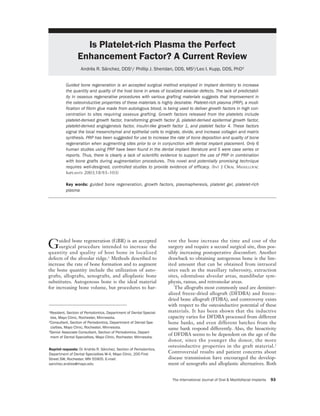
- 5. SÁNCHEZ ET AL Fig 1 Preparation of PRP with general-purpose centrifuges. Platelet-poor plasma (PPP) One unit 5,600 rpm autologous Red blood whole blood cells Erythrocytes 2,400 rpm + buffy coat Platelet-rich plasma (PRP) PLATELET-RICH PLASMA PROCUREMENT platelet-concentrating cell separators are presently TECHNIQUES commercially available and FDA-approved for the production of PRP: the Harvest SmartPrep Platelet Use of General-Purpose Cell Separators Concentrate System (HSPCS) (Harvest Technolo- The clinical procedure for obtaining autologous gies, Plymouth, MA) and the 3i Platelet Concen- PRP requires the use of an ultracentrifuge and gradi- trate Collection System (3i PCCS) (3i/Implant ent density cell separator, which can sequester and Innovations, Palm Beach Gardens, FL). HSPCS concentrate platelets.27 Approximately 450 mL of and 3i PCCS use tailored centrifuge containers to whole blood is drawn from the patient directly into a manipulate the blood cells to achieve the separation standard labeled blood collection bag containing cit- and sequestration of platelets. Similar quantities of rate-phosphate-dextrose anticoagulant.6 The use of fresh whole blood are required in both units. Both EDTA as an anticoagulant is not recommended dur- systems consist of a desktop centrifuge and individ- ing the PRP procurement because it fragments the ual disposable kits that are designated to come into platelets, 29 although it gives a greater yield of contact with blood. Each system uses centrifugal platelets.30 First, the blood is centrifuged in a gen- forces to separate blood cells through centrifuge eral-purpose cell separator, such as the ELMD-500 cycles with short- and long-duration spins. After the (Medtronic Electromedic, Autotransfusion System, first spin (short), HSPCS automatically decants the Parker, CO) at 5,600 rpm to separate the platelet- plasma into the plasma chamber, where during the poor plasma (PPP) from the red blood cells (RBC) second spin (long), the PPP is separated from a and the PRP (also termed “buffy coat,” which con- platelet concentrate. After the first spin in the 3i tains the platelets and the leukocytes). Then the cen- PCCS system, the operator manipulates a valve and trifuge speed is slowed to 2,400 rpm to obtain fur- inserts air into an air bladder, forcing the plasma ther separation of approximately 30 mL of PRP from into the second chamber. After the second spin, the the RBC preparation into a transfer bag (Fig 1). operator forces air into a bladder that separates the The procurement of PRP with this technique can PPP from the PRP. Some performance and opera- be accomplished in 30 minutes, and use of the tional data provided from manufacturers are obtained PRP is recommended within 6 hours after detailed in Table 2. Recently, 2 methods of PRP being drawn from the patient. Platelet counts of procurement, the 3i PCCS system and the Curasan- 500,000 to 1,000,000 in the PRP are usually obtained type PRP kit (Curasan, Kleinostheim, Germany), with this plasmapheresis technique. With this pro- were studied to compare the growth factors levels cessing technique, the remaining erythrocytes and obtained with the different techniques. The authors PPP can be returned to the circulation or discarded. concluded that the PRP obtained with the 3i PCCS system had both a higher platelet count and a Use of Platelet-concentrating Cell Separators higher content of the growth factors investigated.32 Recent advanced technologies permit the procure- In summary, both platelet-concentrating cell sep- ment of PRP using smaller volumes of blood, arators are similar in performance and simplicity, and increasing the platelet concentrations and avoiding they represent a significant advance over the general- the need for RBC and PPP reinfusion. 31 Two purpose cell separators. The noticeable difference The International Journal of Oral & Maxillofacial Implants 97 COPYRIGHT © 2002 BY QUINTESSENCE PUBLISHING CO, INC. PRINTING OF THIS DOCUMENT IS RESTRICTED TO PERSONAL USE ONLY. NO PART OF THIS ARTICLE MAY BE REPRODUCED OR TRANSMITTED IN ANY FORM WITHOUT WRITTEN PERMISSION FROM THE PUBLISHER.
- 6. SÁNCHEZ ET AL Table 2 Comparison Data of 2 Commercially Based on the reported coagulopathies, Landes- Available Platelet-Concentrating Cell berg and coworkers 33 suggested that alternative Separators methods of activating PRP need to be studied and made available to the dental community. Performance data* HSPCS 3i PCCS In a later study, 30 Landesberg and coworkers Average % recovery of platelets 68% 65% described a new method to activate PRP gel with Average platelet count/µL in 4 mL 2,278,000 1,818,000 the ITA gelling agent (Natrex Technologies, Average platelet count/µL in 5 mL 1,823,000 1,454,000 Greenville, NC). They stated that this method Average platelet count/µL in 6 mL 1,519,000 1,212,000 Operational time 15 min 20 min could be used more safely as an alternative to bovine thrombin for gelling the PRP; however, they *All data taken from manufacturer brochures. HSPCS = Harvest SmartPrep Platelet Concentrate System, Harvest did not describe the specific composition and mech- Technologies, Plymouth, MA; 3i PCCS = 3i Platelet Concentrate Col- anism of action of ITA. Marx has used bovine lection System, Implant Innovations, Palm Beach Gardens, FL. thrombin in his patients since 1996 without devel- opment of any coagulopathies; however, he indi- cated that the use of reagents such as recombinant human thrombin or autologous thrombin can avoid the risk or concern about the potential of bovine thrombin to cause coagulopathies. 29 In contrast, between the 2 systems is that the HSPCS requires Kassolis and colleagues27 utilized autologous throm- less time to produce the PRP (15 minutes versus 20 bin to activate the PRP in their patients. minutes) and less operator intervention and training. Based on these data, apparently other methods should be considered in the preparation of PRP gel. Safer methods to consider could include the utiliza- POTENTIAL RISKS WITH THE USE OF PRP tion of recombinant human thrombin, autologous thrombin, or perhaps extra-purified thrombin. The preparation of PRP involves isolation of the PRP, after which gel formation is accelerated using calcium chloride and topical bovine thrombin PRP PREPARATION TECHNIQUE (TBT).6 The use of TBT has been reported to be associated with the development of antibodies to The PRP application described by most authors factors V and XI and thrombin, resulting in the risk requires initiating the coagulation process with a of life-threatening coagulopathies. Landesberg and mixture of 1,000 US units of TBT powder sus- coworkers33 reviewed reports appearing in the liter- pended in 10 mL of sterile saline with 10% calcium ature of serious coagulopathies that were difficult to chloride. The protocol for PRP activation requires treat following exposure to TBT.34–36 TBT prepara- the use of an individual 10-mL syringe for each mix. tions contain factor V, which results in reaction of Each mix draws 6 mL of PRP, 1 mL of the 10% cal- the immune system when challenged with a foreign cium chloride, 1 mL of thrombin, and 1 mL of air protein. The factor V deficiency after thrombin to act as a mixing bubble. The syringe is agitated exposure is thought to be caused by the cross-reac- for a few seconds to initiate clotting and is then tivity of anti-bovine factor V antibodies with human ready to be applied to the bone grafts. Once added factor V.34–37 To date, TBT-induced coagulopathies to the grafts, the fibrin, fibronectin, and other cell have been reported in 32 cases of patients undergo- adhesion molecules establish a network that may ing cardiovascular operations, specifically in those serve as osteoconductors in bone growth.39 patients with repeat exposure to TBT.34 The sever- ity of bleeding varies widely, from no clinical evi- dence to life-threatening diatheses developing 7 to THERAPEUTIC POTENTIAL OF PRP GEL IN 14 days after successive exposure to TBT. IMPLANT DENTISTRY However, the adverse reactions reported could depend upon an increased awareness of the coagu- PRP has been recommended for use in increasing the lopathy, as well as the source and quantity of throm- rate of bone deposition and quality of bone regenera- bin used. Differences in product purity have been tion when augmenting edentulous sites for future documented. One brand of thrombin (Thrombin- implant placement.20,26,27,31 Most reports on PRP in JMI, Jones Medical Industries, St Louis, MO) the literature have proposed its utilization as a bone applies an extra purification step to decrease the fac- graft enhancement material rich in growth media- tor V concentration to less than 0.2 µg/mL.38 tors.5,6,13,30 In this context, PRP may be valuable for 98 Volume 18, Number 1, 2003 COPYRIGHT © 2002 BY QUINTESSENCE PUBLISHING CO, INC. PRINTING OF THIS DOCUMENT IS RESTRICTED TO PERSONAL USE ONLY. NO PART OF THIS ARTICLE MAY BE REPRODUCED OR TRANSMITTED IN ANY FORM WITHOUT WRITTEN PERMISSION FROM THE PUBLISHER.
- 7. SÁNCHEZ ET AL Table 3 Summary of Human Studies Involving the Use of PRP in Combination with Bone Grafts Study Random- Defect Authors type Patients Groups Control ization Length type Graft Biopsy Marx et al5 Prospective 88 44 PRP+graft/ Yes Questionable 4 months Mandibular Autologous Yes 44 graft discontinuity Anitua26 Case series 20 10 PRP, 10 control Yes Questionable 6 months Postextraction None/ Yes Autologous* 3 3 split-mouth Yes No Same Same None Yes Kassolis et al27 Case series 15 No No 12 months Ridge and SA FDBA Yes Rosenberg and Case report 1 No No 9 months SA Alloplastic No Torosian28 Shanamen Case reports 3 No No No RA 1 DFDBA, Yes et al45 1 alloplastic/ DFDBA/ autograft, 1 DFDBA Froum et al46 Case reports 3 Split mouth Yes Questionable 7–9 months SA ABB Yes *In 5 patients in the PRP group. PRP = platelet-rich plasma; SA = sinus augmentation; RA = ridge augmentation; DFDBA = demineralized freeze-dried bone allograft; FDBA = freeze-dried bone allograft; ABB = anorganic bovine bone. use in conjunction with bone autografts, allografts, were case reports (Table 3). Only 3 included con- xenografts, or alloplasts in sinus lifting and in alveo- trols and randomization, but the experimental lar ridge augmentation procedures prior to or in con- design was inadequate. junction with dental implant placement. PRP has Marx and coworkers 5 performed the first and also been recommended for use alone or in combina- most compelling study available on the use of PRP tion with bone grafts and barrier membranes in the in combination with bone grafts. They evaluated treatment of peri-implant defects created as a conse- the effect of PRP on bone graft reconstructions of quence of immediate implant placement or as a result mandibular continuity defects 5 cm or greater aris- of peri-implantitis. ing from tumor extirpations. One group received PRP is thought to accelerate soft tissue healing cancellous cellular marrow grafts with the addition by promoting a more rapid revascularization and re- of PRP, and the other group received a cancellous epithelialization of flaps and cell proliferation. cellular marrow graft alone, serving as the positive Therefore, there may be merit in applying it to the control. In this study, a complete description of the flap margins and to the underlying tissues. Garg technique for procurement and application of PRP and coworkers40 proposed that resorbable barrier was given. The grafts were allowed to consolidate membrane materials be infused with PRP. They and mature for 6 months, and panoramic radio- have proposed that this PRP-based membrane graphs were obtained at 2, 4, and 6 months. These could serve as a short-acting biologic barrier, since radiographs were used to evaluate the age (maturity) all platelets contained in PRP will degranulate of the graft at each interval. From this subjective within 3 to 5 days, and their initial growth activity radiographic assessment, the authors calculated a expires within 10 days. graft maturity index but they did not provide an Nine articles were found documenting the clini- explanation of this index. From the results of this cal use of PRP for humans, but 3 were excluded; 2 questionable index, the authors claimed that the of these involved a case report and a clinical re- bone grafts combined with PRP showed a maturity entry study on the use of PRP in intraosseous peri- index more than twice and slightly less than twice odontal defects,41,42 and the other study was a case the actual maturity at 2 and 4 months, respectively. report on a new technique to expand the alveolar Marx and coworkers5 also performed a mono- ridge using a piezoelectric scalpel.43 In the latter clonal antibody study on the platelets sequestered case, the author did use PRP, but without any com- by the centrifugation process and on the harvested ment on the PRP performance, and focused primar- bone graft material. The data indicated that PDGF ily on the performance of the new device.43 Of the 6 and TGF- from the platelets had been absorbed articles reviewed, only 1 had a prospective design, by the graft, and receptors for PDGF and TGF- and of the 5 remaining, 2 were case series and 3 were present within the autogenous grafts. Similar The International Journal of Oral & Maxillofacial Implants 99 COPYRIGHT © 2002 BY QUINTESSENCE PUBLISHING CO, INC. PRINTING OF THIS DOCUMENT IS RESTRICTED TO PERSONAL USE ONLY. NO PART OF THIS ARTICLE MAY BE REPRODUCED OR TRANSMITTED IN ANY FORM WITHOUT WRITTEN PERMISSION FROM THE PUBLISHER.
- 8. SÁNCHEZ ET AL analyses of core bone specimens of each graft at 6 with better organized trabeculae and greater bone months indicated that TGF- was still present, but regeneration. In all patients treated with PRP, the PDGF was not identified. Marx and coworkers also epithelialization was described based on subjective compared the baseline mean platelet counts with observations as very good to excellent. PRP mean platelet counts and found an increase of Kassolis and colleagues27 presented case reports 338% in the platelet count because of the sequestra- of 15 patients undergoing ridge and sinus floor aug- tion process. The authors also performed a histo- mentation treated with PRP and FDBA. Seventeen morphometry graft assessment at 6 months. A core areas, including 14 sinuses and 3 maxillary ridges, bone biopsy was taken from each grafted site (bone were treated and 36 endosseous implants were graft and bone graft with PRP). These bone speci- placed. Twenty-nine implants were placed at the mens from grafted sites were compared to 10 resec- time of the GBR procedure. Thirty-two of the 36 tion specimens retrieved from the midbody of the implants (89%) were successful and 4 implants were mandible (native bone). The trabecular bone area determined to have failed at the time of uncovering. (TBA) was measured in the 3 groups; a TBA of 38.9 Histologic study of bone retrieved from 2 patients ± 6% was seen in the native bone group, 55.1 ± 8% confirmed the presence of vital bone formation in in the bone graft group, and 74.0 ± 11% in the bone apposition to residual FDBA particles. The authors graft/PRP group. They concluded that the addition suggested that the use of PRP may allow for earlier of PRP accelerated the rate and degree of bone for- implant placement and loading, but in the absence mation and that bone autografts contained platelets of a control group and an adequate study design this and thus were positive for receptors for the growth conclusion is unacceptable. factors. Furthermore, it was noted that the PRP was Rosenberg and Torosian28 presented a case report an autologous preparation obtained at the time of concerned with the use of PRP in combination with the surgery, thus eliminating concerns about disease an alloplastic graft in a sinus floor augmentation transmission, immunogenic reactions, and mislabel- procedure. A 70-year-old male patient underwent a ing of the sample. It was reported that the subjects sinus grafting procedure using PRP in addition to were randomized into 2 groups; however, no expla- an alloplastic bone particulate graft. In this case, the nation of the randomization procedure was given patient was scheduled at 3 months for implant and therefore it is questionable. Current standards placement. One month after implant placement, a of experimental design expect that the method used 4-unit provisional restoration was placed and 5 for randomization be reported.44 months later the definitive prosthesis was con- Anitua26 reported the results of the use of PRP in nected. Based on this case alone, the authors erro- a series of patients who underwent tooth extraction neously concluded that the duration of the treat- because of root fracture or periodontitis. Ten ment was reduced by one half with the use of PRP. patients were treated with PRP. In 5 of these It was not appropriate to draw conclusions regard- patients, autogenous bone was used in combination ing a novel treatment based on the clinical results of with PRP to prevent soft tissue collapse. The the treatment of a single patient because of the high remaining 10 patients served as controls; the extrac- variability among patients and lack of appropriate tion sockets were left to heal without application of study design. PRP. In the same study, another 3 patients, who Recently Shanaman and coworkers45 published 3 needed multiple extractions on both sides of the case reports. Patient 1 underwent GBR prior to mouth, were assigned in a split-mouth fashion to implant placement using e-PTFE plus DFDBA/ PRP and control treatment modalities alternatively. PRP. In patient 2, GBR was accomplished using e- Biopsies of the sites in all 23 patients were taken PTFE plus alloplastic/DFDBA/autogenous bone in between 10 and 16 weeks. Anitua reported total combination with PRP. Patient 3 underwent GBR regeneration of bone in 8 of the 10 patients based using bioabsorbable membrane plus a PRP/DFDBA on periodontal probing, and biopsies of these sites combination. The histologic observations demon- showed compact mature bone with well-organized strated new bone formation, and the authors stated trabeculae and normal morphology. In 2 patients in that the PRP did not appear to enhance the quality the PRP group, partially regenerated bone was of newly formed bone. The authors concluded from found with connective tissue and non-organized the results that PRP used in conjunction with vari- trabeculae. The 10 patients in the control group ous derivatives/substitutes and a barrier membrane showed connective tissue filling the main part of the can support new bone formation in localized ridge defect, and no mature bone was found. In the 3 augmentation. However, they indicated that the patients assigned to the split-mouth design, the sites results in these 3 patients appeared comparable to treated with PRP demonstrated more mature bone, other GBR studies without the use of PRP, but this 100 Volume 18, Number 1, 2003 COPYRIGHT © 2002 BY QUINTESSENCE PUBLISHING CO, INC. PRINTING OF THIS DOCUMENT IS RESTRICTED TO PERSONAL USE ONLY. NO PART OF THIS ARTICLE MAY BE REPRODUCED OR TRANSMITTED IN ANY FORM WITHOUT WRITTEN PERMISSION FROM THE PUBLISHER.
- 9. SÁNCHEZ ET AL conclusion is also null because of the inappropriate • Growth factors released from platelets seem to experimental design. signal the local mesenchymal cells and epithelial More recently, Froum and associates46 presented cells to migrate, divide, and increase collagen and reports of 3 patients undergoing sinus floor aug- matrix formation. mentation treated with PRP + anorganic bovine • PRP is an autologous preparation; thus it elimi- bone (ABB). On the day of surgery, a flip of a coin, nates concerns about disease transmission or an unacceptable method for randomization, deter- immunogenic reaction. mined the experimental side (PRP+ABB) and the • PRP gel seems to improve the handling charac- control side (ABB). In patient 3, miniature test teristics of grafts. implants, 2.0 mm in diameter and 10 mm in length, • Some of the limited number of studies in the were placed through the crestal bone into the sinus dental literature suggest some benefit when PRP grafts. Placement of the permanent implants was is combined with autogenous bone. Specifically, performed at 7 months, 7.5 months, and 11 months, PRP seems to improve the rate of bone forma- respectively, for patients 1, 2, and 3. At the time of tion and the quality of bone formed. implant placement, trephine cores 3 mm in diame- • To avoid possible development of coagulopathies ter and 10 mm in length were harvested from the associated with bovine thrombin, the use of an former site of the lateral window. In addition, the 3 alternative method for PRP activation, other test implants were removed with a trephine and the than TBT, is advisable. cores were sent for histologic and histomorphomet- • Recent advanced technologies permit the pro- ric evaluation. The histomorphometric analysis of curement of PRP using smaller volumes of vital bone in the grafted sinuses demonstrated simi- blood, increasing the platelet concentrations and lar values in the percentages of vital bone between avoiding the need for RBC and PPP reinfusion. the experimental and control groups. The 2 test • Longitudinal studies are needed to determine implants placed in the experimental group sinuses whether the addition of PRP to bone substitutes showed slightly higher percentages of bone-implant would allow earlier implant placement and load- contact (37.6% and 38.8%) than the test implant ing and increase the predictability of regenera- placed in the contralateral control sinus (33.8%). tive procedures. Therefore, it was concluded from these case reports that PRP did not make a significant difference in the production of vital bone or in the bone contact at SUMMARY the implant interface. These conclusions are also apparently invalid because of the lack of appropriate This literature review demonstrates a lack of scien- study design. tific evidence to support the current use of PRP in All the available literature reviewed on PRP con- combination with bone grafts during augmentation sisted of case series and individual reports, which in procedures. Most of the evidence on the clinical some instances involved a quasi-experimental potential of PRP comes from case series and case design. In some instances, the authors used subjec- reports. While it has been recognized that these tive indices to measure the ability of PRP to types of studies represent the starting point, they increase the bone density5 or reached conclusions are not definitive. It has been suggested that this based on subjective observations.26–28,44 Because of treatment-observation-description strategy is at the the absence of adequate controls and because the lowest level in the hierarchy of evidence. The need subjects were not selected randomly, the experimen- for evidence-based clinical decisions regarding tal designs were flawed. treatment recommendations and alternatives is quite apparent. PRP may have strong clinical potential associated with the growth factors it con- CONCLUSIONS tains, but there is a need for well-controlled ran- domized clinical studies to assess the ideal concen- Several conclusions can be drawn from this review. tration of the different growth factors and to determine whether PRP application to different • The platelet sequestration process results in a bone graft substitutes and different barrier mem- high platelet concentrate that upon activation branes provides a beneficial effect. Well-designed releases a cascade of growth factors contained in controlled studies are necessary to determine the the alpha granules. efficacy of the technique. The International Journal of Oral & Maxillofacial Implants 101 COPYRIGHT © 2002 BY QUINTESSENCE PUBLISHING CO, INC. PRINTING OF THIS DOCUMENT IS RESTRICTED TO PERSONAL USE ONLY. NO PART OF THIS ARTICLE MAY BE REPRODUCED OR TRANSMITTED IN ANY FORM WITHOUT WRITTEN PERMISSION FROM THE PUBLISHER.
- 10. SÁNCHEZ ET AL REFERENCES 21. Canalis E, McCarthy TL, Centrella M. Effects of platelet- derived growth factor on bone formation in vitro. J Cell 1. Hammerle CH, Karring T. Guided bone regeneration at Physiol 1989;140:530–537. oral implant sites. Periodontology 2000 1998;17:151–175. 22. Stephan EB, Renjen R, Lynch SE, Dziak R. Platelet-derived 2. Schwartz Z, Somers A, Mellonig JT, et al. Ability of com- growth factor enhancement of a mineral-collagen bone sub- mercial demineralized freeze-dried bone allograft to induce stitute. J Periodontol 2000;71:1887–1892. new bone formation is dependent on donor age but not gen- 23. Lynch SE, Ruiz de Castilla G, Williams RC, et al. The der. J Periodontol 1998;69:470–478. effects of short-term application of a combination of 3. Gibble J, Ness P. Fibrin glue: The perfect operative sealant? platelet-derived and insulin-like growth factors on periodon- Transfusion 1990;30:741–747. tal wound healing. J Periodontol 1991;62:458–467. 4. Hood AG, Hill AG, Reeder GD. Perioperative autologous 24. Lynch SE, Buser D, Hernandez RA, Weber HP, Sitch H, sequestration. III: A new physiologic glue with wound heal- Fox CH, Williams RC. Effects of PDGF/IGF-1 combina- ing properties. Proc Am Acad Cardiovasc Perfusion 1993;14: tion on bone regeneration around titanium dental implants. 126–130. Results of a pilot study on beagle dogs. J Periodontol 1991; 5. Marx RE, Carlson ER, Eichstaedt RN, Schimmele SR, 62:710–716. Strauss JE, Georgeff KR. Platelet-rich plasma: Growth fac- 25. Stefani CM, Machado MA, Sallum EA, Sallum AW, Toledo tor enhancement for bone grafts. Oral Surg Oral Med Oral S, Nociti FH. Platelet-derived growth factor/insulin-like Pathol Oral Radiol Endod 1998;85:638–646. growth factor-1 around implants placed into extraction sock- 6. Whitman DH, Berry RL, Green DM. Platelet gel: An autol- ets: A histometric study in dogs. Implant Dent 2000;9: ogous alternative to fibrin glue with applications in oral and 126–132. maxillofacial surgery. J Oral Maxillofac Surg 1997;55: 26. Anitua E. Plasma rich in growth factors: Preliminary results 1294–1299. of use in the preparation of future sites for implants. Int J 7. Hallmon WW, Carranza FA, Drisko CL, Rapley JW. Peri- Oral Maxillofac Implants1999;14:529–535. odontal Literature Reviews: A summary of current knowl- 27. Kassolis JD, Rosen PS, Reynolds MA. Alveolar ridge and edge. Amer Acad Periodontol 1996;11(9):189–194. sinus augmentation utilizing platelet-rich plasma in combi- 8. Ganio C, Tenewitz FE, Wilson RC, Maules BG. The treat- nation with freeze-dried bone allograft: Case series. J Perio- ment of chronic nonhealing wounds using autologous dontol 2000;71:1654–1661. platelet-derived growth factors. J Foot Ankle Surg 1993;32: 28. Rosenberg ES, Torosian J. Sinus grafting using platelet-rich 263–268. plasma—Initial case presentation. Pract Periodontics Aes- 9. Wirthlin MR. Growth substances: Potential use in perio- thet Dent 2000;12:843–850. dontics. J West Soc Periodontol Periodontal Abstr 1989;37: 29. Marx RE. Quantification of growth factor levels using a sim- 101–125. plified method of platelet-rich plasma preparation [discus- 10. Rudkin GH, Miller TA. Growth factors in surgery: Review. J sion]. J Oral Maxillofac Surg 2000;58:300–301. Plastic Reconstr Surg 1996;97:469–476. 30. Landesberg R, Roy M, Glickman RS. Quantification of 11. Pierce GF, Mustoe TA, Atrock BW, Deuel TF, Tomason A. growth factors levels using a simplified method of platelet- Role of platelet-derived growth factor in wound healing. J rich plasma gel preparation. J Oral Maxillofac Surg 2000; Cell Biochem 1991;45:319–326. 58:297–300. 12. Roberts AB. Transforming growth factor- . In: Canalis E 31. Lozada JL, Caplanis N, Proussaefs P, Willardsen J, Kam- (ed). Skeletal Growth Factors. Baltimore: Lippincott, meyer G. Platelet-rich plasma application in sinus graft Williams & Wilkins, 2000:221–249. surgery: Part I—Background and processing techniques. J 13. Carlson ER. Bone grafting the jaws in the 21st century: The Oral Implantol 2001;27:38–42. use of platelet-rich plasma and bone morphogenetic protein. 32. Weibrich G, Kleis WKG, Hafner G. Growth factors levels Alpha Omegan 2000;93:26–30. in the platelet-rich plasma produced by 2 different methods: 14. Pierce GF, Mustoe TA, Deuel TF. Transforming growth fac- Curasan-type PRP kit versus PCCS PRP system. Int J Oral tor- induces increased directed cellular migration and tis- Maxillofac Implants 2002;17:184–190. sue repair in rats. In: Barbul A, et al (eds). Growth Factors 33. Landesberg R, Moses M, Karpatkin M. Risk of using platelet- and Other Aspects of Wound Healing: Biological and Clini- rich plasma gel. J Oral Maxillofac Surg 1998;56:1116–1117. cal Implications. New York: Alan R. Liss, 1988. 34. Cmolik BL, Spero JA, Magovern GJ. Redo cardiac surgery: 15. Sporn MB, Roberts AB. Peptide growth factors are multi- Late bleeding complications from topical thrombin-induced functional. Nature 1988;332:217–219. factor five deficiency. J Thorac Cardiovasc Surg 1993;105: 16. Folkman J, Klagsbrun M. Angiogenic factors. Science 1987; 222–228. 233:442–447. 35. Muntean W, Zenz W, Finding K. Inhibitor to factor V after 17. Cohen S. Isolation of submaxillary gland protein accelerat- exposure to fibrin sealant during cardiac surgery in a two- ing incisor eruption and eyelid opening in the newborn ani- year-old child. Acta Paediatr 1994;83:84–87. mal. J Biol Chem 1962;237:1555–1559. 36. Spero JA. Bovine thrombin-induced inhibitor of factor V 18. Hom DB, Maisel RH. Angiogenic growth factors: Their and bleeding risk in postoperative neurosurgical patient. A effects and potential in soft tissue wound healing. Ann Otol report of three cases. J Neurosurg 1993;78:817–820. Rhinol Laryngol 1992;101:349–354. 37. Knobl P, Lechner K. Acquired factor V inhibitors. A review. 19. Knighton D, Doucette M, Fiegel VD, Ciresi K, Butler E, Baillieres Clin Hematol 1998;11:305–318. Austin L. The use of platelet-derived wound healing formula 38. Christie RJ, Carrington L, Alving B. Postoperative bleeding in human clinical trials. Prog Clin Biol Res 1988;266: induced by topical bovine thrombin: Report of two cases. 319–329. Surg 1997;121:708–710. 20. Garg AK. The use of platelet-rich plasma to enhance the 39. Marx RE. Biology of PRP [reply/letter to the editor]. J Oral success of bone grafts around dental implants. Dental Maxillofac Surg 2001;59:1120–1121. Implantol Update 2000;11:1–5. 102 Volume 18, Number 1, 2003 COPYRIGHT © 2002 BY QUINTESSENCE PUBLISHING CO, INC. PRINTING OF THIS DOCUMENT IS RESTRICTED TO PERSONAL USE ONLY. NO PART OF THIS ARTICLE MAY BE REPRODUCED OR TRANSMITTED IN ANY FORM WITHOUT WRITTEN PERMISSION FROM THE PUBLISHER.
- 11. SÁNCHEZ ET AL 40. Garg AK, Gargenese D, Peace I. Using platelet-rich plasma 43. Vercelotti T. Piezoelectric surgery in implantology. A case to develop an autologous membrane for growth factor deliv- report—New piezoelectric ridge expansion technique. Int J ery in dental implant therapy. Dent Implantol Update 2000; Periodontics Restorative Dent 2000;20:359–365. 11:1–4. 44. Brunette DM. Introduction. In: Brunette DM. Critical 41. Obarrio JJ, Arauz-Dutari JI, Chamberlain TM, Croston A. Thinking: Understanding and Evaluating Dental Research. The use of autologous growth factors in periodontal surgical Chicago: Quintessence, 1996:1–12. therapy: Platelet gel biotechnology—Case reports. Int J 45. Shanaman R, Filstein MR, Danesh-Meyer MJ. Localized Periodontics Restorative Dent 2000;20:487–497. ridge augmentation using GBR and platelet-rich plasma: 42. Lekovic V, Camargo PM, Weinlaender M, Vasilic N, Ken- Case reports. Int J Periodontics Restorative Dent 2001;21: ney EB. Comparison of platelet-rich plasma, bovine porous 345–355. bone mineral, and guided tissue regeneration versus platelet- 46. Froum SJ, Wallace SS, Tarnow DP, Cho SC. Effect of rich plasma and bovine porous bone mineral in the treat- platelet-rich plasma on bone growth and osseointegration in ment of intrabony defects: A reentry study. J Periodontol human maxillary sinus grafts: Three bilateral case reports. 2002;73:198–205. Int J Periodontics Restorative Dent 2002;22:45–53. The International Journal of Oral & Maxillofacial Implants 103 COPYRIGHT © 2002 BY QUINTESSENCE PUBLISHING CO, INC. PRINTING OF THIS DOCUMENT IS RESTRICTED TO PERSONAL USE ONLY. NO PART OF THIS ARTICLE MAY BE REPRODUCED OR TRANSMITTED IN ANY FORM WITHOUT WRITTEN PERMISSION FROM THE PUBLISHER.